



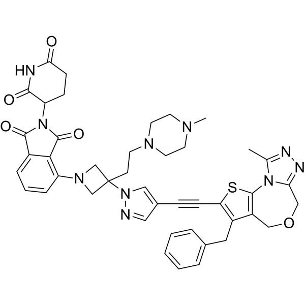
BD-9136

Based on 1 Customer Validation
BD-9136 is a selective BRD4 PROTAC degrader with a DC50 of 1.2 nM, and exhibits a selectivity of ≥1000-fold over BRD2 and BRD3. BD-9136 preferentially forms a ternary complex with the BD1 domain of BRD4, and downregulates the expression of B7-H4 by disrupting the PR-P300-BRD4 axis. BD-9136 depletes BRD4 protein in tumor tissues, inhibits tumor growth, reduces B7-H4 protein expression, increases CD8+ T cell infiltration, and enhances tumor sensitivity to anti-PD-L1. Degradation of BRD4 by BD-9136 rescues the erythroid differentiation block induced by LSD1 inhibition, and transient administration restores erythroid output while retaining HbF induction. BD-9136 causes no adverse effects in mice at effective doses. BD-9136 can be used in studies related to acute myeloid leukemia, acute lymphoblastic leukemia and breast cancer.
(Pink: BRD4 ligand (HY-44103); Blue: Cereblon ligand (HY-103596); Black: linker (HY-168692)).
For research use only. We do not sell to patients.
- Purity : 98.81%
- CAS No.: 3037514-38-5
- Formula: C44H44N10O5S
- Molecular Weight:824.95
-
Storage:
-20°C, protect from light, stored under nitrogen
* In solvent : -80°C, 6 months; -20°C, 1 month (protect from light, stored under nitrogen)

To place orders, for customer services and technical support, please contact: MedChemExpress USA
Tel: 609-228-6898 E-mail: [email protected] [email protected]


-
 Biological Activity
-
Chemical Information
-
Solvent & Solubility
- Protocols
- Purity & Documentation
- References
-
 Help & FAQs


All PROTACs Isoforms
More

Biological Activity
Description
|
BRD4 (BD1) 1.2 nM (DC50) |
B7-H4 |
Cellular Effect
|
Cell Line

|
Type | Value | Description | References |
|---|---|---|---|---|
| HL-60 | IC50 |
5.1 nM
Compound: 8; BD-9136
|
Antiproliferative activity against human HL-60 cells assessed as cell growth inhibition incubated for 4 days by WST-8 assay
Antiproliferative activity against human HL-60 cells assessed as cell growth inhibition incubated for 4 days by WST-8 assay
|
[PMID: 37289649] |
| MCF7 | IC50 |
38.4 nM
Compound: 8; BD-9136
|
Antiproliferative activity against human MCF7 cells assessed as cell growth inhibition incubated for 4 days by WST-8 assay
Antiproliferative activity against human MCF7 cells assessed as cell growth inhibition incubated for 4 days by WST-8 assay
|
[PMID: 37289649] |
| MDA-MB-231 | IC50 |
17.6 nM
Compound: 8; BD-9136
|
Antiproliferative activity against human MDA-MB-231 cells assessed as cell growth inhibition incubated for 4 days by WST-8 assay
Antiproliferative activity against human MDA-MB-231 cells assessed as cell growth inhibition incubated for 4 days by WST-8 assay
|
[PMID: 37289649] |
| MDA-MB-453 | IC50 |
80.9 nM
Compound: 8; BD-9136
|
Antiproliferative activity against human MDA-MB-453 cells assessed as cell growth inhibition incubated for 4 days by WST-8 assay
Antiproliferative activity against human MDA-MB-453 cells assessed as cell growth inhibition incubated for 4 days by WST-8 assay
|
[PMID: 37289649] |
| MOLM-13 | IC50 |
69 nM
Compound: 8; BD-9136
|
Antiproliferative activity against human MOLM-13 cells assessed as cell growth inhibition incubated for 4 days by WST-8 assay
Antiproliferative activity against human MOLM-13 cells assessed as cell growth inhibition incubated for 4 days by WST-8 assay
|
[PMID: 37289649] |
| MV4-11 | IC50 |
11 nM
Compound: 8; BD-9136
|
Antiproliferative activity against human MV4-11 cells assessed as cell growth inhibition incubated for 4 days by WST-8 assay
Antiproliferative activity against human MV4-11 cells assessed as cell growth inhibition incubated for 4 days by WST-8 assay
|
[PMID: 37289649] |
| RS4-11 | IC50 |
4.6 nM
Compound: 8; BD-9136
|
Antiproliferative activity against human RS4-11 cells assessed as cell growth inhibition incubated for 4 days by WST-8 assay
Antiproliferative activity against human RS4-11 cells assessed as cell growth inhibition incubated for 4 days by WST-8 assay
|
[PMID: 37289649] |
| T47D | IC50 |
3.8 nM
Compound: 8; BD-9136
|
Antiproliferative activity against human T47D cells assessed as cell growth inhibition incubated for 4 days by WST-8 assay
Antiproliferative activity against human T47D cells assessed as cell growth inhibition incubated for 4 days by WST-8 assay
|
[PMID: 37289649] |
In Vitro
BD-9136 (0.1-1000 nM; 4 h) efficiently and selectively degrades BRD4 in MDA-MB-231, MDA-MB-453, MCF-7 and T47D human breast cancer cell lines, with a selectivity of ≥1000-fold over BRD2 and BRD3, and DC50 values for BRD4 ranging from <0.1 to 0.6 nM[1].
BD-9136 (for 4 days) potently inhibits the growth of human cancer cell lines MV4;11, MOLM13, HL60, RS4;11, MDA-MB-231, MDA-MB-453, MCF-7 and T47D, with IC50 values ranging from 3.8 to 80.9 nM[1].
Among more than 5700 proteins analyzed in MV4;11 and MDA-MB-231 human cancer cells treated with BD-9136 (30 nM; 3 h), only BRD4 is selectively degraded[1].
BD-9136 (3-10 nM; 7-18 days) dose-dependently reverses the erythroid differentiation arrest induced by CCG-385349 in human CD34+ HSPCs via degrading BRD4 (DC50 = 1.2 nM) and attenuating the induction of RUNX1/PU.1; in contrast, transient administration preserves the fetal hemoglobin induction mediated by CCG-385349 and restores mature erythrocyte production[2].
BD-9136 (100-1000 nM; 48 h) dose-dependently downregulates the expression of B7-H4 in human T-47D breast cancer cells[3].
MedChemExpress (MCE) has not independently confirmed the accuracy of these methods. They are for reference only.
-
Cell Line:MV4;11, MOLM13, HL60, RS4;11
-
Concentration:0, 0.1, 0.3, 1, 3,10, 30, 100, 300, 1000 nM
-
Incubation Time:4 h
-
Result:Induced BRD4 degradation with DC50 values of 4.7 nM (MV4;11, D_max = 96%), 1.5 nM (MOLM13, D_max = 90%), 0.5 nM (HL60, D_max = 99%), and 0.7 nM (RS4;11, D_max = 99%).
Had no significant effect on BRD2 or BRD3 levels at concentrations up to 1000 nM, with DC50 values >1000 nM and D_max values ≤18% for both proteins across all cell lines.
-
Cell Line:MDA-MB-231, MDA-MB-453, MCF-7, T47D
-
Concentration:0, 0.1, 0.3, 1, 3,10, 30, 100, 300, 1000 nM
-
Incubation Time:4 h
-
Result:Induced BRD4 degradation with DC50 values of 0.5 nM (MDA-MB-231, D_max = 95%), 0.6 nM (MDA-MB-453, D_max = 99%), <0.1 nM (MCF-7, D_max = 99%), and 0.2 nM (T47D, D_max = 99%).
Had no significant effect on BRD2 or BRD3 levels at concentrations up to 1000 nM, with DC50 values >1000 nM and D_max values ≤25% for both proteins across all cell lines.
-
Cell Line:T-47D
-
Concentration:0, 100, 200, 1000
-
Incubation Time:48 h
-
Result:Effectively reduced B7-H4 expression in a dose-dependent meffectively reduced B7-H4 expression in a dose-dependent manneranner
In Vivo
BD-9136 (20 mg/kg; i.p.; single administration) achieves sustained BRD4 protein depletion for at least 48 hours in SCID mice bearing MV4;11 or MDA-MB-231 xenografts, with no significant effects on BRD2 and BRD3 proteins[1].
MedChemExpress (MCE) has not independently confirmed the accuracy of these methods. They are for reference only.
-
Animal Model:SCID mice (8-12 weeks, female) were subcutaneously implanted with 5 × 10⁶ MV4;11 cells in 50% Matrigel/PBS. Treatment was initiated when tumors reached 80-200 mm3[1]
-
Dosage:20 mg/kg
-
Administration:i.p.; single dose; daily for 5 days per week for 4 weeks; weekly
-
Result:Reduced BRD4 protein levels in tumor tissue by 80% at 6 h, 87% at 24 h, and 70% at 48 h compared to vehicle control, with no significant effect on BRD3 protein levels and only a modest increase in BRD2 protein at 3 h.
Achieved 92% tumor growth inhibition with 20 mg/kg daily 5-day-per-week schedule.
Achieved 56% tumor growth inhibition with 20 mg/kg weekly schedule.
Caused no significant weight loss or toxicity with either dosing schedule.
-
Animal Model:SCID mice (8-12 weeks, female) were subcutaneously implanted with 5 × 10⁶ MDA-MB-231 cells in 50% Matrigel/PBS. Treatment was initiated when tumors reached 80-200 mm3.[1]
-
Dosage:20 mg/kg
-
Administration:i.p.; single dose; daily for 5 days per week; weekly
-
Result:Profoundly reduced BRD4 protein levels in tumor tissues as early as 3 h, with the effect persisting for at least 48 h; no significant effect on BRD3 protein levels was observed, while BRD2 protein levels were modestly increased at all tested time points.
Achieved 87% tumor growth inhibition with 20 mg/kg daily 5-day-per-week schedule.
Caused no significant weight loss or toxicity with either dosing schedule.
-
Animal Model:WT mice (age-matched) were subcutaneously inoculated with B7-H4⁺ tumor cells (1×10⁷ cells) isolated from primary mammary tumors induced by MPA plus DMBA to establish a syngeneic transplant tumor model. Tumor-bearing mice were treated starting from day 3 post-inoculation.[3]
-
Dosage:20 mg/kg
-
Administration:i.p.; 3 times per week
-
Result:Reduced B7-H4 protein expression in tumor tissues.
Moderately inhibited tumor growth.
When combined with anti-PD-L1, further enhanced tumor growth inhibition.
Increased the percentage of CD8+ T cells among CD45+ cells in tumor tissues.
Increased the frequency of polyfunctional IFN-γ+TNF-α+ CD8+ T cells in the tumor microenvironment and tumor-draining lymph nodes.
- RNA extraction experimental
- Western Blot
- Patient-Derived Xenograft (PDX)
- Patient-Derived Orthotopic Xenograft (PDOX)
- Subcutaneous Cell-Line-Derived Xenograft
- Breast Cancer Modeling
- Orthotopic Cell-Line Xenograft
- Research Protocol for Cancer Immunology
- Protocol For Protein Expression And Purification


Chemical Information
-
CAS No. 3037514-38-5
-
Appearance Solid
-
Molecular Weight 824.95
-
Formula C44H44N10O5S
-
Color Light yellow to yellow
-
SMILES
CC1=NN=C2COCC(C(CC3=CC=CC=C3)=C(S4)C#CC5=CN(N=C5)C6(CCN7CCN(CC7)C)CN(C8=C(C9=CC=C8)C(N(C9=O)C%10CCC(NC%10=O)=O)=O)C6)=C4N21
-
Shipping
Room temperature in continental US; may vary elsewhere.
-
Storage
-20°C, protect from light, stored under nitrogen
* In solvent : -80°C, 6 months; -20°C, 1 month (protect from light, stored under nitrogen)


Solvent & Solubility
In Vitro:
DMSO : 100 mg/mL (121.22 mM; Need ultrasonic; Hygroscopic DMSO has a significant impact on the solubility of product, please use newly opened DMSO)
Please refer to the solubility information to select the appropriate solvent. Once prepared, please aliquot and store the solution to prevent product inactivation from repeated freeze-thaw cycles.
Storage method and period of stock solution: -80°C, 6 months; -20°C, 1 month (protect from light, stored under nitrogen). When stored at -80°C, please use it within 6 months. When stored at -20°C, please use it within 1 month.
Please refer to the solubility information to select the appropriate solvent. Once prepared, please aliquot and store the solution to prevent product inactivation from repeated freeze-thaw cycles.
Storage method and period of stock solution: -80°C, 6 months; -20°C, 1 month (protect from light, stored under nitrogen). When stored at -80°C, please use it within 6 months. When stored at -20°C, please use it within 1 month.
Concentration (start) × Volume (start) = Concentration (final) × Volume (final)


Purity & Documentation
-
Data Sheet (301 KB)
-
SDS (252 KB)
- English - EN (252 KB)
- Français - FR (252 KB)
- Deutsch - DE (252 KB)
- Norwegian - NO (252 KB)
- Español - ES (252 KB)
- Swedish - SV (252 KB)
- Italian - IT (252 KB)
- Korean - KR (252 KB)
- Portuguese - PT (252 KB)
-
Handling Instructions (2659 KB)
References
[1]. Hu J, et al. Precise Conformational Control Yielding Highly Potent and Exceptionally Selective BRD4 Degraders with Strong Antitumor Activity. Journal of medicinal chemistry. 2023 Jun 22;66(12):8222-8237. [Content Brief]
[2]. Wang Y, et al. Novel, potent, and orally bioavailable LSD1 inhibitors induce fetal hemoglobin synthesis in a sickle cell disease mouse model. Blood. 2025 Jul 17;146(3):356-368. [Content Brief]
[3]. Yu J, et al. Progestogen-driven B7-H4 contributes to onco-fetal immune tolerance. Cell. 2024 Aug 22;187(17):4713-4732.e19. [Content Brief]


Complete Stock Solution Preparation Table
Please refer to the solubility information to select the appropriate solvent. Once prepared, please aliquot and store the solution to prevent product inactivation from repeated freeze-thaw cycles.
Storage method and period of stock solution: -80°C, 6 months; -20°C, 1 month (protect from light, stored under nitrogen). When stored at -80°C, please use it within 6 months. When stored at -20°C, please use it within 1 month.
| Optional Solvent | Concentration Solvent Mass | 1 mg | 5 mg | 10 mg | 25 mg |
|---|---|---|---|---|---|
| DMSO | 1 mM | 1.2122 mL | 6.0610 mL | 12.1219 mL | 30.3049 mL |
| 5 mM | 0.2424 mL | 1.2122 mL | 2.4244 mL | 6.0610 mL | |
| 10 mM | 0.1212 mL | 0.6061 mL | 1.2122 mL | 3.0305 mL | |
| 15 mM | 0.0808 mL | 0.4041 mL | 0.8081 mL | 2.0203 mL | |
| 20 mM | 0.0606 mL | 0.3030 mL | 0.6061 mL | 1.5152 mL | |
| 25 mM | 0.0485 mL | 0.2424 mL | 0.4849 mL | 1.2122 mL | |
| 30 mM | 0.0404 mL | 0.2020 mL | 0.4041 mL | 1.0102 mL | |
| 40 mM | 0.0303 mL | 0.1515 mL | 0.3030 mL | 0.7576 mL | |
| 50 mM | 0.0242 mL | 0.1212 mL | 0.2424 mL | 0.6061 mL | |
| 60 mM | 0.0202 mL | 0.1010 mL | 0.2020 mL | 0.5051 mL | |
| 80 mM | 0.0152 mL | 0.0758 mL | 0.1515 mL | 0.3788 mL | |
| 100 mM | 0.0121 mL | 0.0606 mL | 0.1212 mL | 0.3030 mL |


BD-9136 Related Classifications
HY-149878 Related Classifications
 Powered by Bioz