



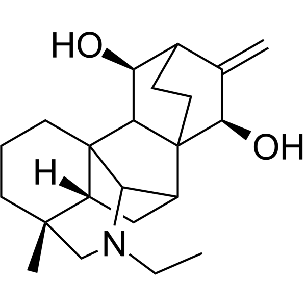
Bullatine A

Based on 1 Customer Validation
Bullatine A, a diterpenoid alkaloid, is a potent P2X7 antagonist. Bullatine A possesses anti-rheumatic, anti-inflammatory and anti-nociceptive effects. Bullatine A inhibits ATP-induced BV-2 cell death/apoptosis and P2X receptor-mediated inflammatory responses. Bullatine A suppresses glioma cell growth by targeting SIRT6. Bullatine A specifically attenuates pain hypersensitivity in rats. Bullatine A attenuates LPS (HY-D1056)-induced systemic inflammatory response by inhibiting the ROS/JNK/NF-κB pathway in mice. Bullatine A improves despair behavior in Chronic chronic social defeat stress (CSDS) mice. Bullatine A can be used for the study of inflammation, glioblastoma (GBM) and depression.
For research use only. We do not sell to patients.
- Purity : 98.0%
- CAS No.: 1354-84-3
- Formula: C22H33NO2
- Molecular Weight:343.50
-
Storage:
4°C, protect from light
* In solvent : -80°C, 6 months; -20°C, 1 month (protect from light)

To place orders, for customer services and technical support, please contact: MedChemExpress USA
Tel: 609-228-6898 E-mail: [email protected] [email protected]


-
 Biological Activity
-
Chemical Information
-
Solvent & Solubility
- Protocols
- Purity & Documentation
- References
-
 Help & FAQs


-
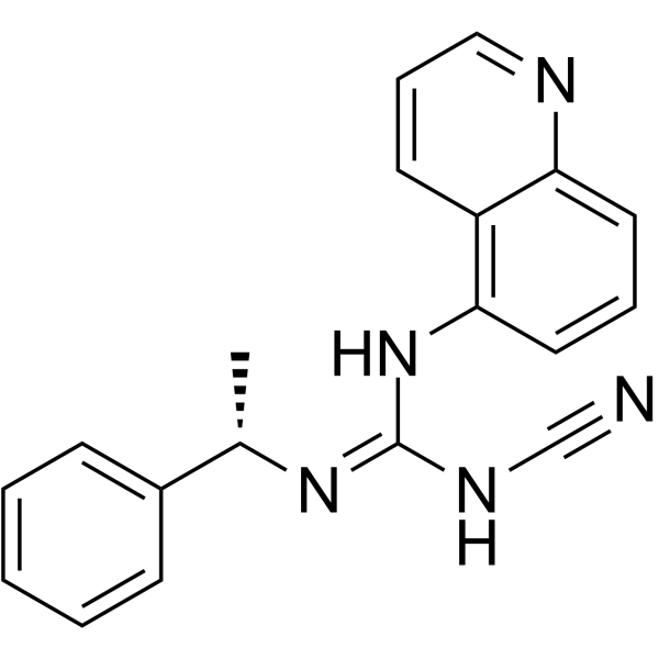
HY-100483
A-804598P2X7 Antagonist -
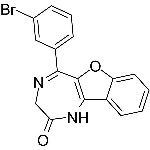
HY-101911
5-BDBDP2X4 Receptor Antagonist -

HY-122575
Aurintricarboxylic acidP2X3 Receptor Antagonist -
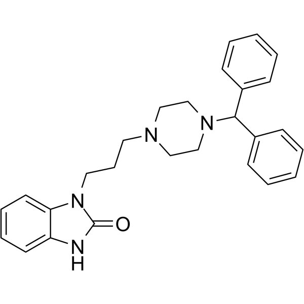
HY-123205
OxatomideH1-histamine/P2X7 Receptor Antagonist -
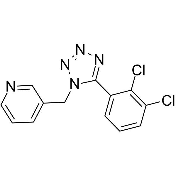
HY-15488
A 438079P2X Receptor Antagonist -
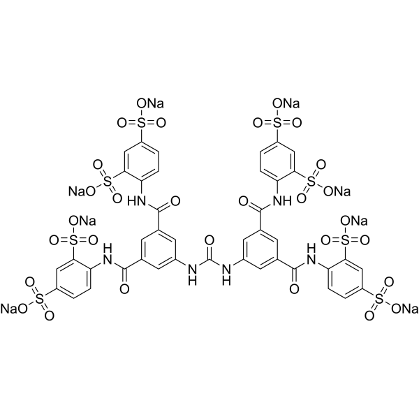
HY-112461A
NF449 octasodiumP2X1 Receptor Antagonist -
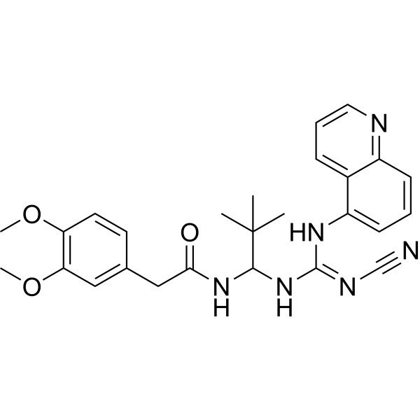
HY-50697
A-740003P2X Receptor Antagonist -
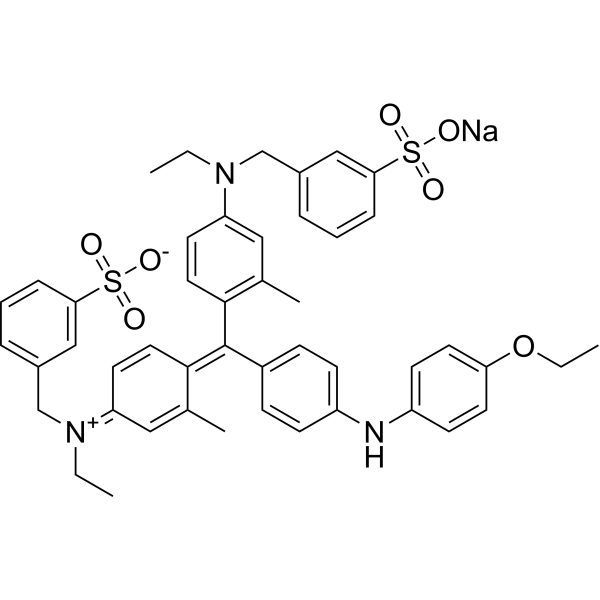
HY-D0014
Brilliant blue G-250Dye/P2×7R Antagonist -
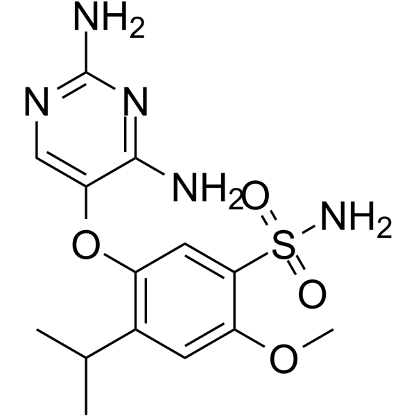
HY-101588
GefapixantP2X3 Antagonist -
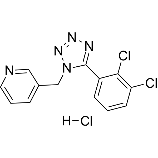
HY-15488A
A 438079 hydrochlorideP2X7 Receptor Antagonist -
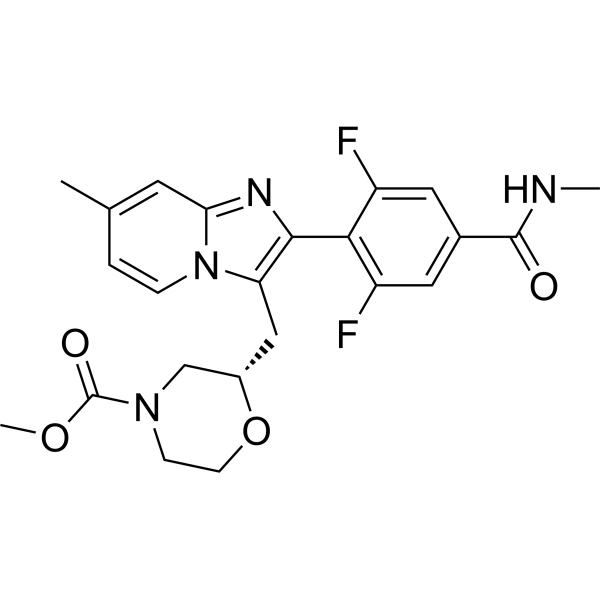
HY-136026
CamlipixantP2X3 Antagonist -
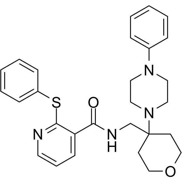
HY-101418
JNJ-47965567P2X7 Antagonist
All P2X Receptor Isoforms
More

Biological Activity
Description
IC50 & Target
[1]|
P2X7 Receptor |
In Vitro
Bullatine A (1-50 μM, 24 h) inhibits ATP-induced BV-2 cell death, down-regulates mRNA levels of IL-6、IL-1β、iNOS, reduces overproduction of NO and IL-6, and selectively inhibits up-regulation of P2X7 receptor mRNA (without effect on P2X4 mRNA) in BV-2 cells[1].
Bullatine A (1-100 μM, 6 h) dose-dependently stimulates prodynorphin expression in primary microglia with an EC50 of 3.2 μM[2].
Bullatine A (10-80 μM, 6 h) significantly inhibits LPS (HY-D1056)-induced mRNA expression of IL-1β, IL-6, iNOS and TNF-α in BV2 microglia and iBMDMs[3].
Bullatine A (80 μM) inhibits LPS-induced IKKα/β、IκBα phosphorylation and NF-κB p65 nuclear translocation, decreases JNK phosphorylation but not p38、ERK1/2 phosphorylation in iBMDMs[3].
Bullatine A (80 μM, 24 h) reduces LPS-induced intracellular ROS generation in iBMDMs[3].
Bullatine A (50-800 nM, 7 days) dose-dependently reduces the colony-forming ability of U87MG and U251 cells[4].
Bullatine A (5-45 μM, 24 h) dose-dependently increases the early and late apoptosis rates of U87MG cells, reduces mitochondrial membrane potential and induces G2/M phase cell cycle arrest in U87MG and U251 cells[4].
Bullatine A (5-45 μM, 24 h) downregulates p-ERK and Myc and dose-dependently inhibits H3K9Ac、H3K56Ac while upregulating SIRT6 in U87MG cells[4].
Bullatine A (50 μM, 24 h) inhibits eATP-induced mitochondrial calcium overload, increased ER-mitochondria colocalization, activation of PERK-elF-2α UPR, lysosome production, elevated NLRP3 inflammasome protein expression, and reduced viability in BV-2 cells[5].
MedChemExpress (MCE) has not independently confirmed the accuracy of these methods. They are for reference only. Further protocols information, click here.
-
Cell Line:ATP-induced BV-2 cells
-
Concentration:1, 10, 50 μM
-
Incubation Time:24 h
-
Result:Up-regulated the ratio of Bcl-2/Bax mRNA.
Down-regulated ATP-induced mRNA levels of IL-6, IL-1β and iNOS in BV-2 cells.
Inhibited ATP-induced up-regulation of P2X7 receptor mRNA without obvious effect on P2X4 mRNA in BV-2 cells.
-
Cell Line:LPS-induced BV2 microglia and iBMDMs
-
Concentration:10, 20, 40, 80 μM
-
Incubation Time:6 h
-
Result:Inhibited LPS (HY-D1056)-induced mRNA expression of IL-1β, IL-6, iNOS and TNF-α in BV2 microglia and iBMDMs.
-
Cell Line:U87MG and U251 cells
-
Concentration:5, 15, 45 μM
-
Incubation Time:24 h
-
Result:Induced G2/M phase cell cycle arrest in U87MG and U251 cells.
-
Cell Line:U87MG cells
-
Concentration:5, 15, 45 μM
-
Incubation Time:24 h
-
Result:Increased the early and late apoptosis rates of U87MG cells.
Upregulated the expression of cleaved caspase-9, cleaved caspase-3 and Bax.
Downregulated the expression of Bcl-2.
-
Cell Line:U87MG cells
-
Concentration:5, 15, 45 μM
-
Incubation Time:24 h
-
Result:Downregulated the expression of p-ERK and Myc proteins in U87MG cells.
Inhibitd the expression of H3K9Ac and H3K56Ac in U87MG cells.
Upregulated the expression of SIRT6.
In Vivo
Bullatine A (0.3-30 mg/kg, s.c., cumulative doses at 1 h intervals ) mitigates mechanical allodynia in Walker 256-induced bone cancer pain rats and Streptozotocin (HY-13753)-induced neuropathic rats[2].
Bullatine A (0.3-30 μg (10 μL), i.t., single dose) inhibits mechanical allodynia in neuropathic rats[2].
Bullatine A (5-20 mg/kg, i.p., twice at 12 h and 1 h prior to LPS injection) attenuates LPS-induced systemic inflammatory response in mice[3].
Bullatine A (10 μg/kg, i.g., once daily, 2 weeks) improves despair behavior in CSDS mice[5].
Bullatine A (10 μg, intra-hippocampal microinjection, every two days, 10 days) partially ameliorates CSDS-induced depressive-like behaviors in hippocampal MAMs[5].
MedChemExpress (MCE) has not independently confirmed the accuracy of these methods. They are for reference only.
-
Animal Model:Spinal nerve ligation-induced neuropathic pain model: adult male Wistar rats were subjected to tight ligation of left L5 and L6 spinal nerves under isoflurane anesthesia[2]
-
Dosage:0.3, 1, 3, 10, 30 mg/kg
-
Administration:s.c. cumulative doses at 1 h intervals
-
Result:Attenuated mechanical allodynia and thermal hyperalgesia in spinal nerve ligation-induced neuropathic pain rats, with ED50 of 1.9 mg/kg and 0.7 mg/kg, Emax of 56.6% MPE and 66.1% MPE respectively.
-
Animal Model:Diabetic neuropathic pain model: adult male Wistar rats were fasted for 16 h, then received a single intravenous injection of Streptozotocin (40 mg/kg)[2]
-
Dosage:0.3, 1, 3, 10, 30 mg/kg
-
Administration:s.c. cumulative doses at 1 h intervals
-
Result:Alleviated mechanical allodynia in streptozotocin-induced diabetic neuropathic pain rats, with ED50 of 1.2 mg/kg and Emax of 65.5% MPE.
-
Animal Model:Complete Freund’s adjuvant (CFA)-induced inflammatory pain model: 100 μL of CFA was injected into the tibiotarsal joint of the left hindpaw of adult male Wistar rats under mild isoflurane anesthesia[2]
-
Dosage:0.3, 1, 3, 10, 30 mg/kg
-
Administration:s.c. cumulative doses at 1 h intervals
-
Result:Reduced mechanical allodynia and thermal hyperalgesia in CFA-induced inflammatory pain rats, with ED50 of 1.4 mg/kg and 0.6 mg/kg, Emax of 50.2% MPE and 60.2% MPE respectively.
-
Animal Model:Bone cancer pain model: Adult female Wistar rats were anesthetized with intraperitoneal pentobarbital (50 mg/kg), and 4 × 1050 Walker 256 carcinoma cells in 10 μL phosphate buffer solution were injected into the medullary cavity of the left tibia[2]
-
Dosage:0.3, 1, 3, 10, 30 mg/kg
-
Administration:s.c. cumulative doses at 1 h intervals
-
Result:Mitigated mechanical allodynia in bone cancer pain rats induced by tibial implantation of Walker 256 carcinoma cells, with ED50 of 0.9 mg/kg and Emax of 45.6% MPE.
-
Animal Model:Spinal nerve ligation-induced neuropathic pain model: adult male Wistar rats were subjected to tight ligation of left L5 and L6 spinal nerves under isoflurane anesthesia[2]
-
Dosage:0.3, 1, 3, 10, 30 μg (10 μL)
-
Administration:i.t. for a single dose
-
Result:Inhibited mechanical allodynia in neuropathic pain rats, with ED50 of 1.1 μg and Emax of 55.5% MPE.
-
Animal Model:C57BL/6 mice (8-10 weeks old, male, 20-25 g) were intraperitoneally injected with 5 mg/kg LPS to induce systemic inflammatory response[3]
-
Dosage:5, 10, 20 mg/kg
-
Administration:i.p., twice at 12 h and 1 h prior to LPS injection
-
Result:Alleviated LPS-induced liver and lung tissue damage with reduced pathological scores.
Reduced serum IL-6 levels in LPS-treated mice.
Down-regulated mRNA expression of IL-1β, IL-6, iNOS and TNF-α in liver and showed similar trend in lung.
Attenuated immune cell infiltration and structural disruption in liver and lung tissues.
-
Animal Model:8-week-old C57BL/6J mice were subjected to chronic social defeat stress (CSDS) for 10 days[5]
-
Dosage:10 μg/kg
-
Administration:i.g. once daily for 2 weeks
-
Result:Shortened immobility time in forced swimming test (FST) at 10 μg/kg dose, improving despair behavior.
Showed no significant effects on social interaction ratio (SIT), total travel distance in open field test (OFT), or sucrose preference rate (SPT).
Failed to inhibit hippocampal microglia activation induced by CSDS.
-
Animal Model:8-week-old C57BL/6J mice underwent hippocampal catheterization surgery, recovered, then were subjected to 10-day CSDS[5]
-
Dosage:10 μg
-
Administration:Intra-hippocampal microinjection every two days for 10 days
-
Result:Increased social interaction ratio (SIT) and shortened immobility time in FST at 10 μg dose, partially ameliorating depressive-like behaviors.
Showed no significant changes in total travel distance in OFT.
Inhibited the increase of Facl-4 protein in hippocampal mitochondrial-associated ER membranes (MAMs) .
Had no significant effect on other MAMs-related proteins (Sigma-1, VDAC, Mfn2).


Chemical Information
-
CAS No. 1354-84-3
-
Appearance Solid
-
Molecular Weight 343.50
-
Formula C22H33NO2
-
Color White to off-white
-
SMILES
O[C@H](C1C2([C@@H]3O)C4C[C@]5([H])[C@@]6(C)CCCC51C4N(CC)C6)C(CC2)C3=C
-
Structure Classification
-
Initial Source
-
Shipping
Room temperature in continental US; may vary elsewhere.
-
Storage
4°C, protect from light
* In solvent : -80°C, 6 months; -20°C, 1 month (protect from light)


Solvent & Solubility
In Vitro:
DMSO : 12.5 mg/mL (36.39 mM; ultrasonic and warming and heat to 80°C; Hygroscopic DMSO has a significant impact on the solubility of product, please use newly opened DMSO)
Please refer to the solubility information to select the appropriate solvent. Once prepared, please aliquot and store the solution to prevent product inactivation from repeated freeze-thaw cycles.
Storage method and period of stock solution: -80°C, 6 months; -20°C, 1 month (protect from light). When stored at -80°C, please use it within 6 months. When stored at -20°C, please use it within 1 month.
Please refer to the solubility information to select the appropriate solvent. Once prepared, please aliquot and store the solution to prevent product inactivation from repeated freeze-thaw cycles.
Storage method and period of stock solution: -80°C, 6 months; -20°C, 1 month (protect from light). When stored at -80°C, please use it within 6 months. When stored at -20°C, please use it within 1 month.
Concentration (start) × Volume (start) = Concentration (final) × Volume (final)

Protocols
-
Mammalian live/dead viability and cytotoxicity staining
Live/dead viability and cytotoxicity staining assays are based on the simultaneous detection of intracellular esterase activity in metabolically active (viable) cells and membrane integrity loss in non-viable cells. In commonly used dual-staining approaches, membrane-permeant fluorogenic substrates are converted by intracellular esterases into fluorescent products in live cells, while impermeant DNA-binding dyes selectively enter cells with compromised plasma membranes and label nucleic acids in dead or dying cells, enabling discrimination between viable and non-viable populations by fluorescence microscopy or flow cytometry.
-
Annexin V plus membrane-impermeant dye apoptosis staining
Annexin V-based apoptosis assays rely on the detection of phosphatidylserine (PS) externalization from the inner leaflet of the plasma membrane to the outer leaflet, an early biochemical hallmark of apoptosis. Fluorescently labeled Annexin V binds PS in a calcium-dependent manner, enabling identification of early apoptotic cells by flow cytometry or fluorescence microscopy. When combined with a membrane-impermeant DNA-binding dye (e. g. , propidium iodide), this approach allows discrimination between viable (Annexin V−/dye−), early apoptotic (Annexin V+/dye−), and late apoptotic or necrotic (Annexin V+/dye+) cell populations by assessing membrane integrity and PS exposure.
-
ROS/oxidative-stress fluorescent staining
ROS/oxidative-stress fluorescent staining uses cell-permeant fluorogenic probes that become fluorescent after oxidation inside cells or tissues; commonly used examples include DCFH-DA/DCFDA for broad cellular oxidant detection, DHE for superoxide-related signal detection, MitoSOX for mitochondrial superoxide-related signal detection, and CellROX probes for oxidative-stress-associated fluorescence readouts. The assay detects probe oxidation rather than a single ROS species unless the probe and analysis method have been chemically validated for that species. DCFH-DA enters cells, is deacetylated by intracellular esterases to DCFH, and produces fluorescent DCF after oxidation, so the readout is used as an operational measure of total cellular oxidative stress rather than a species-specific ROS measurement. DHE and MitoSOX can report superoxide-related oxidation, but red fluorescence alone can include non-specific ethidium-like oxidation products; HPLC or optimized spectral approaches are
-
Somatic Cell Culture
A method of simulating the in vivo environment in vitro to maintain the cell growth, differentation and main functions.
-
CCK-8/WST-8 Cell Proliferation Assay
The CCK-8/WST-8 assay is based on the reduction of the water-soluble tetrazolium salt WST-8 to a water-soluble formazan product by cellular dehydrogenases in metabolically active cells, where the generated formazan amount is proportional to the number of living cells and is quantified by measuring absorbance in the visible range, providing a colorimetric readout for cell viability and proliferation assessment. This class of tetrazolium-based assays improves upon earlier MTT-based systems by producing a water-soluble formazan, eliminating the need for organic solubilization steps and enabling direct spectrophotometric measurement in culture medium.
-
Research Protocol for Inflammation-related Diseases
The NLRP3 inflammasome is a cytosolic innate immune signaling platform that integrates priming signals and danger-signal activation to promote caspase-1 activation, maturation of IL-1β and IL-18, and gasdermin D-mediated pyroptotic cell death. The core experimental logic is to determine whether inflammatory disease phenotypes are driven by increased NLRP3 expression, ASC-containing inflammasome assembly, caspase-1 cleavage, GSDMD cleavage, and extracellular release of IL-1β/IL-18 rather than by nonspecific cell injury alone. The pathway is strongly linked to inflammation-related disease phenotypes because monosodium urate crystals activate NALP3/NLRP3 inflammasome signaling in gout-like crystal inflammation, cholesterol crystals activate NLRP3 inflammasomes in atherogenesis models, and DSS-induced intestinal inflammation has been reported to involve NLRP3 inflammasome activity. However, experimental colitis studies also show context-dependent protective effects of NLRP3 inflammasome co
-
Cell Counting-Based Growth Curve Assay
Cell counting-based growth curve assays quantify cell proliferation by directly measuring changes in viable cell number over time using manual or automated counting methods such as hemocytometer-based counting or instrument-assisted cell enumeration, enabling construction of growth curves that reflect population expansion dynamics in response to culture conditions. A widely used approach is trypan blue exclusion with hemocytometer counting, where membrane-compromised (non-viable) cells take up the dye, allowing discrimination between viable and non-viable cells while simultaneously enabling total cell number quantification. Repeated sampling across time points allows estimation of proliferation rate, growth phases, and comparative growth kinetics between experimental conditions.
-
Cell Viability Determination by MTT Colorimetric Assay
The following protocol uses the MTT colorimetric assay as a classic literature-established method for assessing cell viability/metabolic activity in cultured mammalian cells. MTT[3-(4,5-dimethylthiazol-2-yl)-2,5-diphenyltetrazolium bromide] is reduced by metabolically active cells to a colored formazan product; the amount of formazan is quantified spectrophotometrically and provides an indirect measure of metabolically active viable cells. Importantly, MTT reduction reflects cellular oxidoreductase/metabolic activity rather than an absolute direct count of living cells, so changes in cellular metabolism can alter the signal independently of cell number.
-
Apoptosis
Apoptosis, also called programmed cell death, is generally characterized by distinct morphological characteristics.
-
TUNEL staining for apoptotic DNA fragmentation
TUNEL staining detects DNA strand breaks by using terminal deoxynucleotidyl transferase to add labeled nucleotides to exposed 3′-OH DNA termini, generating either microscopic staining in fixed cells or tissue sections, or fluorescence/cytometric signal in cell suspensions. TUNEL positivity reflects DNA fragmentation but should not be interpreted alone as definitive apoptosis, because TUNEL can also label necrotic, autolytic, mechanically damaged, or DNA-repair-associated DNA breaks.
-
Cell Cytotoxicity Assay
Cytotoxicity assays are usually based on the assessment of cell membrane damage, which can also be indirectly detected by measuring cell viability. Detection methods include MTT assay, CKK-8 assay, LDH assay and ATP assay, etc.
-
Protocol for Cell Counting and Cell Density Analysis
Cell counting and cell-density analysis estimate the number of cells in a known volume or field area. Manual hemocytometer counting uses a chamber of defined geometry to convert counted cells into cells/mL, while automated counters and image-analysis workflows detect cell objects from optical, brightfield, fluorescence, impedance, or digital-image features. Trypan blue viability counting is based on dye exclusion: viable cells with intact membranes exclude dye, while non-viable cells with compromised membranes stain blue. The readout is total cell density, viable-cell density, dead-cell density, and percent viability. Cell density can also be estimated from microscopy images by counting objects per image area, from flow cytometry using calibrated volume or reference particles, or from in situ microscopy in bioreactors after calibration against reference methods such as hemocytometer or flow cytometry.
-
Apoptosis Solutions
Apoptosis is a regulated, generally non-lytic cell-death pathway that removes unwanted, damaged, infected, or abnormal cells through coordinated morphological changes, caspase activation, DNA fragmentation, and membrane remodeling. The intrinsic apoptosis pathway is controlled mainly by mitochondrial outer membrane permeabilization, BCL-2 family proteins, cytochrome c release, apoptosome formation, caspase-9 activation, and downstream executioner caspase-3/7 activation. The extrinsic apoptosis pathway is initiated by death receptors such as Fas, TNFR, and TRAIL receptors, which recruit adaptor proteins and activate caspase-8 before engaging executioner caspases or mitochondrial amplification through BID cleavage. Apoptosis is linked to many phenotypes, including cancer cell killing, tissue homeostasis, immune regulation, neurodegeneration, infection response, and treatment-induced cytotoxicity; unresolved questions include how apoptosis interacts with necroptosis, pyroptosis, ferroptos
-
MTT Cell Proliferation Assay
The MTT assay is a colorimetric endpoint assay for estimating viable cell number, cell growth, cytotoxicity, or cell activation in cultured mammalian cells. Living cells reduce the yellow tetrazolium salt MTT into purple/blue formazan, while dead cells do not generate the same signal; the resulting color can be quantified with a multiwell spectrophotometer. MTT reduction is commonly interpreted as a readout of metabolic activity that often correlates with viable cell number, but it should not be treated as a direct cell-counting method unless the assay is optimized for the cell type and experimental condition. Studies show that MTT reduction can involve mitochondrial and non-mitochondrial reducing systems, and formazan may accumulate in intracellular lipid droplets rather than simply marking mitochondria.


Purity & Documentation
-
Data Sheet (300 KB)
-
SDS (251 KB)
- English - EN (251 KB)
- Français - FR (251 KB)
- Deutsch - DE (251 KB)
- Norwegian - NO (251 KB)
- Español - ES (251 KB)
- Swedish - SV (251 KB)
- Italian - IT (251 KB)
- Korean - KR (251 KB)
- Portuguese - PT (251 KB)
-
Handling Instructions (2659 KB)
References
[1]. Li J, et al. Bullatine A, a diterpenoid alkaloid of the genus Aconitum, could attenuate ATP-induced BV-2 microglia death/apoptosis via P2X receptor pathways. Brain Res Bull. 2013 Aug;97:81-5. [Content Brief]
[2]. Huang Q, et al. Bullatine A stimulates spinal microglial dynorphin A expression to produce anti-hypersensitivity in a variety of rat pain models. J Neuroinflammation. 2016 Aug 30;13(1):214. [Content Brief]
[3]. Liu S, et al. Bullatine A exerts anti-inflammatory effects by inhibiting the ROS/JNK/NF-κB pathway and attenuating systemic inflammatory responses in mice. Pharm Biol. 2022 Dec;60(1):1840-1849. [Content Brief]
[4]. Wang Z, et al. Bullatine A suppresses glioma cell growth by targeting SIRT6. Heliyon. 2024 Dec 24;11(1):e41440. [Content Brief]
[5]. Zhang JR, et al. Bullatine A has an antidepressant effect in chronic social defeat stress mice; Implication of microglial inflammasome. Brain Res Bull. 2023 Apr;195:130-140. [Content Brief]


Complete Stock Solution Preparation Table
Please refer to the solubility information to select the appropriate solvent. Once prepared, please aliquot and store the solution to prevent product inactivation from repeated freeze-thaw cycles.
Storage method and period of stock solution: -80°C, 6 months; -20°C, 1 month (protect from light). When stored at -80°C, please use it within 6 months. When stored at -20°C, please use it within 1 month.
| Optional Solvent | Concentration Solvent Mass | 1 mg | 5 mg | 10 mg | 25 mg |
|---|---|---|---|---|---|
| DMSO | 1 mM | 2.9112 mL | 14.5560 mL | 29.1121 mL | 72.7802 mL |
| 5 mM | 0.5822 mL | 2.9112 mL | 5.8224 mL | 14.5560 mL | |
| 10 mM | 0.2911 mL | 1.4556 mL | 2.9112 mL | 7.2780 mL | |
| 15 mM | 0.1941 mL | 0.9704 mL | 1.9408 mL | 4.8520 mL | |
| 20 mM | 0.1456 mL | 0.7278 mL | 1.4556 mL | 3.6390 mL | |
| 25 mM | 0.1164 mL | 0.5822 mL | 1.1645 mL | 2.9112 mL | |
| 30 mM | 0.0970 mL | 0.4852 mL | 0.9704 mL | 2.4260 mL |


Bullatine A Related Classifications
HY-N5025 Related Classifications
 Powered by Bioz