



AU-24118
Based on 3 publication(s) in Google Scholar
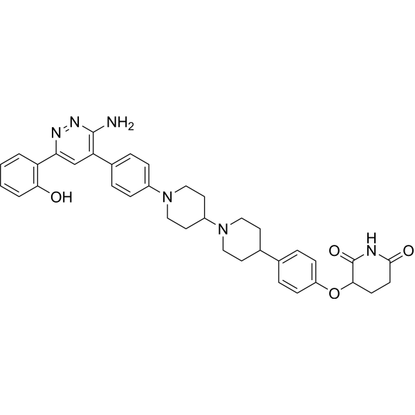
AU-24118 is an orally active PROTAC degrader that recruits cereblon to target the degradation of SMARCA2, SMARCA4 and PBRM1. AU-24118 impairs the chromatin accessibility of oncogenic transcription factors, inhibits colony formation, dislodges chromatin-bound transcription factors, attenuates downstream signal transduction, induces apoptosis, and exerts antiproliferative and cytotoxic effects. AU-24118 can be used in research related to castration-resistant prostate cancer, colorectal cancer, small cell lung cancer and multiple myeloma (including the t (4;14) subtype).
(Pink: SMARCA2 and SMARCA4 and PNRM1 ligand (HY-171774); Blue: Cereblon ligand (HY-171775); Black: linker).
For research use only. We do not sell to patients.
- Purity : 98.82%
- CAS No.: 3084244-26-5
- Formula: C37H40N6O4
- Molecular Weight:632.75
-
Storage:Powder -20°C, 3 years , 4°C, 2 years ; In solvent -80°C, 6 months , -20°C, 1 month

To place orders, for customer services and technical support, please contact: MedChemExpress USA
Tel: 609-228-6898 E-mail: [email protected] [email protected]


-
 Biological Activity
-
Chemical Information
-
Solvent & Solubility
- Protocols
- Purity & Documentation
- References
-
 Help & FAQs


-
HY-138536
PROTAC CBP/P300 Degrader-1PROTAC CBP/P300 Degrader -
HY-145388
AU-15330Anticancer Reagent -
HY-13803
TazemetostatEZH2 Inhibitor -

HY-15371
ForskolinAdenylate Cyclase Activator -
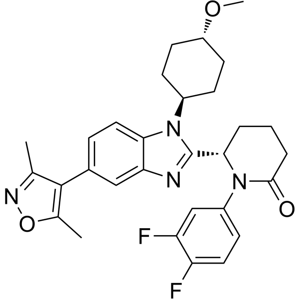
HY-111784
Inobrodibp300/CBP Inhibitor -
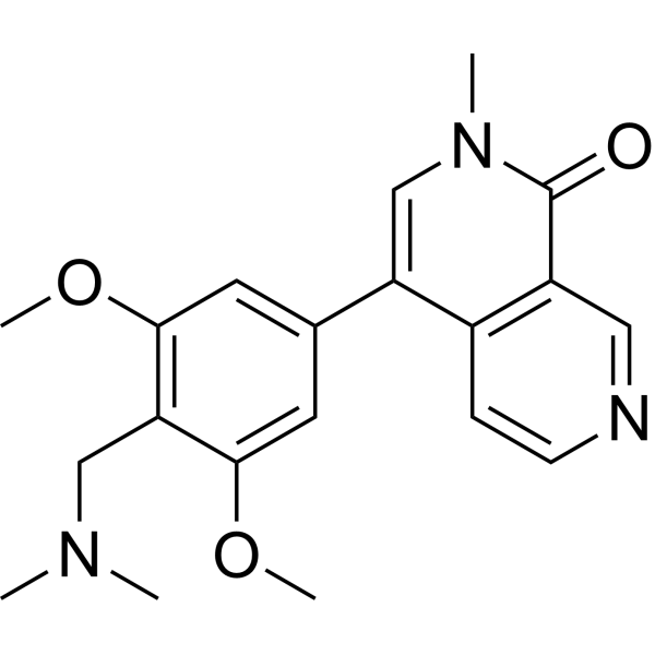
HY-100351
BI-7273BRD9 Inhibitor -
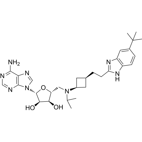
HY-15593
PinometostatDOT1L Inhibitor -
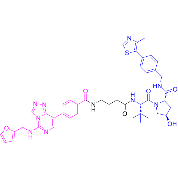
HY-130708
UNC6852PRC2 PROTAC Degrader -
HY-136328
EZM0414 TFASETD2 Inhibitor -
HY-139664
GSK-3685032DNMT1 Inhibitor -
HY-141644
PY-60YAP Activator -

HY-160769
NIBR-LTSiLATS Inhibitor
Publications Citing Use of MedChemExpress (MCE) AU-24118
MoreAll PROTACs Isoforms
MoreAll SWI/SNF Complex Isoforms
More

Biological Activity
Description
IC50 & Target
[1]|
SMARCA2 |
SMARCA4 |
PBRM1 |
In Vitro
AU-24118 (3, 10, 30 nM; 4 h) efficiently degrades SMARCA4 and PBRM1 in VCaP prostate cancer cells[1].
AU-24118 (100 nM; 2 h) selectively degrades SMARCA2, SMARCA4 and PBRM1 in VCaP prostate cancer cells, with no significant off-target degradation of other bromodomain-containing proteins or non-targeted mSWI/SNF subunits[1].
AU-24118 (administered for 5 days) potently inhibits the viability of transcription factor-dependent cancer cell lines (VCaP, 22Rv1, LNCaP, C4-2B), with IC50 values ranging from ~3 nM to ~30 nM; in contrast, non-transcription factor-driven cancer cell lines and normal cell lines (HEK293, PANC-1, MIA PaCa-2) are resistant to it, with IC50 values > 100 nM[1].
AU-24118 (1.5-6 μM; 5 days) exerts selective activity against PBRM1−/− human colorectal cancer cell line HCT116, with an IC50 of 1.179 μM and a selectivity index of 3.2, but shows no genotype-specific activity in PBRM1−/− MC38 or B16F10 cells[2].
AU-24118 (0.75-6 μM; 12 days) exerts comparable inhibitory effects on clonogenic survival in wild-type and PBRM1−/− human colorectal cancer cell line HCT116, with no genotype-specific activity[2].
AU-24118 (administered continuously for 5 days) potently and selectively inhibits the proliferation of SCLC-P cell lines, with IC50 values ranging from 3.01 to 20.59 nM, while it exhibits only extremely low activity against SCLC-A, SCLC-N and SCLC-Y cell lines[3].
Treatment of PBRM1+/+ and PBRM1−/− HCT116 cells with AU-24118 (0-6 μM; 24 h) induces no significant genotype-selective cell cycle changes[2].
AU-24118 (administered for 5 consecutive days) potently inhibits the viability of most human myeloma cell lines, with a median IC50 of 5.9 nM. It shows a trend of enhanced activity in t (4;14)-positive subtypes and exhibits picomolar potency in sensitive cell lines[4].
MedChemExpress (MCE) has not independently confirmed the accuracy of these methods. They are for reference only.
-
Cell Line:VCaP prostate cancer cells
-
Concentration:3, 10, 30 nM
-
Incubation Time:4 h
-
Result:Induced concentration-dependent degradation of SMARCA4 and PBRM1.
-
Cell Line:HCT116 PBRM1+/+ and PBRM1−/−; MC38 PBRM1+/+ and PBRM1−/−; B16F10 PBRM1+/+ and PBRM1−/−
-
Concentration:0, 1.5, 3, 6 μM
-
Incubation Time:5 days
-
Result:Preferentially reduced HCT116 PBRM1−/− cell viability, with IC50 values of 1.179 μM in PBRM1−/− cells and 3.880 μM in PBRM1+/+ cells, but showed no PBRM1-loss selectivity in MC38 or B16F10 cells.
-
Cell Line:NCI-H1048; NCI-H526
-
Concentration:3, 10, 30, 100, 1000 nM
-
Incubation Time:4 h
-
Result:Dose-dependently degraded SMARCA4 and PBRM1 and reduced POU2F3, with marked effects at ≥30 nM.
-
Cell Line:NCI-H1048; NCI-H526
-
Concentration:30 nM
-
Incubation Time:1, 2, 4, 8, 12, 16, 24 h
-
Result:Progressively reduced SMARCA4, PBRM1, and POU2F3 protein levels over time.
Parmacokinetics
In Vivo
AU-24118 (15-20 mg/kg; p.o.; three times weekly; 25 days) has a maximum tolerated dose of 15 mg/kg (p.o., three times weekly) in tumor-free CB17 SCID mice, with minimal toxicity to host organs and only a slight elevation in ALT after prolonged treatment[1].
AU-24118 (1.875-7.5 mg/kg) causes early mortality and rapid body weight loss in hCRBN transgenic mice at doses ≥1.875 mg/kg[4].
AU-24118 (15 mg/kg; p.o.; three times weekly) enhances the anti-tumor efficacy of cisplatin plus etoposide in NCI-H526 SCLC-P xenografts, but concurrent administration causes body weight loss[3].
AU-24118 (15 mg/kg; p.o.; three times weekly; 2 weeks) demonstrates potent anti-tumor efficacy against SCLC-P patient-derived xenografts with no signs of toxicity[3].
AU-24118 (15 mg/kg; p.o.; three times weekly; 3-4 weeks) exhibits potent anti-tumor efficacy against multiple myeloma subcutaneous xenografts, with greater activity than standard-of-care agents pomalidomide and carfilzomib, and no signs of single-agent toxicity[3].
AU-24118 (1 mg/kg; three times per week; 2 weeks) achieves a 40% reduction in serum M-protein in de novo Vk*MYChCRBN multiple myeloma mice with good tolerability[4].
AU-24118 (15 mg/kg; p.o.; three times weekly; until study endpoint) potently inhibits tumor proliferation and extends survival in a disseminated multiple myeloma orthotopic xenograft model, with complete abatement of tumor cells observed in marrow tissue[3].
MedChemExpress (MCE) has not independently confirmed the accuracy of these methods. They are for reference only.
-
Animal Model:CB17 severe combined immunodeficiency (SCID) (male, 6 weeks old, subcutaneous xenograft of VCaP cells, castrated post-tumor growth)[1]
-
Dosage:15 mg/kg
-
Administration:p.o.; three times weekly; 4 weeks
-
Result:Induced regression in every tumor, with individual tumors showing a decrease in volume from baseline (endpoint waterfall plot values below 0%).
Robustly downregulated SMARCA2, SMARCA4, PBRM1, AR, ERG, c-Myc, and PSA, along with increased cleaved PARP levels in day 5 tumors via western blot analysis.
Revealed increased apoptosis via TUNEL staining.
Confirmed SMARCA4 degradation, downregulation of AR, ERG, c-Myc, and reduced Ki-67 proliferation marker levels via IHC.
Caused no significant body weight changes during treatment.
-
Animal Model:CB17 severe combined immunodeficiency (SCID) (male)[1]
-
Dosage:15 mg/kg; 20 mg/kg
-
Administration:p.o.; three times weekly; 25 days
-
Result:Caused noticeable weight loss after 1 week, with morbidity or fatality by week 2 in mice receiving 20 mg/kg.
Allowed all mice receiving up to 15 mg/kg to tolerate treatment well.
Showed no discernible toxicity in spleen, kidney, and liver via H&E staining after 15 mg/kg treatment.
Confirmed successful target degradation in normal tissues via immunohistochemistry.
Resulted in only a slight elevation in alanine transaminases (ALT), with all other blood panel values comparable to vehicle-treated mice and within normal ranges after 25 days of 15 mg/kg treatment.
-
Animal Model:CB17/lcr-Prkdcscid/IcrlcoCrl (SCID) (gender matched to cell line donor, 6 weeks old)[3]
-
Dosage:15 mg/kg
-
Administration:p.o.; three times weekly; 3-4 weeks
-
Result:Caused significant reductions in SCLC-P (NCI-H526 and NCI-H1048) tumor volumes and weights.
Showed no significant effect on SCLC-A (NCI-H69) tumor growth.
Induced degradation of SMARCA2, SMARCA4, and PBRM1, downregulation of POU2F3, POU2AF2, GFI1B, and N-MYC, and increased cleaved PARP in SCLC-P tumors.
Revealed increased apoptotic bodies, necrotic debris, and TUNEL-positive cells in SCLC-P tumors via histopathological analysis.
Demonstrated loss of SMARCA4, POU2F3, and tuft cell marker DCLK1 in SCLC-P tumors via IHC.
Caused no significant changes in body weight or DCLK1-positive tuft cells in normal lung and small intestine tissues.
-
Animal Model:CB17/lcr-Prkdcscid/IcrlcoCrl (SCID) (gender matched to cell line donor, 6 weeks old)[3]
-
Dosage:15 mg/kg
-
Administration:p.o.; three times weekly
-
Result:Showed enhanced tumor growth inhibition when combined with cisplatin and etoposide compared to AU-24118 or chemotherapy alone, despite chemotherapy being stopped at day 8.
Led to 10-20% body weight loss in the combination group, while no weight loss was observed in the AU-24118 single-agent group.
-
Animal Model:NOD.Cg-Prkdcscid Il2rgtm1Wjl/SzJ (6-8 weeks old)[3]
-
Dosage:15 mg/kg
-
Administration:p.o.; three times weekly; 2 weeks
-
Result:Significantly inhibited Lx1322 tumor growth without causing changes in body weight.
Induced degradation of SMARCA2, SMARCA4, and PBRM1, and loss of SMARCA4 protein expression via IHC in tumors.
-
Animal Model:CB17/lcr-Prkdcscid/IcrlcoCrl (SCID) (gender matched to cell line donor, 6 weeks old)[3]
-
Dosage:15 mg/kg
-
Administration:p.o.; three times weekly; 3-4 weeks
-
Result:Significantly decreased tumor volumes and weights in MM1.S, NCI-H929, and Karpas-25 xenograft models.
Achieved complete tumor regression in all MM1.S-treated mice.
Induced degradation of SMARCA2, SMARCA4, and PBRM1, downregulation of c-MYC and POU2AF1, and loss of SMARCA4 and c-MYC protein expression via IHC in MM1.S tumors.
Caused no notable changes in body weight.
-
Animal Model:CB17/lcr-Prkdcscid/IcrlcoCrl (SCID) (gender matched to cell line donor, 6 weeks old)[3]
-
Dosage:15 mg/kg
-
Administration:p.o.; three times weekly; until study endpoint
-
Result:Caused a substantial reduction in bioluminescence signal over time and at endpoint, indicating diminished tumor proliferation.
Significantly extended overall survival of treated mice.
Induced a significant increase in apoptotic cells via TUNEL staining, and confirmed loss of SMARCA4 and c-MYC in treated tumors via IHC.
Revealed near-total absence of plasma cells (tumor cells) in treated marrow, with remnant hematopoietic cells and RBC-filled sinusoids indicative of tumor regression via histopathological analysis.
-
Animal Model:Vk*MYChCRBN (hCRBN-expressing, de novo multiple myeloma model)[4]
-
Dosage:1 mg/kg
-
Administration:three times per week; 2 weeks
-
Result:Reduced serum M-protein by approximately 40% within 2 weeks.
Caused no notable weight loss.
- Detection of Apoptosis
- TUNEL staining for apoptotic DNA fragmentation
- Research Protocol for Epigenomic Data Analysis
- Annexin V plus membrane-impermeant dye apoptosis staining
- Apoptosis Solutions
- Soft Agar Colony Formation Assay
- Colony Formation (Clonogenic) Assay
- Cell Viability Determination by MTT Colorimetric Assay


Chemical Information
-
CAS No. 3084244-26-5
-
Appearance Solid
-
Molecular Weight 632.75
-
Formula C37H40N6O4
-
Color Light yellow to brown
-
SMILES
NC1=C(C=C(N=N1)C2=C(C=CC=C2)O)C3=CC=C(C=C3)N4CCC(CC4)N5CCC(CC5)C6=CC=C(C=C6)OC7CCC(NC7=O)=O
-
Shipping
Room temperature in continental US; may vary elsewhere.
-
Storage
Powder -20°C 3 years 4°C 2 years In solvent -80°C 6 months -20°C 1 month


Publications (3)
-
Journal Impact Factor
-
Most Recent
-
-
Cell
2026 Jun 11;189(12):3541-3552.e18. PMID: 41999746 -


Solvent & Solubility
In Vitro:
DMSO : 8.33 mg/mL (13.16 mM; ultrasonic and warming and heat to 60°C; Hygroscopic DMSO has a significant impact on the solubility of product, please use newly opened DMSO)
Please refer to the solubility information to select the appropriate solvent. Once prepared, please aliquot and store the solution to prevent product inactivation from repeated freeze-thaw cycles.
Storage method and period of stock solution: -80°C, 6 months; -20°C, 1 month. When stored at -80°C, please use it within 6 months. When stored at -20°C, please use it within 1 month.
Please refer to the solubility information to select the appropriate solvent. Once prepared, please aliquot and store the solution to prevent product inactivation from repeated freeze-thaw cycles.
Storage method and period of stock solution: -80°C, 6 months; -20°C, 1 month. When stored at -80°C, please use it within 6 months. When stored at -20°C, please use it within 1 month.
Concentration (start) × Volume (start) = Concentration (final) × Volume (final)
In Vivo:
Select the appropriate dissolution method based on your experimental animal and administration route.
- For the following dissolution methods, please ensure to first prepare a clear stock solution using an In Vitro approach and then sequentially add co-solvents:
- To ensure reliable experimental results, the clarified stock solution can be appropriately stored based on storage conditions. As for the working solution for In Vivo experiments, it is recommended to prepare freshly and use it on the same day.
- The percentages shown for the solvents indicate their volumetric ratio in the final prepared solution. If precipitation or phase separation occurs during preparation, heat and/or sonication can be used to aid dissolution.
Add each solvent one by one: 10% DMSO 40% PEG300 5% Tween-80 45% Saline
Solubility: ≥ 0.83 mg/mL (1.31 mM); Clear solution
This protocol yields a clear solution of ≥ 0.83 mg/mL (saturation unknown).
Taking 1 mL working solution as an example, add 100 μL DMSO stock solution (8.3 mg/mL) to 400 μL PEG300, and mix evenly; then add 50 μL Tween-80 and mix evenly; then add 450 μL Saline to adjust the volume to 1 mL.
Preparation of Saline: Dissolve 0.9 g sodium chloride in ddH₂O and dilute to 100 mL to obtain a clear Saline solution.
Add each solvent one by one: 10% DMSO 90% (20% SBE-β-CD in Saline)
Solubility: ≥ 0.83 mg/mL (1.31 mM); Clear solution
This protocol yields a clear solution of ≥ 0.83 mg/mL (saturation unknown).
Taking 1 mL working solution as an example, add 100 μL DMSO stock solution (8.3 mg/mL) to 900 μL 20% SBE-β-CD in Saline, and mix evenly.
Preparation of 20% SBE-β-CD in Saline (4°C, storage for one week): 2 g SBE-β-CD powder is dissolved in 10 mL Saline, completely dissolve until clear.
For the following dissolution methods, please prepare the working solution directly:
It is recommended to prepare fresh solutions and use them promptly within a short period of time.
The percentages shown for the solvents indicate their volumetric ratio in the final prepared solution. If precipitation or phase separation occurs during preparation, heat and/or sonication can be used to aid dissolution.
Add each solvent one by one: 50% PEG300 50% Saline
Solubility: 12.5 mg/mL (19.76 mM); Suspended solution; Need ultrasonic
In Vivo Dissolution Calculator
Please enter the basic information of animal experiments:
-
-
-
-
Recommended: Prepare an additional quantity of animals to account for potential losses during experiments.
Please enter your animal formula composition:
-
%DMSO +
Recommended: Keep the proportion of DMSO in working solution below 2% if your animal is weak.
-
%+
-
+%Tween-80 + +
-
%Saline +
The co-solvents required include: DMSO, . All of co-solvents are available by MedChemExpress (MCE). , Tween 80. All of co-solvents are available by MedChemExpress (MCE).
Working solution concentration: 0.22 mg/mL
Method for preparing stock solution: mg drug dissolved in μL DMSO. Stock solution concentration: mg/mL.
1. Take μL DMSO stock solution;
2. Add μL .
μL , mix evenly;
3. Then add μL Tween 80, mix evenly;
4. Then add μL
Please ensure that the stock solution in the first step is dissolved to a clear state, and add co-solvents in sequence. You can use ultrasonic heating (ultrasonic cleaner, recommended frequency 20-40 kHz), vortexing, etc. to assist dissolution.


Purity & Documentation
-
Data Sheet (291 KB)
-
SDS (254 KB)
- English - EN (254 KB)
- Français - FR (254 KB)
- Deutsch - DE (254 KB)
- Norwegian - NO (254 KB)
- Español - ES (254 KB)
- Swedish - SV (254 KB)
- Italian - IT (254 KB)
- Korean - KR (254 KB)
- Portuguese - PT (254 KB)
-
Handling Instructions (2659 KB)
References
[1]. He T, et al. Development of an orally bioavailable mSWI/SNF ATPase degrader and acquired mechanisms of resistance in prostate cancer. Proceedings of the National Academy of Sciences of the United States of America. 2024 Apr 09;121(15):e2322563121. [Content Brief]
[2]. Islam MT, et al. Synthetic lethality between PBRM1 deficiency and PARP inhibitors: exploiting G2/M checkpoint arrest in colorectal cancer. Molecular medicine (Cambridge, Mass.). 2026 Jun 06. [Content Brief]
[3]. He T, et al. Targeting the mSWI/SNF complex in POU2F-POU2AF transcription factor-driven malignancies. Cancer cell. 2024 Aug 12;42(8):1336-1351.e9. [Content Brief]


Complete Stock Solution Preparation Table
Please refer to the solubility information to select the appropriate solvent. Once prepared, please aliquot and store the solution to prevent product inactivation from repeated freeze-thaw cycles.
Storage method and period of stock solution: -80°C, 6 months; -20°C, 1 month. When stored at -80°C, please use it within 6 months. When stored at -20°C, please use it within 1 month.
| Optional Solvent | Concentration Solvent Mass | 1 mg | 5 mg | 10 mg | 25 mg |
|---|---|---|---|---|---|
| DMSO | 1 mM | 1.5804 mL | 7.9020 mL | 15.8040 mL | 39.5101 mL |
| 5 mM | 0.3161 mL | 1.5804 mL | 3.1608 mL | 7.9020 mL | |
| 10 mM | 0.1580 mL | 0.7902 mL | 1.5804 mL | 3.9510 mL |


AU-24118 Related Classifications
HY-163410 Related Classifications
 Powered by Bioz