



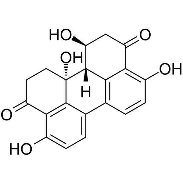
Altertoxin I

Based on 1 Customer Validation
Altertoxin I (Dihydroalterperylenol; ATX-I) is a quinone mycotoxin with immunosuppressive, mutagenic and anticancer activities. Altertoxin I inhibits LPS (HY-D1056)-mediated NF-κB activation and estrogen-dependent alkaline phosphatase activity. Altertoxin I activates the AhR signaling pathway, induces the expression of CYP 1A1/1A2/1B1 and the enzymatic activity of EROD. Altertoxin I can be used in breast cancer research.
Nur für Forschungszwecke. Wir verkaufen nicht an Patienten.
- Reinheit : 99.16%
- CAS. Nr.: 56258-32-3
- Formel: C20H16O6
- Molecular Weight:352.34
-
Speicherung:Powder -20°C, 3 years ; In solvent -80°C, 6 months , -20°C, 1 month

To place orders, for customer services and technical support, please contact: MedChemExpress USA
Tel: 609-228-6898 E-mail: [email protected] [email protected]


-
 Biologische Aktivität
-
Chemical Information
- Protokoll
- Reinheit & Dokumentation
- Verweise
-
 Help & FAQs


Alle Endogenous Metabolite Isoform-spezifische Produkte anzeigen
More

Biologische Aktivität
Beschreibung
In Vitro
Altertoxin I (0.001-20 μM; 20 h) suppresses LPS-induced NF-κB pathway activation in THP1-Lucia™ monocytes with significant effects starting at 1 μM, and does not induce cytotoxicity at concentrations up to 20 μM[1].
Altertoxin I (0.0002-10 μM; 48 h) exerts antiestrogenic effects in Ishikawa cells by suppressing E2-induced alkaline phosphatase activity with significant effects starting at 2 μM, and does not induce cytotoxicity at concentrations up to 10 μM[1].
Altertoxin I (0.01-5 µM; 24 h) dose-dependently induces EROD enzyme activity in MCF-7 human mammary breast cancer cells, with significant activation starting at 2.5 µM[2].
Altertoxin I (0.01-5 µM; 24 h) enhances metabolic activity in MCF-7 human mammary breast cancer cells at 5 µM without reducing cell metabolic activity at lower tested concentrations[2].
Altertoxin I (20 μM; 24 h) exhibits minimal cytotoxicity in A549, HeLa, U2OS, and HepG2 cancer cell lines[3].
MedChemExpress (MCE) has not independently confirmed the accuracy of these methods. They are for reference only.
-
Cell Line:human mammary breast cancer MCF-7 cells
-
Concentration:0.01 µM, 0.1 µM, 1 µM, 2.5 µM, 5 µM
-
Incubation Time:24 h
-
Result:Did not compromise cell metabolic activity at any tested concentration.
Induced a significant enhancement of metabolic activity at 5 µM, reaching approximately 130% of solvent control.
-
Cell Line:A549, HeLa, U2OS, HepG2
-
Concentration:20 μM
-
Incubation Time:24 h
-
Result:Maintained cell viability near control levels across all four tested cancer cell lines.
Showed no significant growth inhibition.


Chemical Information
-
CAS. Nr. 56258-32-3
-
Appearance Solid
-
Molecular Weight 352.34
-
Formel C20H16O6
-
Color Light yellow to yellow
-
SMILES
O=C(C1=C2C3=CC=C1O)CC[C@]2([C@@]([H])(C4=C3C=CC(O)=C45)[C@H](CC5=O)O)O
-
Synonyms
Dihydroalterperylenol; ATX-I
-
Structure Classification
-
Initial Source
Alternaria alternata
-
Versand
Room temperature in continental US; may vary elsewhere.
-
Speicherung
Powder -20°C 3 years In solvent -80°C 6 months -20°C 1 month


Reinheit & Dokumentation
-
Data Sheet (278 KB)
-
SDS (480 KB)
- English - EN (480 KB)
- Français - FR (480 KB)
- Deutsch - DE (480 KB)
- Norwegian - NO (480 KB)
- Español - ES (480 KB)
- Swedish - SV (480 KB)
- Italian - IT (480 KB)
- Korean - KR (480 KB)
- Portuguese - PT (480 KB)
-
Handling Instructions (2659 KB)
Verweise
[1]. Crudo F, et al. Discovery of the Alternaria mycotoxins alterperylenol and altertoxin I as novel immunosuppressive and antiestrogenic compounds in vitro. Archives of toxicology. 2025 Jan;99(1):407-421. [Content Brief]
[2].
Hohenbichler J, et al Alternaria alternata Toxins Synergistically Activate the Aryl Hydrocarbon Receptor Pathway In Vitro. Biomolecules. 2020 Jul 9;10(7):1018.
[Content Brief]
[3]. Xi J, et al. Alterperylenol as a Novel Thioredoxin Reductase Inhibitor Induces Liver Cancer Cell Apoptosis and Ferroptosis. Journal of agricultural and food chemistry. 2022 Dec 21;70(50):15763-15775. [Content Brief]

Calculators
Konzentration (Stammlösung) × Volumen (Stammlösung) = Konzentration (Ziellösung) × Volumen (Ziellösung)


Altertoxin I Related Classifications
HY-N6724 Related Classifications
 Powered by Bioz