



Fluorofenidone
Based on 1 publication(s) in Google Scholar
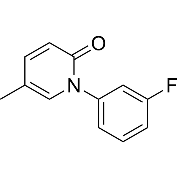
Fluorofenidone (AKF-PD) is an orally active compound with anti-fibrotic, antioxidant, and anti-inflammatory pharmacological effects. Fluorofenidone downregulates the expression of ACSL4, upregulates GPX4 expression and inhibits the NF-κB signaling pathway to alleviate inflammation and fibrosis. Fluorofenidone ameliorates cholestasis and fibrosis by inhibiting hepatic Erk/-Egr-1 signaling and Tgfβ1/Smad pathway in mice. Fluorofenidone demonstrates protective effects against chronic lung injury in mice. Fluorofenidone can be used for the study of chronic obstructive pulmonary disease (COPD), pulmonary interstitial fibrosis (PIF) and non-small cell lung cancer (NSCLC).
Nur für Forschungszwecke. Wir verkaufen nicht an Patienten.
- Reinheit : 99.78%
- CAS. Nr.: 848353-85-5
- Formel: C12H10FNO
- Molecular Weight:203.21
-
Speicherung:Powder -20°C, 3 years , 4°C, 2 years ; In solvent -80°C, 2 years , -20°C, 1 year

To place orders, for customer services and technical support, please contact: MedChemExpress USA
Tel: 609-228-6898 E-mail: [email protected] [email protected]


-
 Biologische Aktivität
-
Chemical Information
-
Lösungsmittel & Löslichkeit
- Protokoll
- Reinheit & Dokumentation
- Verweise
-
 Help & FAQs



Publications Citing Use of MedChemExpress (MCE) Fluorofenidone
More

Biologische Aktivität
Beschreibung
IC50 & Target
|
ACSL4 |
Cellular Effect
|
Cell Line

|
Type | Value | Description | References |
|---|---|---|---|---|
| NIH3T3 | IC50 |
2.75 mM
Compound: AKF-PD
|
Anti-fibrosis activity in mouse NIH/3T3 cells assessed as inhibition of proliferation after 72 hrs by MTT assay
Anti-fibrosis activity in mouse NIH/3T3 cells assessed as inhibition of proliferation after 72 hrs by MTT assay
|
[PMID: 22342629] |
| NIH3T3 | IC50 |
4.2 mM
Compound: Fluorofenidone
|
Antiproliferative activity against mouse NIH/3T3 cells by MTT assay
Antiproliferative activity against mouse NIH/3T3 cells by MTT assay
|
[PMID: 28122457] |
In Vitro
Fluorofenidone (24 h) partially reverses the inhibition of cell viability and reduces the levels of inflammatory factors IL-1β, IL-6, and TNF-α in the culture supernatant induced by cigarette smoke extract (CSE) in BEAS-2B cells[1].
Fluorofenidone (24 h) partially reverses the increase in MDA level and decrease in GSH level in BEAS-2B cell supernatant caused by CSE[1].
Fluorofenidone (24 h) improves CSE-induced mitochondrial damage, downregulates the expression of ACSL4 and upregulates the expression of GPX4 in BEAS-2B cells[1].
Fluorofenidone (200-800 μg/mL, 24 h) inhibits proliferation of A549 and H1299 cells in a dose-dependent manner, with EC50 of 1030 μg/mL (A549) and 1118 μg/mL (H1299)[3].
Fluorofenidone (400-800 μg/mL, 3 h) significantly reduces the percentage of EdU-positive proliferating cells[3].
Fluorofenidone (200-800 μg/mL, 12-24 h) dose- and time-dependently inhibits cell migration and reduces the number of invasive cells in A549 and H1299 cells[3].
Fluorofenidone (200-800 μg/mL) upregulates the expression of epithelial marker E-cadherin and downregulates mesenchymal markers MMP9, vimentin, and SNAIL in A549 and H1299 cells[3].
Fluorofenidone (200-800 μg/mL) suppresses phosphorylation of JNK, ERK, P38 (MAPK pathway) and p-PI3K, p-AKT, p-mTOR (PI3K/AKT/mTOR pathway) in A549 and H1299 cells[3].
MedChemExpress (MCE) has not independently confirmed the accuracy of these methods. They are for reference only. Further protocols information, click here.
In Vivo
Fluorofenidone (0.45% in diet, p.o., 14 days) markedly alleviates hepatic inflammation in 3,5-diethoxycarbonyl-1,4-dihydroxychollidine (DDC)-induced cholestasis model of mice[2].
Fluorofenidone (400 mg/kg, p.o., daily, 3 weeks) combined with Cisplatin (HY-17394) significantly reduces the size, weight and volume of subcutaneous tumors in C57 mice bearing Lewis lung cancer cells[3].
MedChemExpress (MCE) has not independently confirmed the accuracy of these methods. They are for reference only.
-
Animal Model:C57BL/6 J mice (4-week-old, 18-20 g) were used to construct the chronic obstructive pulmonary disease (COPD) model[1]
-
Dosage:5 mg/kg
-
Administration:i.p., once daily, 7 weeks
-
Result:Alleviated CS/LPS-induced lung tissue morphological damage, including reducing interstitial inflammatory cell infiltration, alleviating lung overexpansion, thinning alveolar septa, and lowering lung injury score.
Reduced the levels of inflammatory factors IL-1β, IL-6, and TNF-α in bronchoalveolar lavage fluid (BALF) and serum.
Inhibited lung collagen deposition, decreased lung fibrosis score, downregulated mRNA and protein expressions of fibrosis markers α-SMA, FAP, and COL3A1 in lung tissue, and reduced lung hydroxyproline content.
Downregulated ACSL4 expression and upregulated GPX4 expression in lung tissue, reversed CS/LPS-induced increase in MDA level and decrease in GSH level in lung tissue and serum.
-
Animal Model:3,5-diethoxycarbonyl-1,4-dihydroxychollidine (DDC)-induced cholestasis model of 7-week-old male C57BL/6J mice[2]
-
Dosage:0.45% in diet
-
Administration:p.o., 14 days
-
Result:Reduced serum ALT, AST, ALP, and TBA levels, as well as hepatic BAs levels, and decreased the liver/body weight ratio.
Reduced mRNA levels of hepatic inflammatory cytokines and chemokines, lowered serum levels of these inflammatory factors, and decreased F4/80 expression to reduce macrophage infiltration.
Downregulated mRNA levels of profibrogenic markers, inhibited phosphorylation of Smad2/3, and reduced α-SMA protein level, thus mitigating hepatic fibrosis.
Downregulated expression of Cyp7a1 and Cyp27a1, upregulated expression of hepatic detoxification enzymes, and facilitated nuclear translocation.
Suppressed phosphorylation of Erk1/2, reduced Egr-1 protein level, and blocked the Tgfβ1/Smad pathway.
-
Animal Model:C57 mice (5-week-old, 16-18 g) were used to construct the Lewis lung cancer subcutaneous xenograft model by subcutaneously injecting 5 × 10⁵ Lewis lung cancer cells (C7205) into the axillary region[3]
-
Dosage:400 mg/kg combined with Cisplatin (5 mg/kg, i.p., weekly, 3 weeks)
-
Administration:p.o., daily, 3 weeks
-
Result:Reduced the size, weight and volume of subcutaneous tumors.
Downregulated the expression of mesenchymal marker N-cadherin.


Chemical Information
-
CAS. Nr. 848353-85-5
-
Appearance Solid
-
Molecular Weight 203.21
-
Formel C12H10FNO
-
Color White to off-white
-
SMILES
O=C1C=CC(C)=CN1C2=CC=CC(F)=C2
-
Synonyms
AKF-PD
-
Versand
Room temperature in continental US; may vary elsewhere.
-
Speicherung
Powder -20°C 3 years 4°C 2 years In solvent -80°C 2 years -20°C 1 year


Publications (1)
-
Journal Impact Factor
-
Most Recent
-
Sci Rep
Fluorofenidone alleviates cigarette smoke exposure-induced chronic lung injury by targeting ferroptosis. [Abstract]2024 Dec 30;14(1):32149. PMID: 39738585


Lösungsmittel & Löslichkeit
In Vitro:
DMSO : 140 mg/mL (688.94 mM; Need ultrasonic; Hygroscopic DMSO has a significant impact on the solubility of product, please use newly opened DMSO)
Please refer to the solubility information to select the appropriate solvent. Once prepared, please aliquot and store the solution to prevent product inactivation from repeated freeze-thaw cycles.
Storage method and period of stock solution: -80°C, 2 years; -20°C, 1 year. When stored at -80°C, please use it within 2 years. When stored at -20°C, please use it within 1 year.
Please refer to the solubility information to select the appropriate solvent. Once prepared, please aliquot and store the solution to prevent product inactivation from repeated freeze-thaw cycles.
Storage method and period of stock solution: -80°C, 2 years; -20°C, 1 year. When stored at -80°C, please use it within 2 years. When stored at -20°C, please use it within 1 year.
Konzentration (Stammlösung) × Volumen (Stammlösung) = Konzentration (Ziellösung) × Volumen (Ziellösung)
In Vivo:
Select the appropriate dissolution method based on your experimental animal and administration route.
- For the following dissolution methods, please ensure to first prepare a clear stock solution using an In Vitro approach and then sequentially add co-solvents:
- To ensure reliable experimental results, the clarified stock solution can be appropriately stored based on storage conditions. As for the working solution for In Vivo experiments, it is recommended to prepare freshly and use it on the same day.
- The percentages shown for the solvents indicate their volumetric ratio in the final prepared solution. If precipitation or phase separation occurs during preparation, heat and/or sonication can be used to aid dissolution.
Add each solvent one by one: 10% DMSO 40% PEG300 5% Tween-80 45% Saline
Solubility: ≥ 2.33 mg/mL (11.47 mM); Clear solution
This protocol yields a clear solution of ≥ 2.33 mg/mL (saturation unknown).
Taking 1 mL working solution as an example, add 100 μL DMSO stock solution (23.3 mg/mL) to 400 μL PEG300, and mix evenly; then add 50 μL Tween-80 and mix evenly; then add 450 μL Saline to adjust the volume to 1 mL.
Preparation of Saline: Dissolve 0.9 g sodium chloride in ddH₂O and dilute to 100 mL to obtain a clear Saline solution.
Add each solvent one by one: 10% DMSO 90% (20% SBE-β-CD in Saline)
Solubility: ≥ 2.33 mg/mL (11.47 mM); Clear solution
This protocol yields a clear solution of ≥ 2.33 mg/mL (saturation unknown).
Taking 1 mL working solution as an example, add 100 μL DMSO stock solution (23.3 mg/mL) to 900 μL 20% SBE-β-CD in Saline, and mix evenly.
Preparation of 20% SBE-β-CD in Saline (4°C, storage for one week): 2 g SBE-β-CD powder is dissolved in 10 mL Saline, completely dissolve until clear.
In Vivo Dissolution Calculator
Please enter the basic information of animal experiments:
-
-
-
-
Recommended: Prepare an additional quantity of animals to account for potential losses during experiments.
Please enter your animal formula composition:
-
%DMSO +
Recommended: Keep the proportion of DMSO in working solution below 2% if your animal is weak.
-
%+
-
+%Tween-80 + +
-
%Saline +
The co-solvents required include: DMSO, . All of co-solvents are available by MedChemExpress (MCE). , Tween 80. All of co-solvents are available by MedChemExpress (MCE).
Working solution concentration: 0.22 mg/mL
Method for preparing stock solution: mg drug dissolved in μL DMSO. Stock solution concentration: mg/mL.
1. Take μL DMSO stock solution;
2. Add μL .
μL , mix evenly;
3. Then add μL Tween 80, mix evenly;
4. Then add μL
Please ensure that the stock solution in the first step is dissolved to a clear state, and add co-solvents in sequence. You can use ultrasonic heating (ultrasonic cleaner, recommended frequency 20-40 kHz), vortexing, etc. to assist dissolution.

Protokoll
-
Fibrosis/Collagen Morphometry
Fibrosis and collagen morphometry is based on the quantitative visualization of fibrillar collagen deposition in tissue sections using histochemical stains such as Sirius Red (Picrosirius Red) or Masson's trichrome, followed by image-based or polarization-enhanced analysis to estimate collagen proportional area as a surrogate of extracellular matrix accumulation during fibrotic remodeling. Sirius Red combined with polarized light microscopy enhances detection of collagen fibers due to birefringence properties, enabling more specific visualization of collagen type I and III fibrils compared to conventional bright-field histology, while whole-section or region-restricted digital morphometry reduces field-selection bias in fibrosis assessment. Alternative quantitative approaches include second harmonic generation (SHG) and two-photon excited fluorescence microscopy, which enable label-free detection of fibrillar collagen and have been validated against histological staining and biochemica
-
RNA extraction experimental
By lysing cells, releasing RNA, and removing impurities such as proteins and DNA, high-purity RNA products are finally obtained. The commonly used traditional method is the guanidine isothiocyanate/phenol/chloroform method (Trizol), which is suitable for a variety of animal materials including animal tissues, microorganisms, cultured cells, etc., and most plant materials.
-
Collagen: Sirius Red Staining
Sirius Red or picrosirius red staining is a histochemical method for visualizing collagen-rich extracellular matrix in tissue sections, and collagen fibers are detected as red-stained structures under bright-field microscopy with enhanced birefringence under polarized light. Picrosirius red is useful for assessing total collagen organization, distribution, and fibrosis burden, but polarized color should not be interpreted as a definitive collagen type I versus type III readout because color is affected by fiber orientation, thickness, and packing.
-
Connective Tissue: Masson's Trichrome/Collagen Trichrome Staining
Masson’s Trichrome (collagen/trichrome staining) is a histological technique that differentially stains tissue compartments using sequential acidic dyes to distinguish collagen from muscle and cytoplasmic components based on dye affinity and tissue permeability differences, enabling visualization of fibrosis and connective tissue architecture in histological sections. The classical formulation typically uses Weigert's iron hematoxylin for nuclear staining, Biebrich scarlet-acid fuchsin for cytoplasm and muscle, and aniline blue (or light green variants) for collagen, producing a characteristic blue/green collagen signal contrasted against red cytoplasm and dark nuclei. The staining principle relies on selective displacement of smaller dye molecules by larger anionic dyes in collagen-rich regions under controlled acidified conditions, which enhances collagen-specific dye retention. This property makes the method widely used for fibrosis assessment in organs such as heart, liver, lung, a
-
Research Protocol for Inflammation-related Diseases
The NLRP3 inflammasome is a cytosolic innate immune signaling platform that integrates priming signals and danger-signal activation to promote caspase-1 activation, maturation of IL-1β and IL-18, and gasdermin D-mediated pyroptotic cell death. The core experimental logic is to determine whether inflammatory disease phenotypes are driven by increased NLRP3 expression, ASC-containing inflammasome assembly, caspase-1 cleavage, GSDMD cleavage, and extracellular release of IL-1β/IL-18 rather than by nonspecific cell injury alone. The pathway is strongly linked to inflammation-related disease phenotypes because monosodium urate crystals activate NALP3/NLRP3 inflammasome signaling in gout-like crystal inflammation, cholesterol crystals activate NLRP3 inflammasomes in atherogenesis models, and DSS-induced intestinal inflammation has been reported to involve NLRP3 inflammasome activity. However, experimental colitis studies also show context-dependent protective effects of NLRP3 inflammasome co
-
Ferroptosis Solutions
Ferroptosis is an iron-dependent, non-apoptotic form of regulated cell death characterized by lethal lipid peroxidation and sensitivity to suppression by iron chelators or lipophilic radical-trapping antioxidants. The core pathway links cystine uptake through system Xc−, glutathione availability, GPX4-dependent detoxification of phospholipid hydroperoxides, iron-dependent oxidative reactions, and polyunsaturated-phospholipid metabolism into a cell-death program that is biochemically and morphologically distinct from apoptosis, necrosis, and autophagy. The ferroptosis pathway is experimentally linked to phenotype through chemical and genetic perturbation. Erastin induces ferroptosis by inhibiting cystine uptake through system Xc− and weakening antioxidant defenses, while GPX4 inhibition or depletion causes lipid peroxide accumulation and ferroptotic cancer-cell death. ACSL4 and oxidizable arachidonoyl- or adrenoyl-containing phosphatidylethanolamines shape ferroptosis sensitivity by con
-
Inhalation Toxicity Study
Inhalation toxicity studies expose rodents to a controlled aerosol, vapor, gas, or smoke atmosphere and assess respiratory and systemic toxicity using exposure-atmosphere characterization, clinical observations, body and organ weights, bronchoalveolar lavage fluid, histopathology, blood chemistry, hematology, and, when included, molecular endpoints such as transcriptomics, proteomics, lipidomics, or tissue burden analysis. The primary biological readouts are airway irritation, pulmonary inflammation, cytotoxicity, altered surfactant or lipid homeostasis, impaired particle clearance, and tissue remodeling, reflected by BALF cell differentials, BALF protein, LDH, phosphatase activities, cytokines, lung weight, microscopic respiratory-tract lesions, and retained lung burden.


Reinheit & Dokumentation
-
Data Sheet (282 KB)
-
SDS (393 KB)
- English - EN (393 KB)
- Français - FR (393 KB)
- Deutsch - DE (393 KB)
- Norwegian - NO (393 KB)
- Español - ES (393 KB)
- Swedish - SV (393 KB)
- Italian - IT (393 KB)
- Korean - KR (393 KB)
- Portuguese - PT (393 KB)
-
Handling Instructions (2659 KB)
Verweise
[1]. Wu Y, et al. Fluorofenidone alleviates cigarette smoke exposure-induced chronic lung injury by targeting ferroptosis. Sci Rep. 2024 Dec 30;14(1):32149. [Content Brief]
[2]. Wang H,et al. Fluorofenidone ameliorates cholestasis and fibrosis by inhibiting hepatic Erk/-Egr-1 signaling and Tgfβ1/Smad pathway in mice. Biochim Biophys Acta Mol Basis Dis. 2022 Dec 1;1868(12):166556. [Content Brief]
[3]. Wang S, et al. Fluorofenidone enhances cisplatin efficacy in non-small cell lung cancer: a novel approach to inhibiting cancer progression. Transl Lung Cancer Res. 2024 Nov 30;13(11):3175-3188. [Content Brief]


Complete Stock Solution Preparation Table
Please refer to the solubility information to select the appropriate solvent. Once prepared, please aliquot and store the solution to prevent product inactivation from repeated freeze-thaw cycles.
Storage method and period of stock solution: -80°C, 2 years; -20°C, 1 year. When stored at -80°C, please use it within 2 years. When stored at -20°C, please use it within 1 year.
| Optional Solvent | Concentration Solvent Mass | 1 mg | 5 mg | 10 mg | 25 mg |
|---|---|---|---|---|---|
| DMSO | 1 mM | 4.9210 mL | 24.6051 mL | 49.2102 mL | 123.0254 mL |
| 5 mM | 0.9842 mL | 4.9210 mL | 9.8420 mL | 24.6051 mL | |
| 10 mM | 0.4921 mL | 2.4605 mL | 4.9210 mL | 12.3025 mL | |
| 15 mM | 0.3281 mL | 1.6403 mL | 3.2807 mL | 8.2017 mL | |
| 20 mM | 0.2461 mL | 1.2303 mL | 2.4605 mL | 6.1513 mL | |
| 25 mM | 0.1968 mL | 0.9842 mL | 1.9684 mL | 4.9210 mL | |
| 30 mM | 0.1640 mL | 0.8202 mL | 1.6403 mL | 4.1008 mL | |
| 40 mM | 0.1230 mL | 0.6151 mL | 1.2303 mL | 3.0756 mL | |
| 50 mM | 0.0984 mL | 0.4921 mL | 0.9842 mL | 2.4605 mL | |
| 60 mM | 0.0820 mL | 0.4101 mL | 0.8202 mL | 2.0504 mL | |
| 80 mM | 0.0615 mL | 0.3076 mL | 0.6151 mL | 1.5378 mL | |
| 100 mM | 0.0492 mL | 0.2461 mL | 0.4921 mL | 1.2303 mL |


Fluorofenidone Related Classifications
HY-121246 Related Classifications
 Powered by Bioz