



HDAC-IN-46
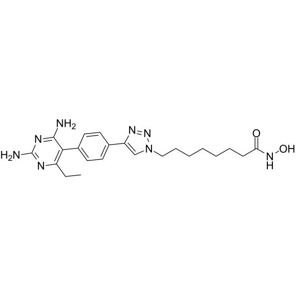
HDAC-IN-46 (compound 12c) is a potent HDAC inhibitor with an IC50 value of 0.21 μM and 0.021 μM for HDAC1 and HDAC6, respectively. HDAC-IN-46 upregulates p-p38, and downregulates Bcl-xL and cyclin D1 in MDA-MB-231 cells. HDAC-IN-46 induces significant G2 phase arrest and apoptosis. HDAC-IN-46 can be used for researching triple-negative breast cancer (TNBC).
Nur für Forschungszwecke. Wir verkaufen nicht an Patienten.
- CAS. Nr.: 2562386-85-8
- Formel: C22H30N8O2
- Molecular Weight:438.53
-
Speicherung:
Please store the product under the recommended conditions in the Certificate of Analysis.

To place orders, for customer services and technical support, please contact: MedChemExpress USA
Tel: 609-228-6898 E-mail: [email protected] [email protected]


-
 Biologische Aktivität
-
Chemical Information
- Protokoll
- Reinheit & Dokumentation
- Verweise
-
 Help & FAQs




Biologische Aktivität
Beschreibung
IC50 & Target
[1]|
HDAC1 0.21 μM (IC50) |
HDAC6 0.021 μM (IC50) |
Cellular Effect
|
Cell Line

|
Type | Value | Description | References |
|---|---|---|---|---|
| A549 | IC50 |
88.46 μM
Compound: 12c
|
Antiproliferative activity in human A549 cells assessed as inhibition of cell viability after 72 hrs by MTS assay
Antiproliferative activity in human A549 cells assessed as inhibition of cell viability after 72 hrs by MTS assay
|
[PMID: 32061484] |
| MCF7 | IC50 |
83.34 μM
Compound: 12c
|
Antiproliferative activity in human MCF7 cells assessed as inhibition of cell viability after 72 hrs by MTS assay
Antiproliferative activity in human MCF7 cells assessed as inhibition of cell viability after 72 hrs by MTS assay
|
[PMID: 32061484] |
| MDA-MB-231 | IC50 |
21.4 μM
Compound: 12c
|
Antiproliferative activity in human MDA-MB-231 cells assessed as inhibition of cell viability after 72 hrs by MTS assay
Antiproliferative activity in human MDA-MB-231 cells assessed as inhibition of cell viability after 72 hrs by MTS assay
|
[PMID: 32061484] |
In Vitro
HDAC-IN-46 (compound 12c) has antiproliferative activity against MDA-MB-231, A549 and MCF-7 with IC50s of 88.46±10.5 μM, 83.34 ± 15.5 μM and 21.4±3.7 μM, respectively[1].
HDAC-IN-46 (5, 12.5 and 25 μM; 24 h) causes concentration-dependent upregulation of p-p38 and downregulation of Bcl-xL and cyclin D1 in MDA-MB-231 cells[1].
HDAC-IN-46 (12.5 and 25 μM; 48 h) induces significant G2 phase arrest and apoptosis[1].
MedChemExpress (MCE) has not independently confirmed the accuracy of these methods. They are for reference only.
-
Cell Line:MDA-MB-231 cells
-
Concentration:5, 12.5 and 25 μM
-
Incubation Time:24 h
-
Result:Caused concentration-dependent upregulation of p-p38 and downregulation of Bcl-xL and cyclin D1.
-
Cell Line:MDA-MB-231 cells
-
Concentration:12.5 and 25 μM
-
Incubation Time:48 h
-
Result:Induced significant G2 phase arrest and apoptosis.


Chemical Information
-
CAS. Nr. 2562386-85-8
-
Molecular Weight 438.53
-
Formel C22H30N8O2
-
SMILES
O=C(NO)CCCCCCCN1N=NC(C2=CC=C(C3=C(CC)N=C(N)N=C3N)C=C2)=C1
-
Versand
Room temperature in continental US; may vary elsewhere.
-
Speicherung
Please store the product under the recommended conditions in the Certificate of Analysis.


Reinheit & Dokumentation
Verweise

Calculators
Konzentration (Stammlösung) × Volumen (Stammlösung) = Konzentration (Ziellösung) × Volumen (Ziellösung)


HDAC-IN-46 Related Classifications
HY-150597 Related Classifications
 Powered by Bioz