



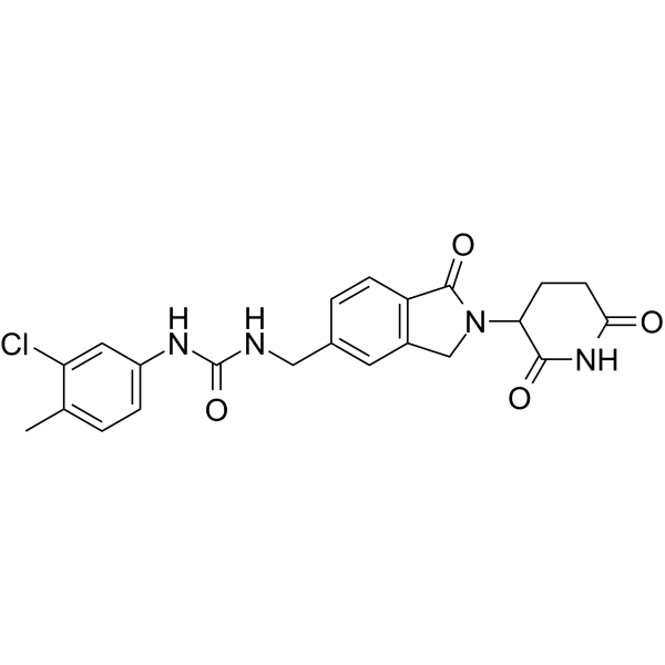
CC-885
Based on 21 publication(s) in Google Scholar
CC-885, a cereblon (CRBN) modulator, acts as a molecular glue degrader targeting GSPT1 with a Kd of 350 nM. CC-885 binds to CRBN to alter the substrate specificity of the CRL4 E3 ligase, promoting the ubiquitination and proteasomal degradation of GSPT1, BNIP3L, PLK1, CDK4, IKZF1/3, GSPT2, WIZ, CHD7, GOLM1, CK1α and ETS1. CC-885 induces apoptosis, cell cycle arrest, transcriptional downregulation and antiproliferation in cancer cells. CC-885 is applicable to research related to relapsed/refractory acute myeloid leukemia, MYC-driven tumors, metastatic castration-resistant prostate cancer, multiple myeloma, hepatocellular carcinoma, glioblastoma, VHL-deficient clear cell renal cell carcinoma and non-small cell lung cancer.
For research use only. We do not sell to patients.
- Purity : 99.26%
- CAS No.: 1010100-07-8
- Formula: C22H21ClN4O4
- Molecular Weight:440.88
-
Storage:Powder -20°C, 3 years ; In solvent -80°C, 6 months , -20°C, 1 month

To place orders, for customer services and technical support, please contact: MedChemExpress USA
Tel: 609-228-6898 E-mail: [email protected] [email protected]


-
 Biological Activity
-
Chemical Information
-
Solvent & Solubility
- Protocols
- Purity & Documentation
- References
-
 Help & FAQs


-
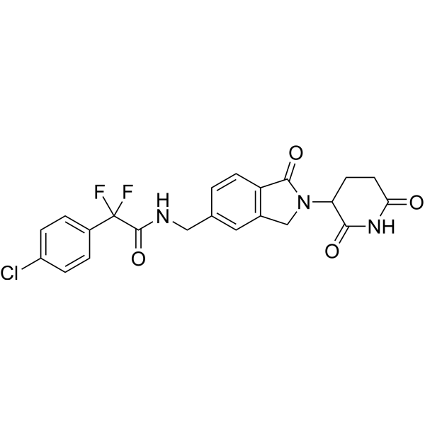
HY-130800
EragidomideGSPT1 Molecular Glue Degrader -
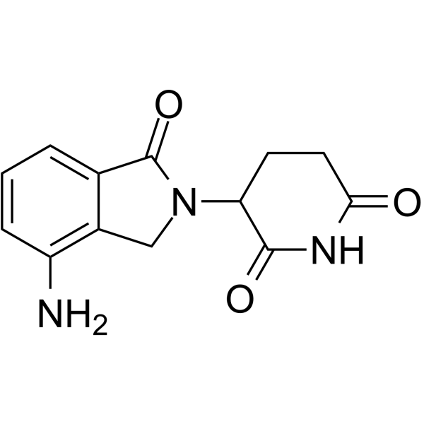
HY-A0003
LenalidomideImmunomodulator -
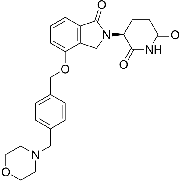
HY-101291
IberdomideCELMoD -
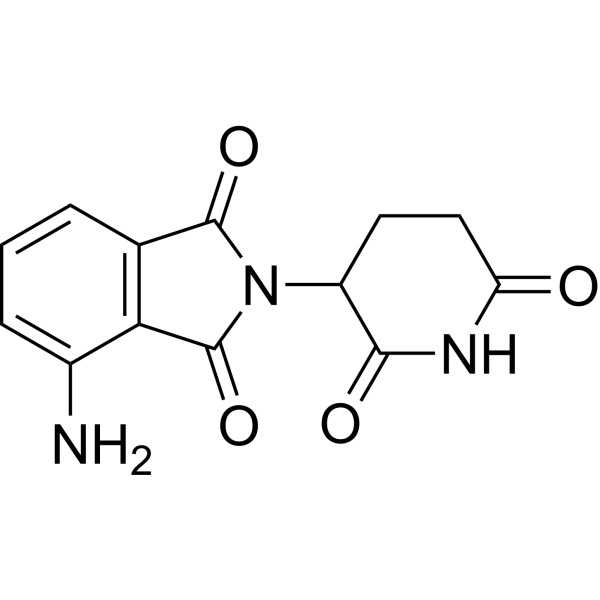
HY-10984
PomalidomideImmunomodulatory Agent -
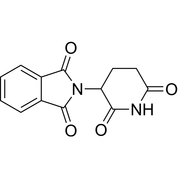
HY-14658
ThalidomideCereblon Inhibitor -
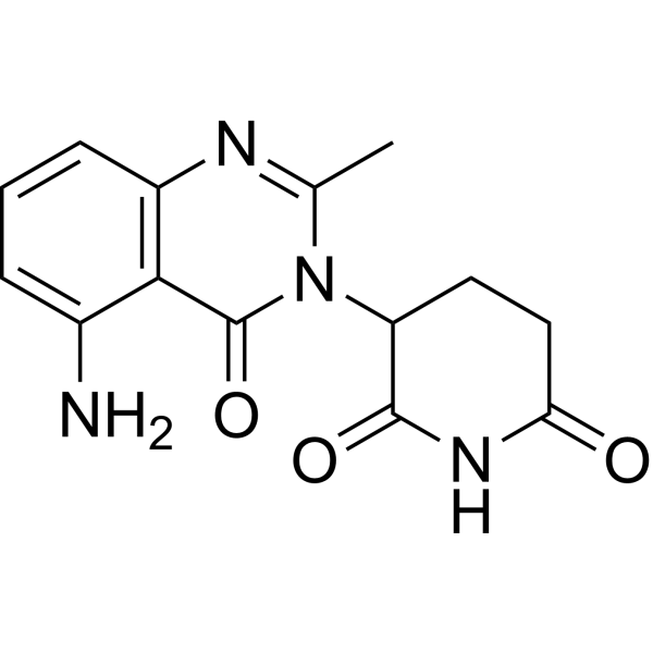
HY-100507
AvadomideCereblon Modulator -
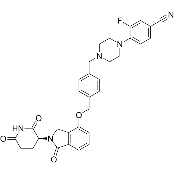
HY-129395
MezigdomideCereblon E3 Ubiquitin Ligase Modulating Agent -
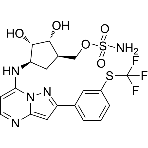
HY-100487
TAK-243UAE/E1 Inhibitor -
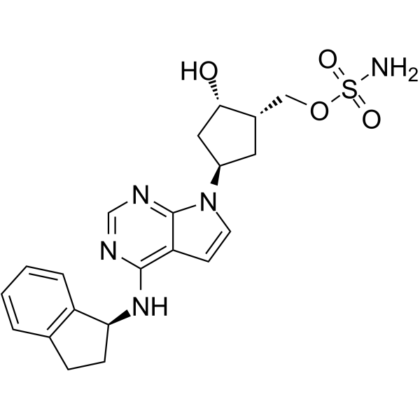
HY-70062
PevonedistatNAE Inhibitor -

HY-13259
MG-132Proteasome Inhibitor -
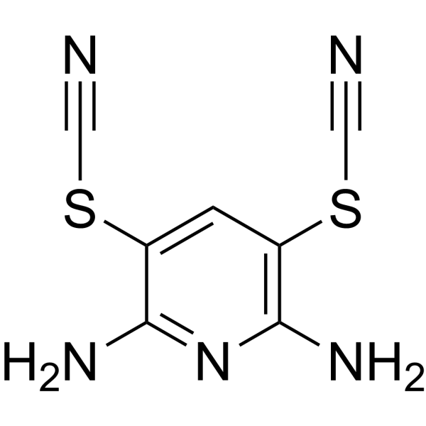
HY-13814
PR-619DUB Inhibitor -
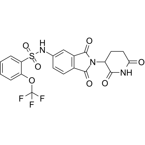
HY-132199
SJ6986GSPT1/2 Molecular Glue Degrader
Publications Citing Use of MedChemExpress (MCE) CC-885
More- Nature. 2026 Oct;658(8134):241-249.
- Cell. 2023 Jul 20;186(15):3227-3244.e20. [Abstract]
- Cancer Discov. 2026 Apr 19. [Abstract]
- Nat Commun. 2025 Nov 19;16(1):10157. [Abstract]
- Nat Commun. 2023 Dec 19;14(1):8437. [Abstract]
- Nat Commun. 2022 Sep 10;13(1):5324. [Abstract]
- Mol Cell. 2026 Jun 18;86(12):2341-2357.e10. [Abstract]
- Nat Chem Biol. 2024 Sep;20(9):1227-1236. [Abstract]
- J Clin Invest. 2022 Aug 15;132(16):e153514. [Abstract]
- Acta Pharmacol Sin. 2020 Sep;41(9):1246-1254. [Abstract]
- Cell Chem Biol. 2020 Jul 16;27(7):866-876.e8. [Abstract]
- Leukemia. 2025 Sep;39(9):2163-2173. [Abstract]
- Mol Ther Oncolytics. 2020 Jun 23;18:215-225. [Abstract]
- Bioorg Chem. 2022 Feb:119:105505. [Abstract]
- Cell Signal. 2025 Jun:130:111665. [Abstract]
- J Proteome Res. 2022 Aug 5;21(8):1842-1856. [Abstract]
- Dis Model Mech. 2025 Nov 7:dmm.052495. [Abstract]
- Biochem Biophys Res Commun. 2021 Apr 16:549:150-156. [Abstract]
- bioRxiv. 2026 Feb 16.
- bioRxiv. 2025 Sep 2:2022.10.31.514544. [Abstract]
- bioRxiv. 2024 Jan 28.
 Customer Validation & Images

-
WB
-
WB
-
WB
-
WB
-
RT-PCR
All Eukaryotic Release Factor (eRF) Isoforms
More

Biological Activity
Description
IC50 & Target
[1]|
Cereblon |
eRF3a/GSPT1 350 nM (Kd) |
Cellular Effect
|
Cell Line

|
Type | Value | Description | References |
|---|---|---|---|---|
| NB-4 | IC50 |
0.00006 μM
|
Antiproliferative activity against human NB-4 AML cells assessed as inhibition of cell proliferation incubated for 48-72 h by cell proliferation assay.
Antiproliferative activity against human NB-4 AML cells assessed as inhibition of cell proliferation incubated for 48-72 h by cell proliferation assay.
|
27338790 |
| U-937 | IC50 |
0.0016 μM
|
Antiproliferative activity against human U-937 AML cells assessed as inhibition of cell proliferation incubated for 48-72 h by cell proliferation assay.
Antiproliferative activity against human U-937 AML cells assessed as inhibition of cell proliferation incubated for 48-72 h by cell proliferation assay.
|
27338790 |
| NB-4 | DC50 |
<1 nM
|
GSPT1 degradation in human NB4 cells, achieving >90% maximal degradation at 1 nM.
GSPT1 degradation in human NB4 cells, achieving >90% maximal degradation at 1 nM.
|
40570036 |
| NB-4 | IC50 |
0.06 nM
|
Antiproliferative activity against human NB4 acute myeloid leukemia cells.
Antiproliferative activity against human NB4 acute myeloid leukemia cells.
|
40570036 |
| MOLM-13 | IC50 |
0.7 nM
|
Antiproliferative activity against human MOLM-13 acute myeloid leukemia cells.
Antiproliferative activity against human MOLM-13 acute myeloid leukemia cells.
|
40570036 |
| HL-60 | IC50 |
0.5 nM
|
Antiproliferative activity against human HL-60 acute myeloid leukemia cells.
Antiproliferative activity against human HL-60 acute myeloid leukemia cells.
|
40570036 |
| PBMC | IC50 |
1.1 nM
|
Toxicity against normal human PBMC cells.
Toxicity against normal human PBMC cells.
|
40570036 |
| THLE-2 | IC50 |
11 nM
|
Toxicity against normal human THLE-2 cells.
Toxicity against normal human THLE-2 cells.
|
40570036 |
| 769-P | IC50 |
23 nM
|
Cytotoxicity against VHL-mutant renal cell carcinoma 769-P cells assessed as reduction in cell viability incubated for 72 hrs by CCK-8 assay.
Cytotoxicity against VHL-mutant renal cell carcinoma 769-P cells assessed as reduction in cell viability incubated for 72 hrs by CCK-8 assay.
|
42089431 |
| 786-0 | IC50 |
0.99 nM
|
Cytotoxicity against VHL-mutant renal cell carcinoma 786-O cells assessed as reduction in cell viability incubated for 72 hrs by CCK-8 assay.
Cytotoxicity against VHL-mutant renal cell carcinoma 786-O cells assessed as reduction in cell viability incubated for 72 hrs by CCK-8 assay.
|
42089431 |
| A498 | IC50 |
11.3 nM
|
Cytotoxicity against VHL-mutant renal cell carcinoma A498 cells assessed as reduction in cell viability incubated for 72 hrs by CCK-8 assay.
Cytotoxicity against VHL-mutant renal cell carcinoma A498 cells assessed as reduction in cell viability incubated for 72 hrs by CCK-8 assay.
|
42089431 |
| CAKI-1 | IC50 |
837 nM
|
Cytotoxicity against VHL-wild-type renal cell carcinoma Caki-1 cells assessed as reduction in cell viability incubated for 72 hrs by CCK-8 assay.
Cytotoxicity against VHL-wild-type renal cell carcinoma Caki-1 cells assessed as reduction in cell viability incubated for 72 hrs by CCK-8 assay.
|
42089431 |
| TK-10 | IC50 |
847000 nM
|
Cytotoxicity against VHL-wild-type renal cell carcinoma TK10 cells assessed as reduction in cell viability incubated for 72 hrs by CCK-8 assay.
Cytotoxicity against VHL-wild-type renal cell carcinoma TK10 cells assessed as reduction in cell viability incubated for 72 hrs by CCK-8 assay.
|
42089431 |
In Vitro
CC-885 induces secondary downregulation of proteins including MYC, PLK1, and CDK4 via transcriptional collapse following GSPT1 degradation, while directly recruiting GSPT2, WIZ, and CHD7 to CRBN, in human MOLT-4 and 293T cells[1].
CC-885 (1-10 μM; 24 h) promotes the ubiquitination of CDK4 and dose-dependently enhances the interaction between CDK4 and CRBN in 293T cells, with maximum interaction observed at 10 μM for 24 h[2].
CC-885 (5-20 h) selectively degrades endogenous GSPT1 in CrbnV/V MEFs within 5 h, with sustained degradation observed up to 20 h, while wild-type Crbn+/+ MEFs are resistant to cereblon E3 ligase modulator CC-885-induced GSPT1 degradation[1].
CC-885 induces potent cytotoxicity in CrbnV/V MEFs, which correlates with GSPT1 degradation, while wild-type and CrbnI/I MEFs are resistant to CC-885-induced cytotoxicity[1].
CC-885 (10 μM; 8 h) induces CRBN-dependent ubiquitination of wild-type ETS1, but not the ETS1Y158A mutant, in 769-P and 293T cells[8].
CC-885 (1 μM (293T cells); 5 nM (786-O cells); 12 h (786-O cells)) enhances the physical interaction between CRBN and transcriptionally active ETS1 p51/p42 isoforms, but not the p27 isoform, in 293T and 786-O cells[8].
CC-885 requires the ETS1 135-242aa domain for CRBN binding and degradation of ETS1 in 293T cells[8].
CC-885 (6.25-50 nM; 24-72 h) inhibits the viability of RPMI8226, MM1S, and U266 multiple myeloma cells in a dose- and time-dependent manner[2].
CC-885 (0.5-10 μM; 12-24 h) induces post-translational degradation of CDK4 via ubiquitination in MM1S, RPMI8226, and U266 multiple myeloma cells in a CRBN-, CRL-, p97-, and proteasome-dependent manner, with maximum degradation observed at 10 μM for 12 h in MM1S cells[2].
CC-885 (62.5-1000 nM; 24-72 h) potently reduces cell viability in HCCLM3, HepG2, and Huh7 human hepatocellular carcinoma cell lines in a dose- and time-dependent manner, with HCCLM3 cells exhibiting the greatest sensitivity[3].
CC-885 (100-400 nM; 12 h initial treatment, followed by 14 days of culture in regular medium) suppresses the clonogenic potential of HCCLM3 human hepatocellular carcinoma cells in a dose-dependent manner[3].
CC-885 (0.01-20 μM) dose-dependently reduces GOLM1 protein levels in HCCLM3 human hepatocellular carcinoma cells, with significant suppression starting at 0.1 μM and near-complete depletion at 20 μM[3].
CC-885 (1 μM; 4-20 h) gradually reduces GOLM1 protein levels in HCCLM3 human hepatocellular carcinoma cells over time, with significant suppression by 4 h and near-maximal depletion by 20 h[3].
CC-885 (1 μM) does not alter GOLM1 mRNA levels in HCCLM3 human hepatocellular carcinoma cells, confirming post-translational regulation of GOLM1[3].
CC-885 (1 μM; 20 h) reduces GOLM1 protein levels in Huh7 and HepG2 human hepatocellular carcinoma cells after 20 h of incubation, confirming the effect is consistent across multiple HCC cell lines[3].
CC-885 (1 μM CC-885 + 10 μM MG132) increases the ubiquitination of GOLM1 in HCCLM3 human hepatocellular carcinoma cells, facilitating proteasomal degradation of GOLM1[3].
CC-885 (0.001-1 μM; 24 h for A549 cells, 1 μM; 1-24 h for HEK293T cells) induces post-transcriptional, dose- and time-dependent degradation of BNIP3L in A549 cells and HEK293T cells stably expressing Flag-tagged BNIP3L, and reduces the half-life of BNIP3L protein[4].
CC-885 (5-200 nM; 16 h) dose-dependently enhances GSPT1 ubiquitination in U87 glioblastoma cells, with maximum activity at 100 nM[5].
CC-885 (0-10 μM; 72 h) potently inhibits viability of VHL-mutant ccRCC cell lines (769-P, 786-O, A498) with IC50 values ranging from 0.99 nM to 23 nM, while showing minimal activity against VHL-wild-type ccRCC cell lines (Caki-1, TK10, ACHN)[8].
CC-885 (100 nM (Caki-1 cells); 1 nM (786-O cells); 14 days) suppresses colony formation in VHL-deficient 786-O ccRCC cells and VHL-knockdown Caki-1 ccRCC cells, with VHL status modulating the magnitude of the effect[8].
CC-885(1 μM; 8 h) downregulates ETS1 mRNA expression in 786-O ccRCC cells[8].
CC-885 (5-200 nM, 10 nM, 100 nM, 1 μM; 8 h, 12 h, 24 h) selectively degrades transcriptionally active ETS1 p51 and p42 isoforms in VHL-deficient ccRCC cells and 293T cells in a dose-dependent manner, while sparing the dominant-negative p27 isoform[8].
CC-885 (500 nM; 24 h) reduces viability of A549 NSCLC cells expressing wild-type or mutant PLK1, and (50 nM-1 μM; 24 h) synergistically enhances volasertib-induced viability reduction when used with 50 nM volasertib[9].
CC-885 (1-50 nM; 1 week) synergistically enhances Volasertib (HY-12137)-induced reduction of clonogenic survival in A549 NSCLC cells[9].
CC-885 (10-50 nM; 48 h) synergistically enhances Volasertib-induced cell death in A549 NSCLC cells[9].
CC-885 (10 μM; 12 h) or (2 μM; 24 h) induces CRBN- and proteasome-dependent degradation of PLK1 in A549 and NCI-H1299 NSCLC cells, and this effect requires the C-terminal 19 amino acids of PLK1[9].
CC-885 (1 μM; 24 h) inhibits activation of PLK1 substrates TCTP and Myt1 in A549 NSCLC cells in a CRBN-dependent manner, and (50 nM; 24 h) synergizes with Volasertib to enhance this inhibition[9].
CC-885 (0.1-10 μM; 2 h) promotes CRBN-PLK1 binding in A549 NSCLC cells, and (1 μM; 12 h) induces CRBN- and p97-dependent ubiquitination and degradation of PLK1[9].
CC-885 (100 nM; 24 h) induces G1 phase cell cycle arrest in RPMI8226 and MM1S multiple myeloma cells at a concentration of 100 nM for 24 h[2].
CC-885 (100-500 nM; 24 h) inhibits E2F luciferase activity in 293T cells in a dose-dependent manner at concentrations of 100 and 500 nM for 24 h, and in MM1S multiple myeloma cells at 1 μM for 24 h, it reduces expression of E2F downstream target genes and decreases RB phosphorylation at Ser780[2].
CC-885 (250-1000 nM) inhibits DNA synthesis in HCCLM3 human hepatocellular carcinoma cells[3].
CC-885 (50-100 nM; 72 h) induces dose-dependent cell death in RPMI8226 and MM1S multiple myeloma cells at concentrations of 50 and 100 nM for 72 h[2].
CC-885 (0.1-10 μM) induces apoptosis in HCCLM3 human hepatocellular carcinoma cells, as shown by increased cleaved caspase-3 and BAX levels, and decreased Bcl-2 and full-length caspase-3 levels[3].
CC-885 (2.5-10 μM; 48 h) increases early and late apoptosis rates in HCCLM3 human hepatocellular carcinoma cells after 48 h of incubation[3].
CC-885 (250-1000 nM; 24-48 h post-wounding) inhibits the migration of HCCLM3 human hepatocellular carcinoma cells in a dose-dependent manner, as measured by reduced wound closure at 24 h and 48 h[3].
CC-885 (250-1000 nM; 24-48 h) reduces the invasive potential of HCCLM3 human hepatocellular carcinoma cells in a dose-dependent manner[3].
CC-885 (1 μM; 24 h) modulates gene expression in HCCLM3 human hepatocellular carcinoma cells after 24 h of incubation, activating key pathways including apoptosis, IL6/JAK/STAT3 signaling, inflammatory response, and TNFA signaling via NFKB[3].
CC-885 (200 nM; 12 h) induces CRBN-dependent downregulation of 36 proteins (including BNIP3L) in A549 cells and enriches the negative macromitophagy pathway, with BNIP3L degradation not occurring in CRBN−/− HEK293T cells[4].
CC-885 (1 μM; 24 h pre-incubation followed by 4 h CCCP treatment) abrogates CCCPHY-100941)-induced mitophagy in HCT116 cells by reducing colocalization of autophagosomes and mitochondria[4].
MedChemExpress (MCE) has not independently confirmed the accuracy of these methods. They are for reference only.
-
Cell Line:human multiple myeloma cell lines RPMI8226, MM1S, U266
-
Concentration:6.25-50 nM (monotherapy); 1 μM (combination with CDK4 knockdown or palbociclib)
-
Incubation Time:24-72 h (monotherapy); 24 h (combination with CDK4 knockdown or palbociclib)
-
Result:Induced dose- and time-dependent growth inhibition in RPMI8226, MM1S, and U266 cells.
Reduced cell viability to a greater extent in MM1S cells pre-silenced for CDK4 than in control shRNA cells when treated at 1 μM for 24 h.
Produced greater growth inhibition in MM1S cells when co-used with 1 μM palbociclib for 24 h than either agent alone.
-
Cell Line:human multiple myeloma cell lines RPMI8226, MM1S
-
Concentration:100 nM
-
Incubation Time:24 h
-
Result:Induced cell cycle arrest in the G1 phase compared with DMSO treatment in both RPMI8226 and MM1S cells.
-
Cell Line:human multiple myeloma cell lines RPMI8226, MM1S
-
Concentration:50-100 nM
-
Incubation Time:72 h
-
Result:Induced dose-dependent cell death in both RPMI8226 and MM1S cells.
-
Cell Line:human multiple myeloma cell lines MM1S, RPMI8226, U266, CRISPR-edited CRBN-deficient MM1S cells; 293T cells transfected with Flag-CDK4
-
Concentration:0.5-10 μM (MM1S dose-response; 12 h); 1 μM (MM1S time-response; 2-24 h); 1 μM (RPMI8226, U266, MM1S; 24 h); 10 μM (293T Flag-CDK4; 24 h); 100 nM (MM1S cycloheximide chase pre-treatment); 1 μM (MM1S with proteasome inhibitor/CRL inhibitor/p97 inhibitor; 24 h); 1-10 μM (293T Flag-CDK4 with CRL inhibitor; 24 h)
-
Incubation Time:12 h (MM1S dose-response); 2-24 h (MM1S time-response); 24 h (RPMI8226, U266, MM1S, 293T Flag-CDK4, MM1S with proteasome inhibitor/CRL inhibitor/p97 inhibitor, 293T Flag-CDK4 with CRL inhibitor); 2 h (MM1S cycloheximide chase pre-treatment, followed by 2-10 h cycloheximide treatment)
-
Result:Reduced CDK4 protein expression in MM1S cells in a dose- and time-dependent manner.
Reduced endogenous CDK4 protein in RPMI8226 and U266 cells without altering CDK4 mRNA levels.
Reduced exogenous Flag-CDK4 protein in 293T cells.
Had its induced CDK4 degradation reversed by proteasome inhibitor, CRL inhibitor, and p97 inhibitor in MM1S cells.
Shortened the half-life of CDK4 protein in MM1S cells via cycloheximide chase assays.
Failed to induce CDK4 degradation in CRBN-deficient MM1S cells, while re-expression of CRBN restored this effect.
-
Cell Line:293T cells transfected with Flag-CDK4
-
Concentration:10 μM (ubiquitination assay); 1-10 μM (CRBN interaction assay)
-
Incubation Time:24 h (ubiquitination assay, with additional 6 h proteasome inhibitor treatment); 24 h (CRBN interaction assay with NEDD8-activating enzyme inhibitor)
-
Result:Induced ubiquitination of Flag-CDK4 in 293T cells.
Promoted the interaction between Flag-CDK4 and CRBN in a dose-dependent manner in 293T cells.
-
Cell Line:HCCLM3, HepG2, Huh7 human hepatocellular carcinoma cell lines
-
Concentration:62.5-1000 nM
-
Incubation Time:24 h, 48 h, 72 h
-
Result:Caused a significant, dose-dependent reduction in cell viability across all three HCC cell lines at all three incubation time points.
Showed the highest sensitivity in HCCLM3 cells compared to HepG2 and Huh7 cells.
-
Cell Line:HCCLM3 human hepatocellular carcinoma cell line
-
Concentration:100-400 nM
-
Incubation Time:12 h initial treatment, followed by 14 days of culture in regular medium
-
Result:Caused a pronounced, concentration-dependent decline in the number of colonies formed by HCCLM3 cells, indicating reduced long-term cell survival and proliferation.
-
Cell Line:HCCLM3 human hepatocellular carcinoma cell line
-
Concentration:250-1000 nM
-
Incubation Time:24 h, 48 h post-wounding
-
Result:Significantly impeded the migratory capacity of HCCLM3 cells, with greater inhibition observed at higher concentrations at both 24 h and 48 h post-treatment.
-
Cell Line:HCCLM3 human hepatocellular carcinoma cell line
-
Concentration:250-1000 nM
-
Incubation Time:24-48 h
-
Result:Resulted in a substantial reduction in the number of invaded HCCLM3 cells, with higher concentrations leading to greater inhibitory effects.
-
Cell Line:HCCLM3 human hepatocellular carcinoma cell line
-
Concentration:1 μM
-
Incubation Time:4-20 h
-
Result:Caused a gradual and sustained decrease in GOLM1 protein levels, with significant reductions observed after 4 h and minimal levels reached by 20 h.
-
Cell Line:Huh7, HepG2 human hepatocellular carcinoma cell lines
-
Concentration:1 μM
-
Incubation Time:20 h
-
Result:Significantly reduced GOLM1 protein levels in both Huh7 and HepG2 cells, indicating the effect is not cell line-specific.
-
Cell Line:HCCLM3 human hepatocellular carcinoma cell line
-
Concentration:2.5-10 μM
-
Incubation Time:48 h
-
Result:Significantly elevated both early and late apoptosis rates in HCCLM3 cells.
-
Cell Line:human non-small cell lung cancer A549 cells, human embryonic kidney HEK293T cells stably expressing Flag-tagged BNIP3L
-
Concentration:0.001-1 μM (A549 cells); 1 μM (HEK293T cells); 1 μM (HEK293T cells, pre-incubation)
-
Incubation Time:24 h (A549 cells); 1-24 h (HEK293T cells); 2 h pre-incubation, followed by 0.5-8 h CHX treatment (HEK293T cells)
-
Result:Decreased endogenous BNIP3L protein levels in A549 cells in a dose-dependent manner.
Decreased Flag-BNIP3L protein levels in HEK293T cells in a time-dependent manner, with reductions detectable at 6 h.
Significantly decreased the half-life of Flag-BNIP3L protein compared to CHX treatment alone.
Left BNIP3L mRNA levels in A549 cells unaltered after treatment.
-
Cell Line:VHL-wild-type renal cell carcinoma (ccRCC) cell lines (Caki-1, TK10, ACHN); VHL-mutant ccRCC cell lines (786-O, 769-P, A498)
-
Concentration:0-10 μM
-
Incubation Time:72 h
-
Result:Exhibited selective cytotoxicity, with significantly lower IC50 values in VHL-mutant cell lines: 769-P (23 nM), 786-O (0.99 nM), A498 (11.3 nM).
Showed much higher IC50 values in VHL-wild-type cell lines: Caki-1 (837 nM), TK10 (847000 nM), and no IC50 was detected in ACHN cells.
Reduced sensitivity in VHL-reconstituted 786-O or 769-P cells, which was rescued by CRBN overexpression.
Enhanced sensitivity in VHL-knockdown Caki-1 or TK10 cells, an effect reversed by CRBN knockout.
-
Cell Line:786-O ccRCC cells
-
Concentration:1 μM
-
Incubation Time:8 h
-
Result:Reduced relative ETS1 mRNA expression.
Showed no significant effect on HBB mRNA expression.
Increased GNPTAB mRNA expression.
Increased ISYNA1 mRNA expression.
-
Cell Line:786-O, 769-P, 293T cells
-
Concentration:100 nM (786-O cells, 24 h); 5-200 nM (786-O, 769-P, 293T cells, 24 h); 10 nM (786-O cells, 12 h); 1 μM (786-O cells, 8 h)
-
Incubation Time:8 h (786-O cells, 1 μM); 12 h (786-O cells, 10 nM); 24 h (786-O, 769-P, 293T cells, 5-200 nM, 100 nM)
-
Result:Dose-dependently reduced endogenous ETS1 p51 and p42 isoform protein levels in 786-O and 769-P cells, while leaving the p27 isoform unaffected; proteasome inhibitor MG132 co-treatment rescued ETS1 degradation.
Selectively degraded FLAG-tagged ETS1 p51 and p42 isoforms in 293T cells, but not p27.
Reduced ETS1 protein levels in 786-O cells, with no significant effect on GSPT1 levels.
-
Cell Line:A549 non-small-cell lung cancer (NSCLC) cells
-
Concentration:500 nM (alone); 50 nM (in combination with 50 nM volasertib); 1 μM (in combination with 50 nM volasertib)
-
Incubation Time:24 h
-
Result:Reduced cell viability in A549 cells stably expressing wild-type PLK1, PLK1-F183L, and PLK1-L59W when used alone at 500 nM for 24 h.
Synergistically enhanced volasertib-induced cell viability reduction in A549 cells when combined with 50 nM volasertib at 50 nM for 24 h.
Reduced A549 cell viability compared to volasertib alone when combined with 50 nM volasertib at 1 μM for 24 h.
-
Cell Line:A549 NSCLC cells
-
Concentration:10 nM (in combination with 50 nM volasertib); 50 nM (in combination with 50 nM volasertib)
-
Incubation Time:48 h
-
Result:Increased A549 cell death compared to volasertib alone when combined with 50 nM volasertib at 10 nM for 48 h.
Amplified the cell death effect further when combined with 50 nM volasertib at 50 nM for 48 h.
-
Cell Line:A549 NSCLC cells, NCI-H1299 NSCLC cells, 293T cells
-
Concentration:1 μM; 2 μM; 100 nM; 0, 0.5, 1, 5, 10 μM
-
Incubation Time:12 h, 24 h (1 μM); 0, 4, 8, 10, 12, 24 h (2 μM); 2 h pretreatment, then 0, 2, 4, 8, 10 h co-treatment with cycloheximide (100 nM); 12 h (0, 0.5, 1, 5, 10 μM)
-
Result:Induced dose-dependent reduction of PLK1 protein in A549 cells at 0, 0.5, 1, 5, 10 μM for 12 h.
Induced time-dependent reduction of PLK1 protein in A549 cells at 2 μM over 0-24 h.
Reduced PLK1 protein in A549 cells stably expressing wild-type PLK1, PLK1-F183L, and PLK1-L59W at 1 μM for 12 h, with degradation blocked by pretreatment with 10 μM proteasome inhibitor MG132 or 1 μM NEDD8-activating enzyme inhibitor MLN4924.
Reduced the half-life of PLK1 in cycloheximide chase assays at 100 nM.
Reduced PLK1 and GSPT1 protein in A549 CRBN+/+ cells, but not in CRBN−/− cells, at 1 μM for 24 h; effect restored in CRBN−/− cells transfected with wild-type CRBN, but not IMiD-resistant CRBN-YW/AA.
Reduced PLK1 and GSPT1 protein in NCI-H1299 cells with control shRNA, but not in cells with CRBN-targeting shRNAs, at 1 μM for 24 h.
Reduced full-length PLK1, PLK1-1-305, and PLK1-1-480 protein, but not PLK1-1-584 (lacking the C-terminal 19 amino acids), in 293T cells at 1 μM for 12 h.
-
Cell Line:A549 NSCLC cells
-
Concentration:1 μM; 50 nM (in combination with 50 nM volasertib)
-
Incubation Time:24 h
-
Result:Reduced phosphorylation of TCTP (Ser46) and Myt1 (Ser83) in A549 CRBN+/+ cells, but not in CRBN−/− cells or cells pretreated with MG132 or MLN4924, at 1 μM for 24 h.
Caused greater reduction of phosphorylated TCTP and Myt1 than either agent alone when combined with 50 nM volasertib at 50 nM for 24 h.
In Vivo
CC-885 (5 mg/kg; i.p.; single dose) does not induce toxicity in double mutant humanized CrbnV380E/Gspt1G574N C57BL/6 mice due to abrogated GSPT1 degradation[1].
CC-885 (30 mg/kg; i.p.; daily; 4 weeks) inhibits tumor growth and reduces tumor weight in a BALB/cA nude mouse multiple myeloma xenograft model[2].
CC-885 (5-20 mg/kg; i.p.; twice daily; 21 days) dose-dependently suppresses orthotopic hepatocellular carcinoma tumor growth in BALB/c mice, with near-complete inhibition at the 20 mg/kg dose, and reduces GOLM1 protein levels in tumor tissues[3].
CC-885 (25 mg/kg; i.p.; once every 3 days) reduces tumor weight in 786-O VHL-deficient ccRCC xenograft models, with enhanced efficacy in CRBN-overexpressing tumors[8].
CC-885 (20 mg/kg; i.p.; three times per week; 4 weeks) inhibits NSCLC xenograft tumor growth, and produces a marked reduction in tumor volume and weight when combined with Volasertib (HY-12137)[9].
CC-885 (50-100 mg/kg; i.p.; once every alternate day on days 16, 18, 20, 22; total 4 doses) significantly reduces glioblastoma tumor growth, lowers tumor cell proliferation markers, increases tumor cell apoptosis, and prolongs survival in nude mice with intracranial U87 glioblastoma xenografts[5].
MedChemExpress (MCE) has not independently confirmed the accuracy of these methods. They are for reference only.
-
Animal Model:C57BL/6 (humanized CrbnV380E, gender/age/weight not specified, CRISPR-Cas9 knock-in model)[1]
-
Dosage:5 mg/kg
-
Administration:i.p.; single dose
-
Result:Induced acute morbidity characterized by decline in body temperature, profound lethargy, and subsequent death.
Caused selective depletion of endogenous GSPT1 in tissues.
Triggered intestinal distension with turbid yellow exudate, complete paralytic ileus, crypt abscesses, villus disruption, and widespread crypt cell apoptosis.
Induced systemic inflammation with elevated neutrophils and monocytes, lymphopenia via severe CD19+ B cell loss, splenic white pulp atrophy and apoptosis.
Caused hepatic injury with icteric liver parenchyma, altered plasma transaminase (ALT, AST), bilirubin (DBIL, TBIL), and albumin (ALB) levels, and hepatic congestion/stasis.
-
Animal Model:C57BL/6 (double mutant: humanized CrbnV380E + degradation-resistant Gspt1G574N, gender/age/weight not specified, knock-in model)[1]
-
Dosage:5 mg/kg
-
Administration:i.p.; single dose
-
Result:Failed to induce GSPT1 degradation in spleen or liver.
Fully protected mice against lethality, lethargy, and physical deterioration.
Preserved normal intestinal morphology with no paralytic ileus or fluid accumulation.
Maintained normal neutrophil, lymphocyte, and platelet counts, and protected bone marrow B-cell populations.
Preserved baseline serum levels of ALT, AST, and bilirubin, indicating intact hepatic function.
-
Animal Model:BALB/cA (male, subcutaneous multiple myeloma xenograft via injection of 1×10⁷ RPMI8226 cells)[2]
-
Dosage:30 mg/kg
-
Administration:i.p.; daily; 4 weeks
-
Result:Inhibited tumor growth.
Reduced final tumor volumes.
Reduced tumor weights.
-
Animal Model:BALB/c (orthotopic hepatocellular carcinoma model via injection of 1×10⁶ EGFP-expressing HCCLM3 cells into the liver)[3]
-
Dosage:5 mg/kg; 10 mg/kg; 20 mg/kg
-
Administration:i.p.; twice daily; 21 days
-
Result:Reduced bioluminescence intensity to approximately 70% of control levels at 5 mg/kg.
Reduced bioluminescence intensity to 20% of control levels at 10 mg/kg.
Reduced bioluminescence intensity to nearly undetectable levels at 20 mg/kg.
Reduced GOLM1 protein levels in mouse liver tumor tissues.
-
Animal Model:BALB/c-nu/nu nude mice (8-week-old male; intracranial transplantation of 1×10⁶ U87 glioblastoma cells expressing iRFP720)[5]
-
Dosage:50 mg/kg; 100 mg/kg
-
Administration:i.p.; once every alternate day on days 16, 18, 20, 22; total 4 doses
-
Result:Reduced tumor-associated iRFP720 fluorescence intensity compared to controls.
Significantly prolonged survival periods compared to controls.
Reduced GSPT1 protein expression in tumor tissue compared to controls.
Induced tumor cell swelling, multinucleation, and increased polymorphism.
Significantly reduced Ki-67 proliferation index in tumor tissue compared to controls.
Significantly reduced phosphorylated ERK1/2 in tumor tissue compared to controls.
Significantly increased relative cleaved caspase-3 positive area in tumor tissue compared to controls.
-
Animal Model:Immunocompromised mice[8]
-
Dosage:25 mg/kg
-
Administration:i.p.; once every 3 days
-
Result:Reduced final tumor weights in empty vector group, VHL overexpression group, CRBN overexpression group, and VHL+CRBN overexpression group, with the greatest reduction observed in the CRBN overexpression group.
-
Animal Model:BALB/cA nude mice[9]
-
Dosage:20 mg/kg
-
Administration:i.p.; three times per week; 4 weeks
-
Result:Inhibited NSCLC xenograft tumor growth.
Produced a marked reduction in tumor volume and weight when combined with Volasertib.


Chemical Information
-
CAS No. 1010100-07-8
-
Appearance Solid
-
Molecular Weight 440.88
-
Formula C22H21ClN4O4
-
Color White to off-white
-
SMILES
O=C(NCC1=CC2=C(C(N(C(CC3)C(NC3=O)=O)C2)=O)C=C1)NC4=CC=C(C)C(Cl)=C4
-
Shipping
Room temperature in continental US; may vary elsewhere.
-
Storage
Powder -20°C 3 years In solvent -80°C 6 months -20°C 1 month


Publications (21)
-
Journal Impact Factor
-
Most Recent
-
-
Cell
Mechanisms of readthrough mitigation reveal principles of GCN1-mediated translational quality control. [Abstract]2023 Jul 20;186(15):3227-3244.e20. PMID: 37339632
CC-885 purchased from MedChemExpress. Usage Cited in: Cell. 2023 Jul 20;186(15):3227-3244.e20. [Abstract]
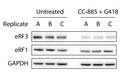
Immunoblot analysis of eRF3 upon treatment of HEK293T cells with CC-885 (10 nM) and G418 (20 μg/mL) for 4 h to induce readthrough (n = 3).
-
Cancer Discov
TNG961 is a selective oral HBS1L molecular glue degrader for the treatment of FOCAD-deleted cancers. [Abstract]2026 Apr 19. PMID: 42001523 -
Nat Commun
2025 Nov 19;16(1):10157. PMID: 41258141 -
Nat Commun
Direct-to-biology, automated, nano-scale synthesis, and phenotypic screening-enabled E3 ligase modulator discovery. [Abstract]2023 Dec 19;14(1):8437. PMID: 38114468
CC-885 purchased from MedChemExpress. Usage Cited in: Nat Commun. 2023 Dec 19;14(1):8437. [Abstract]
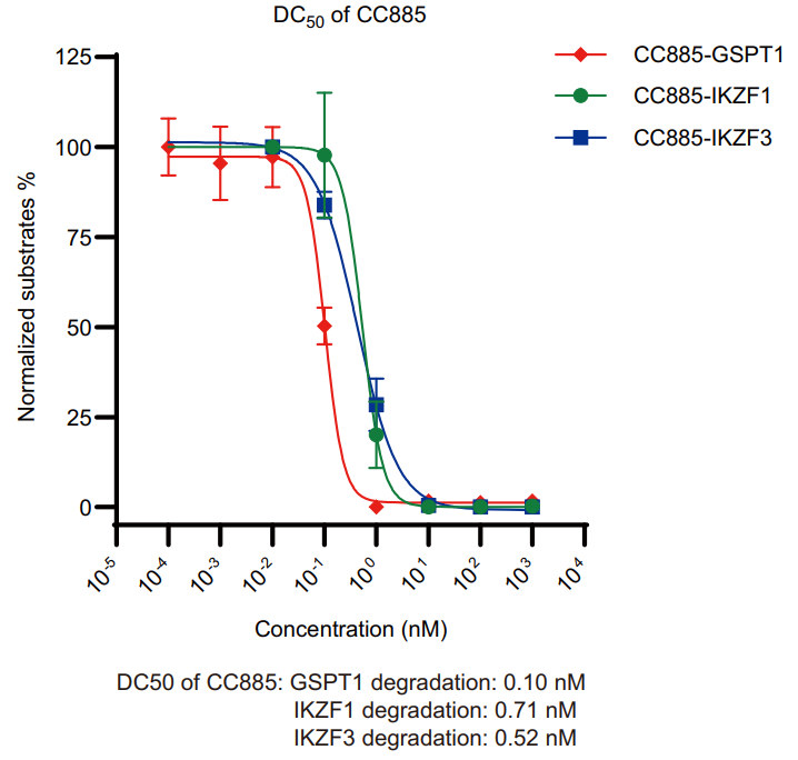
Degradation of neo-substrates by CC-885 (0.00001-10000 nM) in MM.1 S cells after 16 h treatment. Graph bars represent mean values ± SD. n = 3 independent biological replicates. Immunoblot experiment was performed to check protein level. Quantitative protein analysis was performed by software Image J. Source data are provided as a Source Data file.
CC-885 purchased from MedChemExpress. Usage Cited in: Nat Commun. 2023 Dec 19;14(1):8437. [Abstract]
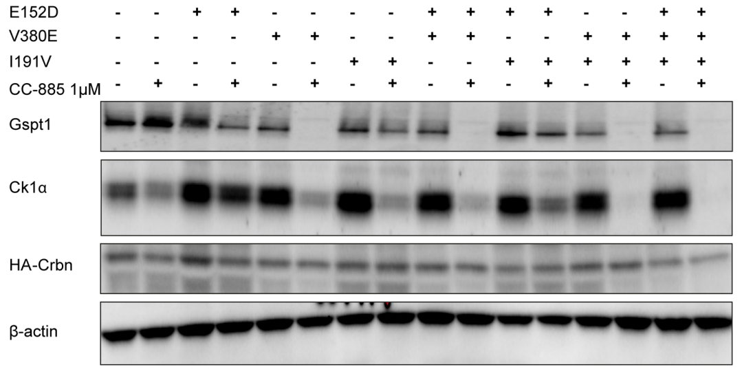
Western blot using lysates from Ba/F3 cells overexpressing Crbn constructs with combinations of humanization mutations E152D, V380E, and I391V as indicated. Protein lysates were harvested following 6 hours of treatment with 1 μM CC-885.
-
Nat Commun
Functional genomics uncovers the transcription factor BNC2 as required for myofibroblastic activation in fibrosis. [Abstract]2022 Sep 10;13(1):5324. PMID: 36088459
CC-885 purchased from MedChemExpress. Usage Cited in: Nat Commun. 2022 Sep 10;13(1):5324. [Abstract]
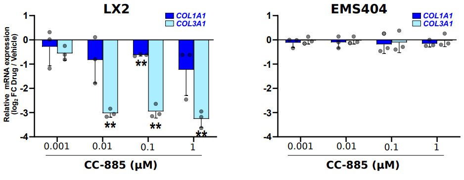
RT-qPCR data showing expression of COL1A1 and COL3A1 upon treatment of LX2 cells (left panel) or EMS404 cells (right panel) with increasing concentrations of CC-885 (0.001-1 μM) or vehicle for 24h (n=3 biologically independent experiments). Log2 FC between drug and vehicle-treated cells are shown. The bar graph shows means ±SD.
-
Mol Cell
2026 Jun 18;86(12):2341-2357.e10. PMID: 42214329 -
Nat Chem Biol
2024 Sep;20(9):1227-1236. PMID: 38514884
CC-885 purchased from MedChemExpress. Usage Cited in: Nat Chem Biol. 2024 Sep;20(9):1227-1236. [Abstract]
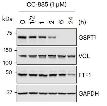
GSPT1, VCL and ETF1 proteins in 293T cells treated with CC-885 (1 μM; 0.5-24 h) over the indicated time course were analyzed by immunoblotting.
-
J Clin Invest
Degradation of GSPT1 causes TP53-independent cell death in leukemia while sparing normal hematopoietic stem cells. [Abstract]2022 Aug 15;132(16):e153514. PMID: 35763353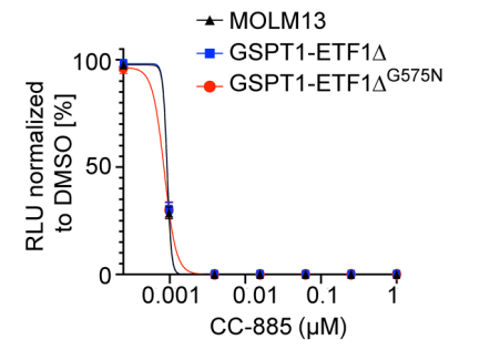
CC-885 purchased from MedChemExpress. Usage Cited in: J Clin Invest. 2022 Aug 15;132(16):e153514. [Abstract]
Cell viability in MOLM13 cells expressing GSPT1-ETF1Δ (drug sensitive and drug resistant; G575N). For all CellTiter-Glo luminescent assays, cell viability was assessed 72 hours after treatment with CC-885 (0.001-1 μM, graphs represent combined data from 3 biological replicates performed in technical triplicate; symbols represent mean ± SEM).
-
Acta Pharmacol Sin
The novel cereblon modulator CC-885 inhibits mitophagy via selective degradation of BNIP3L. [Abstract]2020 Sep;41(9):1246-1254. PMID: 32210356 -
Cell Chem Biol
A Cell-Based Target Engagement Assay for the Identification of Cereblon E3 Ubiquitin Ligase Ligands and Their Application in HDAC6 Degraders. [Abstract]2020 Jul 16;27(7):866-876.e8. PMID: 32413286 -
Leukemia
Targeting of IRAK4 and GSPT1 enhances therapeutic efficacy in AML via c-Myc destabilization. [Abstract]2025 Sep;39(9):2163-2173. PMID: 40670672 -
Mol Ther Oncolytics
A Cereblon Modulator CC-885 Induces CRBN- and p97-Dependent PLK1 Degradation and Synergizes with Volasertib to Suppress Lung Cancer. [Abstract]2020 Jun 23;18:215-225. PMID: 32728610 -
Bioorg Chem
2022 Feb:119:105505. PMID: 34838332 -
Cell Signal
Targeted degradation of GOLM1 by CC-885 via CRL4-CRBN E3 ligase inhibits hepatocellular carcinoma progression. [Abstract]2025 Jun:130:111665. PMID: 39986359 -
J Proteome Res
2022 Aug 5;21(8):1842-1856. PMID: 35848491 -
Dis Model Mech
Recovery of α-L-Fucosidase in Fucosidosis nonsense variants by readthrough stimulation and release factor degradation. [Abstract]2025 Nov 7:dmm.052495. PMID: 41200823 -
Biochem Biophys Res Commun
Cereblon modulator CC-885 induces CRBN-dependent ubiquitination and degradation of CDK4 in multiple myeloma. [Abstract]2021 Apr 16:549:150-156. PMID: 33676183 -
-
bioRxiv
2025 Sep 2:2022.10.31.514544. PMID: 40950106 -


Solvent & Solubility
In Vitro:
DMSO : 66.67 mg/mL (151.22 mM; Need ultrasonic; Hygroscopic DMSO has a significant impact on the solubility of product, please use newly opened DMSO)
Please refer to the solubility information to select the appropriate solvent. Once prepared, please aliquot and store the solution to prevent product inactivation from repeated freeze-thaw cycles.
Storage method and period of stock solution: -80°C, 6 months; -20°C, 1 month. When stored at -80°C, please use it within 6 months. When stored at -20°C, please use it within 1 month.
Please refer to the solubility information to select the appropriate solvent. Once prepared, please aliquot and store the solution to prevent product inactivation from repeated freeze-thaw cycles.
Storage method and period of stock solution: -80°C, 6 months; -20°C, 1 month. When stored at -80°C, please use it within 6 months. When stored at -20°C, please use it within 1 month.
Concentration (start) × Volume (start) = Concentration (final) × Volume (final)
In Vivo:
Select the appropriate dissolution method based on your experimental animal and administration route.
- For the following dissolution methods, please ensure to first prepare a clear stock solution using an In Vitro approach and then sequentially add co-solvents:
- To ensure reliable experimental results, the clarified stock solution can be appropriately stored based on storage conditions. As for the working solution for In Vivo experiments, it is recommended to prepare freshly and use it on the same day.
- The percentages shown for the solvents indicate their volumetric ratio in the final prepared solution. If precipitation or phase separation occurs during preparation, heat and/or sonication can be used to aid dissolution.
Add each solvent one by one: 10% DMSO 40% PEG300 5% Tween-80 45% Saline
Solubility: 2.25 mg/mL (5.10 mM); Suspended solution; Need ultrasonic
This protocol yields a suspended solution of 2.25 mg/mL. Suspended solution can be used for oral and intraperitoneal injection.
Taking 1 mL working solution as an example, add 100 μL DMSO stock solution (22.5 mg/mL) to 400 μL PEG300, and mix evenly; then add 50 μL Tween-80 and mix evenly; then add 450 μL Saline to adjust the volume to 1 mL.
Preparation of Saline: Dissolve 0.9 g sodium chloride in ddH₂O and dilute to 100 mL to obtain a clear Saline solution.
In Vivo Dissolution Calculator
Please enter the basic information of animal experiments:
-
-
-
-
Recommended: Prepare an additional quantity of animals to account for potential losses during experiments.
Please enter your animal formula composition:
-
%DMSO +
Recommended: Keep the proportion of DMSO in working solution below 2% if your animal is weak.
-
%+
-
+%Tween-80 + +
-
%Saline +
The co-solvents required include: DMSO, . All of co-solvents are available by MedChemExpress (MCE). , Tween 80. All of co-solvents are available by MedChemExpress (MCE).
Working solution concentration: 0.22 mg/mL
Method for preparing stock solution: mg drug dissolved in μL DMSO. Stock solution concentration: mg/mL.
1. Take μL DMSO stock solution;
2. Add μL .
μL , mix evenly;
3. Then add μL Tween 80, mix evenly;
4. Then add μL
Please ensure that the stock solution in the first step is dissolved to a clear state, and add co-solvents in sequence. You can use ultrasonic heating (ultrasonic cleaner, recommended frequency 20-40 kHz), vortexing, etc. to assist dissolution.


Purity & Documentation
-
Data Sheet (325 KB)
-
SDS (393 KB)
- English - EN (393 KB)
- Français - FR (393 KB)
- Deutsch - DE (393 KB)
- Norwegian - NO (393 KB)
- Español - ES (393 KB)
- Swedish - SV (393 KB)
- Italian - IT (393 KB)
- Korean - KR (393 KB)
- Portuguese - PT (393 KB)
-
Handling Instructions (2659 KB)
References
[2]. Zhao M, et al. Cereblon modulator CC-885 induces CRBN-dependent ubiquitination and degradation of CDK4 in multiple myeloma. Biochemical and biophysical research communications. 2021 Apr 16;549:150-156. [Content Brief]
[3]. He J, et al. Targeted degradation of GOLM1 by CC-885 via CRL4-CRBN E3 ligase inhibits hepatocellular carcinoma progression. Cellular signalling. 2025 Jun;130:111665. [Content Brief]
[4]. Hao BB, et al. The novel cereblon modulator CC-885 inhibits mitophagy via selective degradation of BNIP3L. Acta pharmacologica Sinica. 2020 Sep;41(9):1246-1254. [Content Brief]
[6]. Matyskiela ME, et al. A novel cereblon modulator recruits GSPT1 to the CRL4(CRBN) ubiquitin ligase. Nature. 2016 Jul 14;535(7611):252-7. [Content Brief]
[9]. Li L, et al. A Cereblon Modulator CC-885 Induces CRBN- and p97-Dependent PLK1 Degradation and Synergizes with Volasertib to Suppress Lung Cancer. Molecular therapy oncolytics. 2020 Sep 25;18:215-225. [Content Brief]


Complete Stock Solution Preparation Table
Please refer to the solubility information to select the appropriate solvent. Once prepared, please aliquot and store the solution to prevent product inactivation from repeated freeze-thaw cycles.
Storage method and period of stock solution: -80°C, 6 months; -20°C, 1 month. When stored at -80°C, please use it within 6 months. When stored at -20°C, please use it within 1 month.
| Optional Solvent | Concentration Solvent Mass | 1 mg | 5 mg | 10 mg | 25 mg |
|---|---|---|---|---|---|
| DMSO | 1 mM | 2.2682 mL | 11.3410 mL | 22.6819 mL | 56.7048 mL |
| 5 mM | 0.4536 mL | 2.2682 mL | 4.5364 mL | 11.3410 mL | |
| 10 mM | 0.2268 mL | 1.1341 mL | 2.2682 mL | 5.6705 mL | |
| 15 mM | 0.1512 mL | 0.7561 mL | 1.5121 mL | 3.7803 mL | |
| 20 mM | 0.1134 mL | 0.5670 mL | 1.1341 mL | 2.8352 mL | |
| 25 mM | 0.0907 mL | 0.4536 mL | 0.9073 mL | 2.2682 mL | |
| 30 mM | 0.0756 mL | 0.3780 mL | 0.7561 mL | 1.8902 mL | |
| 40 mM | 0.0567 mL | 0.2835 mL | 0.5670 mL | 1.4176 mL | |
| 50 mM | 0.0454 mL | 0.2268 mL | 0.4536 mL | 1.1341 mL | |
| 60 mM | 0.0378 mL | 0.1890 mL | 0.3780 mL | 0.9451 mL | |
| 80 mM | 0.0284 mL | 0.1418 mL | 0.2835 mL | 0.7088 mL | |
| 100 mM | 0.0227 mL | 0.1134 mL | 0.2268 mL | 0.5670 mL |


CC-885 Related Classifications
HY-101488 Related Classifications
 Powered by Bioz