



Brazilein
Based on 2 publication(s) in Google Scholar
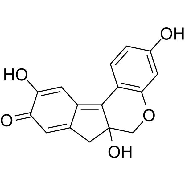
Brazilein is a compound with anti-inflammatory and neuroprotective activities, with an IC50 of 500 μM against guinea pig Na+,K+-ATPase. Brazilein reduces iNOS mRNA expression, thereby inhibiting nitric oxide production in immune cells. Brazilein suppresses inflammatory responses by reducing the mRNA expression of TNF-α and IL-6, but has no effect on IL-1β expression. Brazilein reduces the cerebral infarction volume and improves the neurological function scores of rats with cerebral ischemia-reperfusion injury. Brazilein induces apoptosis of splenic lymphocytes in mice. Brazilein inhibits humoral immune responses in mice, and causes thymus and spleen atrophy as well as body weight loss in mice. Brazilein also possesses antimalarial and antibacterial activities. Brazilein is also a red dye. Brazilein can be used in studies related to the infection, nervous system, cardiovascular system and inflammatory diseases.
Nur für Forschungszwecke. Wir verkaufen nicht an Patienten.
- Reinheit : 99.9%
- CAS. Nr.: 600-76-0
- Formel: C16H12O5
- Molecular Weight:284.26
-
Speicherung:Powder -20°C, 3 years ; In solvent -80°C, 6 months , -20°C, 1 month

To place orders, for customer services and technical support, please contact: MedChemExpress USA
Tel: 609-228-6898 E-mail: [email protected] [email protected]


-
 Biologische Aktivität
-
Chemical Information
-
Lösungsmittel & Löslichkeit
- Protokoll
- Reinheit & Dokumentation
- Verweise
-
 Help & FAQs


-
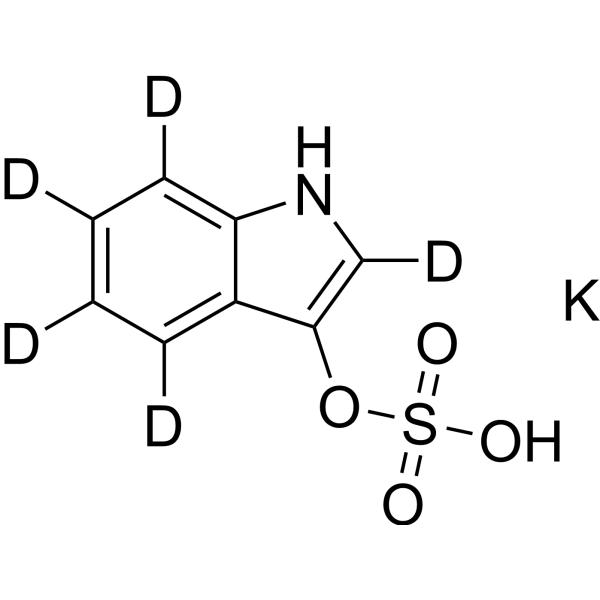
HY-132429S
Potassium 1H-indol-3-yl sulfate-d5Stable Isotope -

HY-N10544
δ-CadineneApoptosis Inducer -
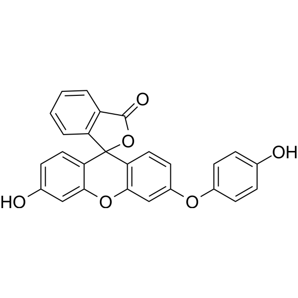
HY-111330
Hydroxyphenyl FluoresceinROS Probe -
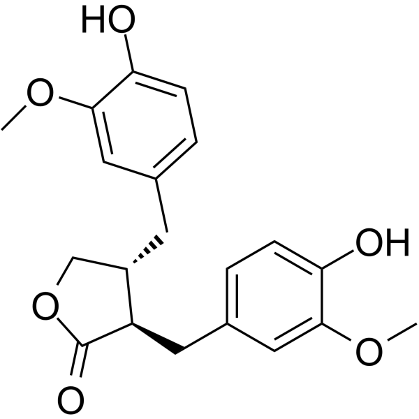
HY-N3312
MatairesinolInflammatory Pathway Inhibitor, Oxidative Stress Inhibitor, Cancer Pathway Inhibitor -

HY-DY1035
Hydroxyphenyl Fluorescein (solution)ROS Probe -
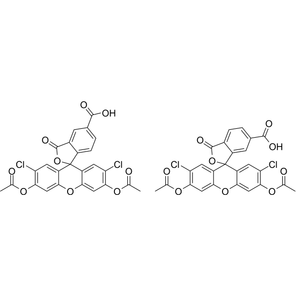
HY-D1078
5(6)-Carboxy-2',7'-dichlorofluorescein diacetateROS Probe -
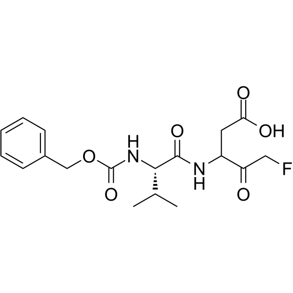
HY-10397A
MX1013Caspase Inhibitor -
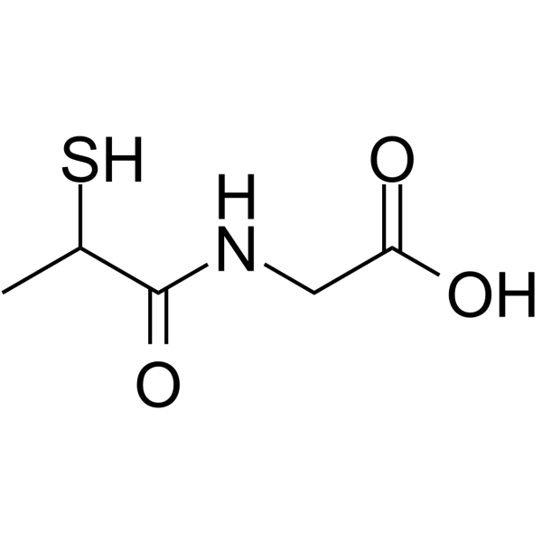
HY-B0373
TioproninAntioxidant -
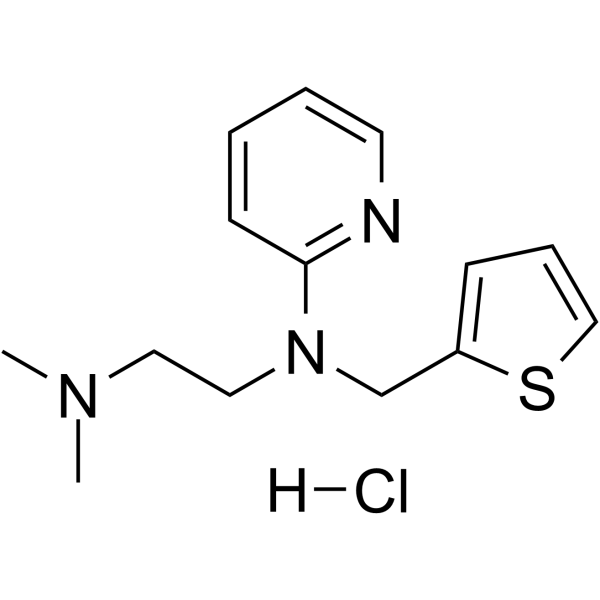
HY-B1483
Methapyrilene hydrochlorideH1-Receptor Antihistamine -

HY-115566
S1PL-IN-1Sphingosine-1-phosphate Lyase Inhibitor -

HY-N3312R
Matairesinol (Standard)Anti-allergic -

HY-117160
4-trans-Hydroxy-glibenclamide
Publications Citing Use of MedChemExpress (MCE) Brazilein
MoreAlle Parasite Isoform-spezifische Produkte anzeigen
More

Biologische Aktivität
Beschreibung
IC50 & Target
[1]|
IL-6 |
iNOS |
Cellular Effect
|
Cell Line

|
Type | Value | Description | References |
|---|---|---|---|---|
| A549 | IC50 |
34.08 μM
Compound: 2, CS-2
|
Cytotoxicity against human A549 cells after 3 days by MTT assay
Cytotoxicity against human A549 cells after 3 days by MTT assay
|
[PMID: 21800859] |
| A549 | IC50 |
9.68 μg/mL
Compound: 1, Brazilein
|
Cytotoxicity against human A549 cells
Cytotoxicity against human A549 cells
|
[PMID: 20036537] |
| Ca9-22 | IC50 |
30.39 μM
Compound: 2, CS-2
|
Cytotoxicity against human Ca9-22 cells after 3 days by MTT assay
Cytotoxicity against human Ca9-22 cells after 3 days by MTT assay
|
[PMID: 21800859] |
| Ca9-22 | IC50 |
8.63 μg/mL
Compound: 1, Brazilein
|
Cytotoxicity against human Ca9-22 cells
Cytotoxicity against human Ca9-22 cells
|
[PMID: 20036537] |
| Hep 3B2 | IC50 |
12.04 μM
Compound: 2, CS-2
|
Cytotoxicity against human Hep3B cells after 3 days by MTT assay
Cytotoxicity against human Hep3B cells after 3 days by MTT assay
|
[PMID: 21800859] |
| Hep 3B2 | IC50 |
3.42 μg/mL
Compound: 1, Brazilein
|
Cytotoxicity against human Hep3B cells
Cytotoxicity against human Hep3B cells
|
[PMID: 20036537] |
| HepG2 | IC50 |
11.09 μM
Compound: 2, CS-2
|
Cytotoxicity against human HepG2 cells after 3 days by MTT assay
Cytotoxicity against human HepG2 cells after 3 days by MTT assay
|
[PMID: 21800859] |
| HepG2 | IC50 |
3.15 μg/mL
Compound: 1, Brazilein
|
Cytotoxicity against human HepG2 cells
Cytotoxicity against human HepG2 cells
|
[PMID: 20036537] |
| MCF7 | IC50 |
13.94 μM
Compound: 2, CS-2
|
Cytotoxicity against human MCF7 cells after 3 days by MTT assay
Cytotoxicity against human MCF7 cells after 3 days by MTT assay
|
[PMID: 21800859] |
| MCF7 | IC50 |
3.96 μg/mL
Compound: 1, Brazilein
|
Cytotoxicity against human MCF7 cells
Cytotoxicity against human MCF7 cells
|
[PMID: 20036537] |
| MDA-MB-231 | IC50 |
2.36 μg/mL
Compound: 1, Brazilein
|
Cytotoxicity against human MDA-MB-231 cells
Cytotoxicity against human MDA-MB-231 cells
|
[PMID: 20036537] |
| MDA-MB-231 | IC50 |
8.31 μM
Compound: 2, CS-2
|
Cytotoxicity against human MDA-MB-231 cells after 3 days by MTT assay
Cytotoxicity against human MDA-MB-231 cells after 3 days by MTT assay
|
[PMID: 21800859] |
| Neutrophil | IC50 |
14.2 μM
Compound: 1, Brazilein
|
Antiinflammatory activity in human neutrophils assessed as inhibition of fMLP/cytochalasin B-induced elastase release
Antiinflammatory activity in human neutrophils assessed as inhibition of fMLP/cytochalasin B-induced elastase release
|
[PMID: 20036537] |
| Neutrophil | IC50 |
4 μM
Compound: 1, Brazilein
|
Antiinflammatory activity in human neutrophils assessed as inhibition of fMLP/cytochalasin B-induced superoxide anion generation
Antiinflammatory activity in human neutrophils assessed as inhibition of fMLP/cytochalasin B-induced superoxide anion generation
|
[PMID: 20036537] |
In Vitro
Brazilein (3.5-28 μM; 16 h) inhibits LPS (HY-D1056)-induced NO production in BV2 and RAW 264.7 cells, with IC50 values of 18.77 μM and 264.18 μM[1].
Brazilein (10-50 μM; 4 h) dose-dependently inhibits LPS-induced mRNA expression of iNOS, TNF-α and IL-6 in BV2 mouse microglia, but exerts no effect on the mRNA expression of IL-1β[1].
Brazilein (1-10 μg/mL; 72 h) dose-dependently inhibits Concanavalin A (HY-P2149)-induced proliferation of mouse splenic T lymphocytes, with an inhibition rate of 56.18% at 10 μg/mL (72 h), 27.25% at 5 μg/mL (72 h), and 10.67% at 1 μg/mL (72 h)[3].
Brazilein (1-10 μg/mL; 72 h) dose-dependently inhibits LPS-induced proliferation of mouse splenic B lymphocytes in vitro, with an inhibition rate of 68.38% at 10 μg/mL (72 h), 54.41% at 5 μg/mL (72 h), 46.69% at 2.5 μg/mL (72 h), and 26.11% at 1 μg/mL (72 h)[3].
Brazilein (1-10 μg/mL; 72 h) induces apoptosis of mitogen-stimulated mouse splenic lymphocytes in a dose-dependent manner in vitro[3].
MedChemExpress (MCE) has not independently confirmed the accuracy of these methods. They are for reference only.
-
Cell Line:LPS-stimulated BV2 mouse microglial cells
-
Concentration:10 μM, 20 μM, 30 μM, 40 μM, 50 μM
-
Incubation Time:4 h (co-incubated with LPS)
-
Result:Inhibited LPS-induced iNOS, TNF-α and IL-6 mRNA expression in a dose-dependent manner.
Showed no significant effect on LPS-induced IL-1β mRNA expression at any tested concentration relative to LPS-only controls.
In Vivo
Brazilein (1-48 mg/kg; intravenous injection; continuous infusion at 0.4 mL/kg/min) dose-dependently alters the electrocardiographic parameters of anesthetized guinea pigs, but does not induce ventricular arrhythmias even at the lethal dose of 48 mg/kg, which causes death via dyspnea[2].
Brazilein (12.5-50 mg/kg/day; intraperitoneal injection; 7 days) exhibits dose-dependent in vivo immunosuppressive activity in healthy mice[3].
MedChemExpress (MCE) has not independently confirmed the accuracy of these methods. They are for reference only.
-
Animal Model:Wistar rats (male, 260-280 g, transient focal cerebral ischemia/reperfusion model)[1]
-
Dosage:2.5 mg/kg; 5 mg/kg; 10 mg/kg
-
Administration:i.v.; single dose at reperfusion onset
-
Result:Reduced brain infarct volume.
Improved neurological scores.
Significantly decreased ischemia-induced TNF-α and IL-6 mRNA expression in ipsilateral cerebral cortex (5 mg/kg), with no significant effect on IL-1β mRNA expression.
Maintained physiological variables (rectal temperature, mean arterial blood pressure, blood gases, pH) within normal limits, with no significant difference from vehicle controls.
-
Animal Model:CD-1 (ICR) (male, adult, 25-30 g)[3]
-
Dosage:12.5-50 mg/kg/day
-
Administration:i.p.; daily; 7 consecutive days
-
Result:Decreased thymus weight/body weight ratio.
Decreased spleen weight/body weight ratio.
Reduced splenic PFC count.
Caused significant atrophy and weight loss of thymus and spleen at 25 and 50 mg/kg/day relative to untreated controls.
Significantly reduced splenic PFC counts at 25 and 50 mg/kg/day relative to untreated controls.
- RT-PCR
- RNA extraction experimental
- Research Protocol for Infectious Diseases
- Detection of Apoptosis
- TUNEL staining for apoptotic DNA fragmentation
- Annexin V plus membrane-impermeant dye apoptosis staining
- Apoptosis Solutions
- Research Protocol for Inflammation-related Diseases
- Real Time qPCR (Q-PCR)
- Pyroptosis Solutions


Chemical Information
-
CAS. Nr. 600-76-0
-
Appearance Solid
-
Molecular Weight 284.26
-
Formel C16H12O5
-
Color Brown to reddish brown
-
SMILES
O=C1C=C2C(C=C1O)=C3C4=C(OCC3(C2)O)C=C(O)C=C4
-
Structure Classification
-
Initial Source
-
Versand
Room temperature in continental US; may vary elsewhere.
-
Speicherung
Powder -20°C 3 years In solvent -80°C 6 months -20°C 1 month


Publications (2)
-
Journal Impact Factor
-
Most Recent
-
Free Radic Biol Med
Brazilin and its oxidized form "brazilein", the natural fluorescently traceable agents, toxify Plasmodium falciparum to pyknotic death via disrupting its iron homeostasis. [Abstract]2026 Feb 28:248:367-385. PMID: 41771360 -
ACS Omega
Identification of New Modulators and Inhibitors of Palmitoyl-Protein Thioesterase 1 for CLN1 Batten Disease and Cancer. [Abstract]2024 Feb 28;9(10):11870-11882. PMID: 38496939


Lösungsmittel & Löslichkeit
In Vitro:
DMF : 11 mg/mL (38.70 mM; Need ultrasonic and warming)
Please refer to the solubility information to select the appropriate solvent. Once prepared, please aliquot and store the solution to prevent product inactivation from repeated freeze-thaw cycles.
Storage method and period of stock solution: -80°C, 6 months; -20°C, 1 month. When stored at -80°C, please use it within 6 months. When stored at -20°C, please use it within 1 month.
Please refer to the solubility information to select the appropriate solvent. Once prepared, please aliquot and store the solution to prevent product inactivation from repeated freeze-thaw cycles.
Storage method and period of stock solution: -80°C, 6 months; -20°C, 1 month. When stored at -80°C, please use it within 6 months. When stored at -20°C, please use it within 1 month.
Konzentration (Stammlösung) × Volumen (Stammlösung) = Konzentration (Ziellösung) × Volumen (Ziellösung)


Reinheit & Dokumentation
-
Data Sheet (286 KB)
-
SDS (251 KB)
- English - EN (251 KB)
- Français - FR (251 KB)
- Deutsch - DE (251 KB)
- Norwegian - NO (251 KB)
- Español - ES (251 KB)
- Swedish - SV (251 KB)
- Italian - IT (251 KB)
- Korean - KR (251 KB)
- Portuguese - PT (251 KB)
-
Handling Instructions (2659 KB)
Verweise
[1]. Shen J, et al. Brazilein protects the brain against focal cerebral ischemia reperfusion injury correlating to inflammatory response suppression. Eur J Pharmacol. 2007;558(1-3):88-95. [Content Brief]
[2]. Zhao YN, et al. Study on cardioactive effects of brazilein. Pharmacology. 2006;76(2):76-83. [Content Brief]
[3]. Ye M, et al. Brazilein, an important immunosuppressive component from Caesalpinia sappan L. Int Immunopharmacol. 2006;6(3):426-432. [Content Brief]
[4]. Kulaphisit M, et al. Brazilin and its oxidized form "brazilein", the natural fluorescently traceable agents, toxify Plasmodium falciparum to pyknotic death via disrupting its iron homeostasis. Free Radic Biol Med. 2026 May;248:367-385. [Content Brief]


Complete Stock Solution Preparation Table
Please refer to the solubility information to select the appropriate solvent. Once prepared, please aliquot and store the solution to prevent product inactivation from repeated freeze-thaw cycles.
Storage method and period of stock solution: -80°C, 6 months; -20°C, 1 month. When stored at -80°C, please use it within 6 months. When stored at -20°C, please use it within 1 month.
| Optional Solvent | Concentration Solvent Mass | 1 mg | 5 mg | 10 mg | 25 mg |
|---|---|---|---|---|---|
| DMF | 1 mM | 3.5179 mL | 17.5895 mL | 35.1791 mL | 87.9477 mL |
| 5 mM | 0.7036 mL | 3.5179 mL | 7.0358 mL | 17.5895 mL | |
| 10 mM | 0.3518 mL | 1.7590 mL | 3.5179 mL | 8.7948 mL | |
| 15 mM | 0.2345 mL | 1.1726 mL | 2.3453 mL | 5.8632 mL | |
| 20 mM | 0.1759 mL | 0.8795 mL | 1.7590 mL | 4.3974 mL | |
| 25 mM | 0.1407 mL | 0.7036 mL | 1.4072 mL | 3.5179 mL | |
| 30 mM | 0.1173 mL | 0.5863 mL | 1.1726 mL | 2.9316 mL |


Brazilein Related Classifications
HY-N10424 Related Classifications
 Powered by Bioz