



Punicic acid
Based on 1 publication(s) in Google Scholar
Punicic acid is a bioactive compound of pomegranate seed oil. Punicic acid is an isomer of conjugated α-linolenic acid and ω-5 polyunsaturated fatty acids. Punicic acid has anti-inflammatory and antioxidant activities and can inhibit the expression of inflammatory mediators such as tumor necrosis factor α (TNF-α). Punicic acid can also reduce the formation of β-amyloid deposits and hyperphosphorylation of tau by increasing the expression of GLUT4 protein and inhibiting the overactivation of calpain, and is used to prevent and treat neurodegenerative diseases. In addition, punicic acid also has breast cancer inhibitor properties that depend on lipid peroxidation and PKC pathways.
For research use only. We do not sell to patients.
- Purity : 98.5%
- CAS No.: 544-72-9
- Formula: C18H30O2
- Molecular Weight:278.43
-
Storage:
Solution, -20°C, 2 years

To place orders, for customer services and technical support, please contact: MedChemExpress USA
Tel: 609-228-6898 E-mail: [email protected] [email protected]


-
 Biological Activity
-
Chemical Information
- Protocols
- Purity & Documentation
- References
-
 Help & FAQs


-
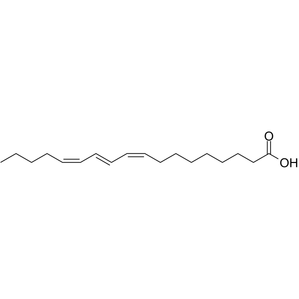
HY-27787
α-Eleostearic acidApoptosis Inducer -
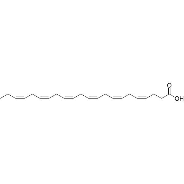
HY-B2167
Docosahexaenoic acidomega-3 Fatty Acid -
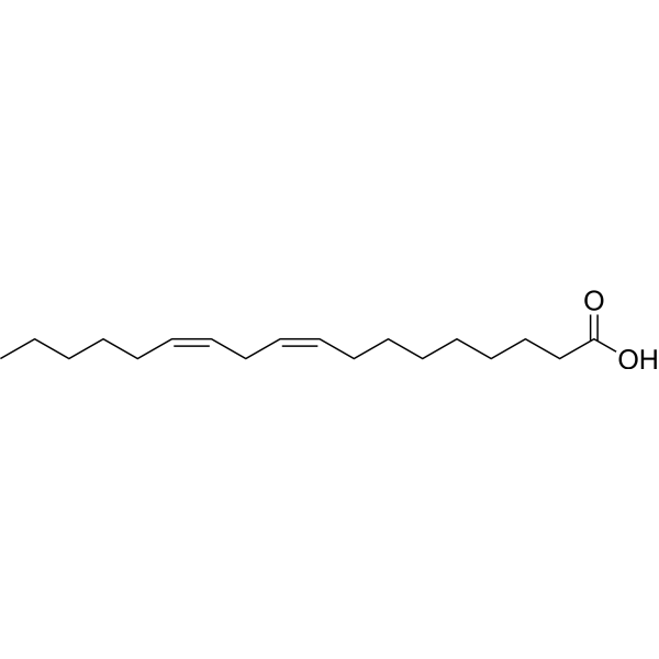
HY-N0729
Linoleic acidHuman Endogenous Metabolite -

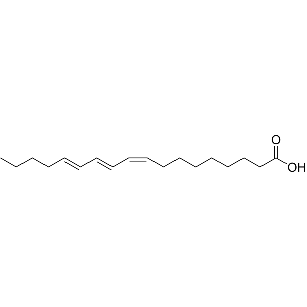
HY-131405
β-Eleostearic acidApoptosis Inducer -
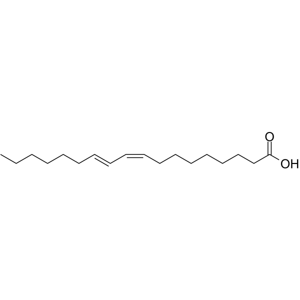
HY-113162
Bovinic acidAnti-inflammatory Agent -
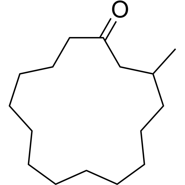
HY-N0633
MusconeTNF Receptor Inhibitor -
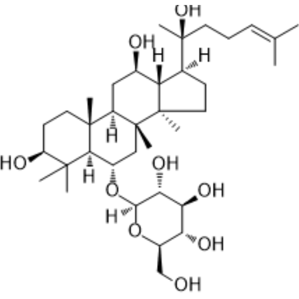
HY-N0604
Ginsenoside Rh1Endogenous Metabolite -
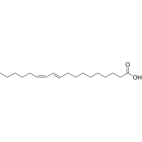
HY-116538
(10E,12Z)-Octadeca-10,12-dienoic acidPPARα Activator -
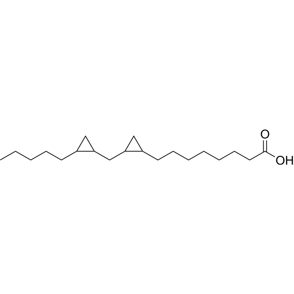
HY-108599
DCP-LAPKC Activator -

HY-W347368
9Z,11E,13Z-Octadecatrienoicacid, methylesterEndogenous Metabolite -
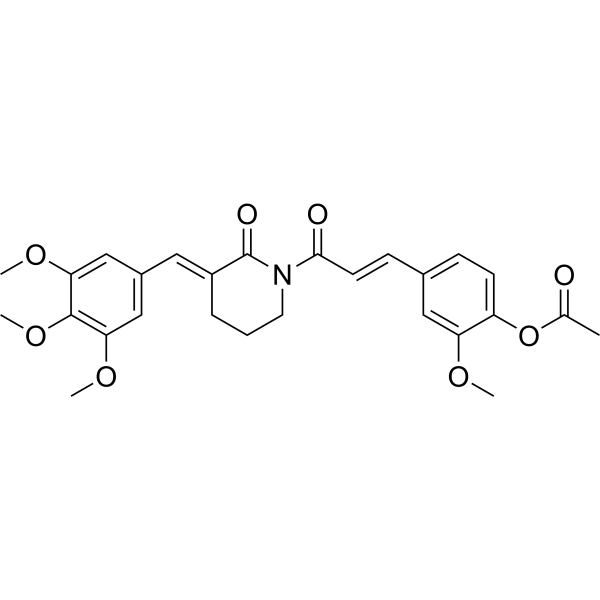
HY-148552
Anti-inflammatory agent 35Anti-inflammatory Agent -
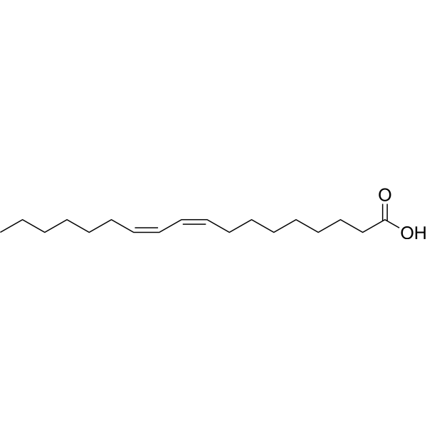
HY-113102B
9(Z),11(Z)-Octadecadienoic acidLinoleic Acid Isomer
Publications Citing Use of MedChemExpress (MCE) Punicic acid
More

Biological Activity
Description
Cellular Effect
|
Cell Line

|
Type | Value | Description | References |
|---|---|---|---|---|
| FaDu | IC50 |
9 μM
Compound: Punicic acid
|
Cytotoxicity against human FaDu cells assessed as reduction in cell growth measured after 24 hrs by MTT assay
Cytotoxicity against human FaDu cells assessed as reduction in cell growth measured after 24 hrs by MTT assay
|
[PMID: 38306267] |
| HCT-116 | IC50 |
5 nM
Compound: Punicic acid
|
Cytotoxicity against human HCT-116 cells assessed as reduction in cell viability measured after 48 hrs by MTT assay
Cytotoxicity against human HCT-116 cells assessed as reduction in cell viability measured after 48 hrs by MTT assay
|
[PMID: 38306267] |
| HCT-116 | IC50 |
5 μM
Compound: 60; Pun A
|
Antiproliferative activity against human HCT-116 cells assessed as reduction in cell viability measured after 72 hrs by presto blue reagent based analysis
Antiproliferative activity against human HCT-116 cells assessed as reduction in cell viability measured after 72 hrs by presto blue reagent based analysis
|
[PMID: 38142509] |
| T98G | IC50 |
9.85 μL/mL
Compound: 161
|
Growth inhibition of human T98 GBM cells
Growth inhibition of human T98 GBM cells
|
[PMID: 35786935] |
In Vitro
Punicic acid (10 μM, 4 min) inhibits TNF-α induced neutrophil ROS overproduction without affecting fMLP or PMA (HY-18739) induced responses[2]. Punicic acid (0-40 μM, 30 min) reduces TNF-α induced p47phox phosphorylation starting from 10 mM concentration[2]. Punicic acid (10 μM, 30 min) inhibits TNF-α induced NADPH oxidase activation[2]. Punicic acid (0-40 μM; 48 h) exhibits a concentration-dependent antiproliferative effect on MDA-ERα7 and MDA-wt cells[4]. Punicic acid (40 μM; 48 h) induces apoptosis of MDA-ERα7 and MDA-wt cells and increases the percentage of cells in the G2/M phase. The ability to induce apoptosis of MDA-ERα7 and MDA-wt cells is completely blocked in the presence of 20 μM α-tocotrienol (HY-129459)[4]. Punicic acid (5-80 μM; 48 h) is partially blocked in the presence of BIM (4 μM) on MDA-ERα7 and MDA-wt cells[4].
MedChemExpress (MCE) has not independently confirmed the accuracy of these methods. They are for reference only. Further protocols information, click here.
-
Cell Line:Neutrophils
-
Concentration:0, 10, 20, 40 µM
-
Incubation Time:30 min
-
Result:Reduced TNFa-induced p47phox phosphorylation starting from 10 µM concentration. Inhibited p38MAPK phosphorylation at 20 µM similarly to p47phox inhibition of Ser345 phosphorylation, but almost no phospho-Ser345 and phosphoP38MAPK were detected.
-
Cell Line:MDA-ERα7 and MDA-wt cells
-
Concentration:40 µM
-
Incubation Time:48 h
-
Result:The apoptosis rate of BRCA cells is 86-91%.
-
Cell Line:MDA-ERα7 and MDA-wt cells
-
Concentration:0-40 µM
-
Incubation Time:48 h
-
Result:The proliferation rate of MDA-ERα7 was only 4.5%, while that of MDA-wt was 8.5% at a dose of 40 µM.
-
Cell Line:MDA-ERα7 and MDA-wt cells
-
Concentration:40 µM
-
Incubation Time:48 h
-
Result:The percent of MDA-ERα7 cells in the G2/M phase increased from 27.3 to 41.3% and the percent of MDA-wt cells in the G2/M phase increased from 12.3 to 20.3%.
In Vivo
MedChemExpress (MCE) has not independently confirmed the accuracy of these methods. They are for reference only.
-
Animal Model:TNBS-induced Colitis male Wistar rat[1]
-
Dosage:400 μg
-
Administration:i.g.
-
Result:Reduced the hyperactivation of neutrophils in the colon of TNBS-treated rats and inhibited the release of MPO and ROS by neutrophils, thereby reducing lipid peroxidation and tissue damage.


Chemical Information
-
CAS No. 544-72-9
-
Appearance Liquid
-
Molecular Weight 278.43
-
Formula C18H30O2
-
Color Colorless to light yellow
-
SMILES
CCCC/C=C\C=C\C=C/CCCCCCCC(O)=O
-
Synonyms
Trichosanic acid
-
Structure Classification
-
Initial Source
-
Shipping
Room temperature in continental US; may vary elsewhere.
-
Storage
Solution, -20°C, 2 years


Publications (1)
-
Journal Impact Factor
-
Most Recent
-
Sci Rep
Influence of vegetable oils and their constituents on in vitro human keratinocyte and fibroblast proliferation and migration. [Abstract]2025 Jul 24;15(1):26898. PMID: 40707519

Protocols
-
RNA extraction experimental
By lysing cells, releasing RNA, and removing impurities such as proteins and DNA, high-purity RNA products are finally obtained. The commonly used traditional method is the guanidine isothiocyanate/phenol/chloroform method (Trizol), which is suitable for a variety of animal materials including animal tissues, microorganisms, cultured cells, etc., and most plant materials.
-
Research Protocol for Inflammation-related Diseases
The NLRP3 inflammasome is a cytosolic innate immune signaling platform that integrates priming signals and danger-signal activation to promote caspase-1 activation, maturation of IL-1β and IL-18, and gasdermin D-mediated pyroptotic cell death. The core experimental logic is to determine whether inflammatory disease phenotypes are driven by increased NLRP3 expression, ASC-containing inflammasome assembly, caspase-1 cleavage, GSDMD cleavage, and extracellular release of IL-1β/IL-18 rather than by nonspecific cell injury alone. The pathway is strongly linked to inflammation-related disease phenotypes because monosodium urate crystals activate NALP3/NLRP3 inflammasome signaling in gout-like crystal inflammation, cholesterol crystals activate NLRP3 inflammasomes in atherogenesis models, and DSS-induced intestinal inflammation has been reported to involve NLRP3 inflammasome activity. However, experimental colitis studies also show context-dependent protective effects of NLRP3 inflammasome co
-
Ferroptosis Solutions
Ferroptosis is an iron-dependent, non-apoptotic form of regulated cell death characterized by lethal lipid peroxidation and sensitivity to suppression by iron chelators or lipophilic radical-trapping antioxidants. The core pathway links cystine uptake through system Xc−, glutathione availability, GPX4-dependent detoxification of phospholipid hydroperoxides, iron-dependent oxidative reactions, and polyunsaturated-phospholipid metabolism into a cell-death program that is biochemically and morphologically distinct from apoptosis, necrosis, and autophagy. The ferroptosis pathway is experimentally linked to phenotype through chemical and genetic perturbation. Erastin induces ferroptosis by inhibiting cystine uptake through system Xc− and weakening antioxidant defenses, while GPX4 inhibition or depletion causes lipid peroxide accumulation and ferroptotic cancer-cell death. ACSL4 and oxidizable arachidonoyl- or adrenoyl-containing phosphatidylethanolamines shape ferroptosis sensitivity by con
-
Amyloid: Congo Red Amyloid Staining
Congo red amyloid staining is a histochemical method used to detect extracellular amyloid deposits in tissue sections based on the affinity of Congo red dye for β-pleated sheet-rich protein aggregates. When bound to amyloid, Congo red produces characteristic apple-green birefringence under polarized light microscopy, which is widely regarded as a diagnostic feature of amyloid deposition in histopathology. The diagnostic principle relies on the combination of dye binding (congophilia) and optical anisotropy under polarized illumination, which distinguishes amyloid from most non-amyloid eosinophilic extracellular deposits in routine histological evaluation. Amyloid identification by Congo red staining remains a cornerstone in diagnostic pathology despite the availability of adjunct methods such as immunohistochemistry and mass spectrometry, particularly because of its ability to localize deposits directly within tissue architecture. The specificity of Congo red-positive deposits is incre
-
Breast Cancer Modeling
Breast cancer is a heterogeneous cancer, and it has been distinguished into four subtypes: luminal A, luminal B, HER2-positive and basal-like. Molecular mutations, epigenetic alterations, hormone exposure and immune microenvironment are related to the progression of breast cancer.


Purity & Documentation
-
Data Sheet (272 KB)
-
SDS (419 KB)
- English - EN (419 KB)
- Français - FR (419 KB)
- Deutsch - DE (419 KB)
- Norwegian - NO (419 KB)
- Español - ES (419 KB)
- Swedish - SV (419 KB)
- Italian - IT (419 KB)
- Korean - KR (419 KB)
- Portuguese - PT (419 KB)
-
Handling Instructions (2659 KB)
References
[1]. Shabbir MA, et al. Punicic acid: A striking health substance to combat metabolic syndromes in humans. Lipids Health Dis. 2017;16(1):99. [Content Brief]
[2]. Boussetta T, et al. Punicic acid a conjugated linolenic acid inhibits TNFα-induced neutrophil hyperactivation and protects from experimental colon inflammation in rats[J]. PloS one, 2009, 4(7): e6458. [Content Brief]
[3]. Guerra-Vázquez, et al. Punicic acid and its role in the prevention of neurological disorders: A review. Foods 11.3 (2022): 252. [Content Brief]
[4]. Grossmann, et al. Punicic acid is an ω-5 fatty acid capable of inhibiting breast cancer proliferation. International journal of oncology 36.2 (2010): 421-426. [Content Brief]

Calculators
Concentration (start) × Volume (start) = Concentration (final) × Volume (final)


Punicic acid Related Classifications
HY-139066 Related Classifications
 Powered by Bioz