



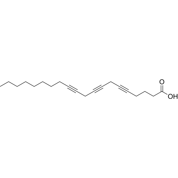
Eicosatetraynoic acid

Based on 1 Customer Validation
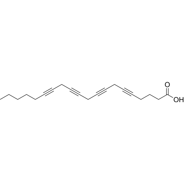
Eicosatetraynoic acid (ETYA) is a non-metabolizable analog of Arachidonic acid (HY-109590) and also an inhibitor of the lipoxygenase (LOX)/cyclooxygenase (COX) pathway (ID50 = 8 μM and 4 μM). Eicosatetraynoic acid acts as a suicide substrate to inhibit the production of inflammatory mediators such as leukotrienes and prostaglandins. Eicosatetraynoic acid acts directly on cell membranes and membrane proteins to exert a wide range of effects, including blocking potassium channels, increasing cell membrane fluidity, elevating intracellular calcium levels, inhibiting DNA synthesis in tumor cells, inducing differentiation of certain cells, and specifically inhibiting the assembly and replication of orthopoxviruses. Eicosatetraynoic acid alleviates acute lung injury induced by chemicals such as phosgene.
For research use only. We do not sell to patients.
- Purity : 99.9%
- CAS No.: 1191-85-1
- Formula: C20H24O2
- Molecular Weight:296.40
-
Storage:Powder -20°C, 3 years
* The compound is unstable in solutions, freshly prepared is recommended.

To place orders, for customer services and technical support, please contact: MedChemExpress USA
Tel: 609-228-6898 E-mail: [email protected] [email protected]


-
 Biological Activity
-
Chemical Information
-
Solvent & Solubility
- Protocols
- Purity & Documentation
- References
-
 Help & FAQs


-
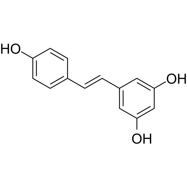
HY-16561
ResveratrolPolyphenolic Phytoalexin -
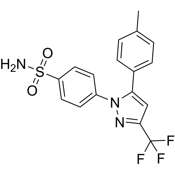
HY-14398
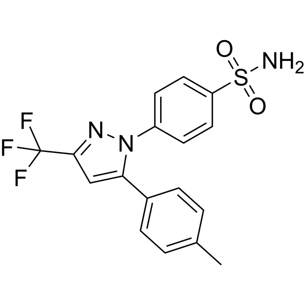
CelecoxibCOX-2inhibitor -
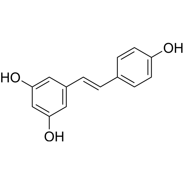
HY-16561R
Resveratrol (Standard) -
HY-14398R
Celecoxib (Standard)COX-2inhibitor -
HY-124252
5,8,11-Eicosatriynoic acidLipoxygenase Inhibitor -

HY-16561G
Resveratrol (GMP)Polyphenolic Phytoalexin -
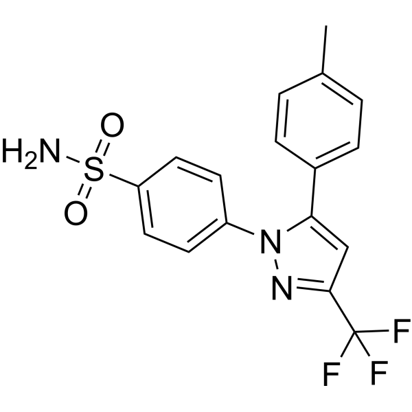
HY-14398G
Celecoxib (GMP)COX-2 Inhibitor -
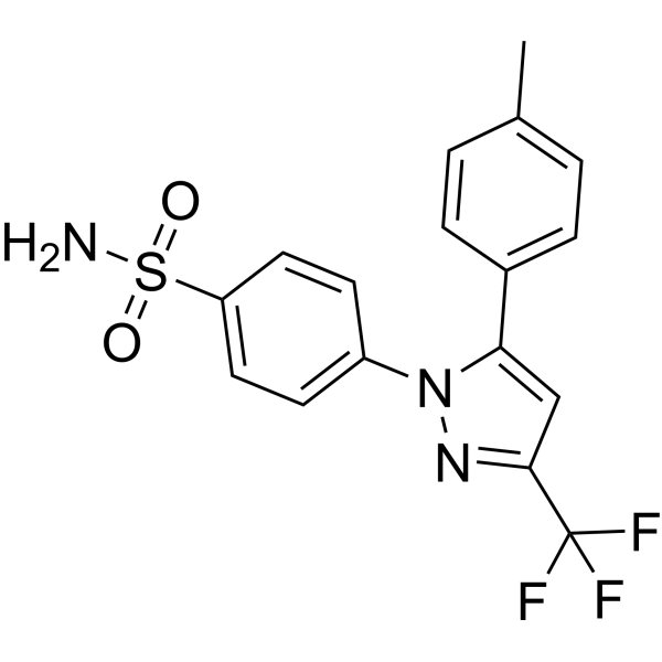
HY-14398GL
Celecoxib (GMP Like)COX-2inhibitor
All DNA/RNA Synthesis Isoforms
More

Biological Activity
Description
IC50 & Target
[2]|
COX 8 μM (ID50) |
In Vitro
Eicosatetraynoic acid dose-dependently reduces MDA production in cultured human umbilical vein endothelial cells co-incubated with low-density lipoprotein[1].
Eicosatetraynoic acid (40 μM; 4-72 h) reversibly inhibits DNA synthesis and blocks the proliferation of PC3, U937 and A172 cells without inducing significant cytotoxicity[2].
Eicosatetraynoic acid (40 μM; 3-7 d) induces partial monocyte-like differentiation in U937 cells, and induces partial glial-like differentiation in A172 cells after 72 h of incubation; in addition, the differentiation of U937 cells progresses gradually over 5 to 7 days[2].
Incubation with eicosatetraynoic acid (40 μM; 72 h) for 72 h induces cell type-specific ultrastructural changes, including severe oxidative stress-related mitochondrial damage in PC3 cells, milder damage in A172 cells, and immature monocyte morphology in U937 cells[2].
Eicosatetraynoic acid (40 μM) rapidly regulates multiple signal transduction pathways in PC3 and U937 cells, including increasing membrane fluidity and intracellular Ca2+ levels, inhibiting O2 uptake, reversibly blocking DNA synthesis, downregulating c-myc, inhibiting eicosanoid synthesis, and altering the localization of protein kinase C[2].
Eicosatetraynoic acid non-selectively inhibits 12-lipoxygenase (ID50 = 4 μM) and fatty acid cyclooxygenase (ID50 = 8 μM) in washed human platelets[3].
Eicosatetraynoic acid (79-316 μM) specifically and effectively blocks the replication of orthopoxviruses (vaccinia, vaccinia vaccine, and ectromelia viruses)[4].
MedChemExpress (MCE) has not independently confirmed the accuracy of these methods. They are for reference only. Further protocols information, click here.
-
Cell Line:PC3 prostate, U937 monoblastoid, A172 glioblastoma cells
-
Concentration:40 μM
-
Incubation Time:4 h, 72 h (DNA synthesis and cell number measurements); 4 h, 72 h (viability assays)
-
Result:Inhibited DNA synthesis in all three cell lines.
Reduced PC3 cell proliferation over 72 h, with control cells increasing from 1.3 million to 3.1 million while treated cells remained at 0.99 million, and maintained >95% viability.
Showed reversible inhibition of DNA synthesis at 4 h and 72 h, with no induced change in Cr51 release from prelabelled PC3 and U937 cells, and kept PC3/A172 cells attached to substrates.
-
Cell Line:U937 monoblastoid, A172 glioblastoma cells
-
Concentration:40 μM
-
Incubation Time:72 h; 5 to 7 days (extended U937 cell cultures)
-
Result:Induced reduced nuclear-to-cytoplasmic ratio, increased cytoplasm, irregular plasma membranes, increased nonspecific acid phosphatase activity, latex bead ingestion, and increased expression of the monocyte-associated surface epitope leu M3 (cd14) in U937 cells.
Caused spindle-shaped morphology, increased glial fibrillary acidic protein (GFAP) expression, and prominent bundles of 8 nm glial filaments in A172 cells.
Produced a progressive increase in differentiated cell functions in U937 cells during extended 5 to 7 day culture.
In Vivo
Eicosatetraynoic acid (300 μM; topical administration) significantly reduces pock formation on the chorioallantoic membranes of chicken embryos infected with the vaccinia virus CPV-BR.D1[4].
MedChemExpress (MCE) has not independently confirmed the accuracy of these methods. They are for reference only.
-
Animal Model:Hartley (male, 600-700 g, phosgene-induced acute lung injury)[1]
-
Dosage:100 µM (i.p. post-exposure); 50 µM (perfusate, every 40 mins starting 60 mins post-exposure)
-
Administration:i.p.; single dose (5 mins post-phosgene exposure); perfusate; every 40 minutes (starting 60 mins post-exposure initiation)
-
Result:Prevented phosgene-induced acute lung injury in male Hartley guinea pigs by maintaining lung GSH levels, reducing pulmonary edema, and increasing the GSH/TBARS protection ratio to 12.
-
Animal Model:12-day-old chick embryo[4]
-
Dosage:300 μM
-
Administration:topical (alongside virus infection)
-
Result:Severely reduced the number of pocks detected on chorioallantoic membranes.


Chemical Information
-
CAS No. 1191-85-1
-
Appearance Solid
-
Molecular Weight 296.40
-
Formula C20H24O2
-
Color Light yellow to brown
-
SMILES
CCCCCC#CCC#CCC#CCC#CCCCC(O)=O
-
Synonyms
ETYA
-
Shipping
Room temperature in continental US; may vary elsewhere.
-
Storage
Powder -20°C 3 years * The compound is unstable in solutions, freshly prepared is recommended.


Solvent & Solubility
In Vitro:
DMSO : 2 mg/mL (6.75 mM; Need ultrasonic; Hygroscopic DMSO has a significant impact on the solubility of product, please use newly opened DMSO)
Ethanol : < 1 mg/mL (insoluble)
Please refer to the solubility information to select the appropriate solvent. The compound is unstable in solutions, freshly prepared is recommended.
Please refer to the solubility information to select the appropriate solvent. The compound is unstable in solutions, freshly prepared is recommended.
Concentration (start) × Volume (start) = Concentration (final) × Volume (final)

Protocols
-
EdU Incorporation Assay (Click Chemistry-Based DNA Synthesis Measurement)
The EdU incorporation assay measures DNA synthesis by adding the thymidine analog 5-ethynyl-2′-deoxyuridine to cells or tissues, where it is incorporated into newly synthesized DNA during S phase. Incorporated EdU is detected by copper-catalyzed azide-alkyne cycloaddition, in which a fluorescent azide covalently reacts with the ethynyl group on EdU, allowing S-phase cells to be detected by fluorescence microscopy, flow cytometry, or high-content imaging. EdU detection does not require DNA denaturation or anti-BrdU antibody access, which preserves sample structure and improves compatibility with immunostaining and multiparameter cytometry compared with BrdU-based detection. EdU can be cytotoxic in a cell-type- and exposure-dependent manner, so pulse duration, concentration, and continuous-labeling designs should be validated for each cell type.
-
BrdU Incorporation Assay
Bromodeoxyuridine (BrdU) incorporation assay is based on the principle that BrdU, a thymidine analog, is incorporated into newly synthesized DNA during the S phase of the cell cycle, thereby serving as a marker of DNA replication and cellular proliferation. Incorporated BrdU can be detected using anti-BrdU antibodies following DNA denaturation, enabling visualization or quantification of proliferating cells through immunochemical detection methods such as immunofluorescence or immunohistochemistry.
-
Calcium Spark Assay
Calcium sparks are localized, transient increases in intracellular calcium concentration ([Ca2+]i) that occur in cardiac myocytes and represent elementary events underlying excitation-contraction coupling. These events are generated by the coordinated opening of clusters of ryanodine receptors (RyRs) on the sarcoplasmic reticulum membrane, leading to a brief release of Ca2+ into the cytosol. The detection and analysis of calcium sparks provide insights into the mechanisms of calcium handling and signaling in cardiac cells. Imaging techniques using fluorescent calcium indicators such as Fluo-3 are employed to visualize these subcellular calcium transients with high spatial and temporal resolution. The protocol is based on established methodologies described in primary literature for both experimental measurement and automated analysis of calcium sparks.
-
Ca2+ Staining Technique
Ca2+ staining is an experimental technique that utilizes specific fluorescent probes (such as Fluo-4 AM, Fura-2, etc.) to qualitatively or quantitatively detect dynamic changes in intracellular Ca2+ concentrations; this is achieved by monitoring the changes in fluorescent signals generated when these probes bind to free intracellular calcium ions. The underlying principle relies primarily on the presence of chelating groups within the probe's molecular structure that possess high affinity for calcium ions.
-
Inhalation Toxicity Study
Inhalation toxicity studies expose rodents to a controlled aerosol, vapor, gas, or smoke atmosphere and assess respiratory and systemic toxicity using exposure-atmosphere characterization, clinical observations, body and organ weights, bronchoalveolar lavage fluid, histopathology, blood chemistry, hematology, and, when included, molecular endpoints such as transcriptomics, proteomics, lipidomics, or tissue burden analysis. The primary biological readouts are airway irritation, pulmonary inflammation, cytotoxicity, altered surfactant or lipid homeostasis, impaired particle clearance, and tissue remodeling, reflected by BALF cell differentials, BALF protein, LDH, phosphatase activities, cytokines, lung weight, microscopic respiratory-tract lesions, and retained lung burden.


Purity & Documentation
-
Data Sheet (275 KB)
-
SDS (254 KB)
- English - EN (254 KB)
- Français - FR (254 KB)
- Deutsch - DE (254 KB)
- Norwegian - NO (254 KB)
- Español - ES (254 KB)
- Swedish - SV (254 KB)
- Italian - IT (254 KB)
- Korean - KR (254 KB)
- Portuguese - PT (254 KB)
-
Handling Instructions (2659 KB)
References
[1]. Sciuto AM, et al. Posttreatment with ETYA protects against phosgene-induced lung injury by amplifying the glutathione to lipid peroxidation ratio. Inhal Toxicol. 2000;12(4):347-356. [Content Brief]
[2]. Anderson KM, et al. ETYA, a pleotropic membrane-active arachidonic acid analogue affects multiple signal transduction pathways in cultured transformed mammalian cells. Clin Biochem. 1992;25(1):1-9. [Content Brief]
[3].
Hammarström S. Selective inhibition of platelet n-8 lipoxygenase by 5,8,11-eicosatriynoic acid. Biochim Biophys Acta. 1977 Jun 22;487(3):517-9.
[Content Brief]
[4]. Palumbo GJ, et al. Inhibitors of the lipoxygenase pathway specifically block orthopoxvirus replication. Virology. 1991;180(1):457-463. [Content Brief]
[5]. Kehl SJ, et al. Eicosatetraynoic acid (ETYA), a non-metabolizable analogue of arachidonic acid, blocks the fast-inactivating potassium current of rat pituitary melanotrophs. Can J Physiol Pharmacol. 2001;79(4):338-345. [Content Brief]


Complete Stock Solution Preparation Table
Please refer to the solubility information to select the appropriate solvent. The compound is unstable in solutions, freshly prepared is recommended.
| Optional Solvent | Concentration Solvent Mass | 1 mg | 5 mg | 10 mg | 25 mg |
|---|---|---|---|---|---|
| DMSO | 1 mM | 3.3738 mL | 16.8691 mL | 33.7382 mL | 84.3455 mL |
| 5 mM | 0.6748 mL | 3.3738 mL | 6.7476 mL | 16.8691 mL |


Eicosatetraynoic acid Related Classifications
HY-124108 Related Classifications
 Powered by Bioz
Keywords
- Eicosatetraynoic acid
- 1191-85-1
- ETYA
- COX
- Lipoxygenase
- Orthopoxvirus
- Potassium Channel
- DNA/RNA Synthesis
- Drug Derivative
- lipoxygenase/cyclooxygenase pathway
- prostate cancer
- prostaglandin
- arachidonic acid
- histiocytic lymphoma
- BS-C-1 African green monkey kidney cells
- PC3 cells
- leukotriene
- glioblastoma
- orthopoxvirus
- Inhibitor
- inhibitor
- inhibit