



Golimumab

Based on 1 Customer Validation
Golimumab (CNTO-148) is a potent human IgG1 TNFα antagonist monoclonal antibody. Golimumab has anti-inflammation activitity and inhibits IL-6 and IL-1β production. Golimumab acts via targeting and neutralizing TNF to prevent inflammation and destruction of cartilage and bone. Golimumab has the anticancer activity and induces cell apoptosis. Golimumab can be used for rheumatoid arthritis, Crohn's disease and cancer research.
For research use only. We do not sell to patients.
- Purity : 99.81%
- CAS No.: 476181-74-5
- Molecular Weight:146.94 kDa
-
Storage:
Please store the product under the recommended conditions in the Certificate of Analysis.


To place orders, for customer services and technical support, please contact: MedChemExpress USA
Tel: 609-228-6898 E-mail: [email protected] [email protected]


-
 Biological Activity
-
Chemical Information
- Protocols
- Purity & Documentation
- References
-
 Help & FAQs


-

HY-P9908
AdalimumabTNF-α Inhibitor -

HY-P9970
InfliximabTNF-α Inhibitor -

HY-P9953
Certolizumab pegolTNF-α Monoclonal Antibody -

HY-108847
EtanerceptTNF Inhibitor -

HY-P99796
OzoralizumabAnti-TNFα Antibody -

HY-P9902
PembrolizumabPD-1 Inhibitor -

HY-P9913
RituximabCD20 Inhibitor -

HY-P9931
GuselkumabIL-23 Neutralizing Antibody -

HY-P990007
TulisokibartAnti-TNFSf15/TL1A Antibody -

HY-108852
BasiliximabAnti-IL-2R mAb -

HY-P9924
IxekizumabIL-17A Antagonist -

HY-P9906
BevacizumabVEGF-A Inhibitor
All Caspase Isoforms
More

Biological Activity
Description
Isotype
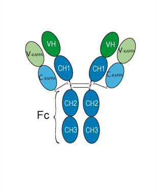
Human IgG1 kappa
Recommend Isotype Controls
Species Reactivity
Human
IC50 & Target
[1]|
IL-1 |
IL-6 |
CD40 |
In Vitro
Golimumab (CNTO-148) (0.1-10 μg /mL; 24-72 hours; transmembrane TNFα–transfected Jurkat cells) induces reductions in the viability of transmembrane TNFα–expressing cells[1].
Golimumab (CNTO-148) (10 μg /mL; 48 hours; transmembrane TNFα–transfected Jurkat cells) induces apoptosis, increase the levels of active caspase-3 and induces apoptotic DNA fragmentation[1].
MedChemExpress (MCE) has not independently confirmed the accuracy of these methods. They are for reference only. Further protocols information, click here.
-
Cell Line:Transmembrane TNFα–transfected Jurkat cells
-
Concentration:0.1, 1 and 10 μg/mL
-
Incubation Time:24, 48 and 72 hours
-
Result:Reduced cell viability in a dose- and time-dependent manner.
-
Cell Line:Transmembrane TNFα–transfected Jurkat cells
-
Concentration:10 μg /mL
-
Incubation Time:48 hours
-
Result:Had the percentage of apoptotic cells for 30.29%.
-
Cell Line:Transmembrane TNFα–transfected Jurkat cells
-
Concentration:10 μg /mL
-
Incubation Time:48 hours
-
Result:Increased the levels of active caspase-3 and induces apoptotic DNA fragmentation.
In Vivo
Golimumab (CNTO-148) (1-10 mg/kg; i.p.;daily; for 4 weeks; Tg197 transgenic mouse model) relieves TNFα-induced arthritis in a mouse model of human[3].
MedChemExpress (MCE) has not independently confirmed the accuracy of these methods. They are for reference only.
-
Animal Model:Swiss-albino healthy male mice[2]
-
Dosage:24 mg/kg
-
Administration:Subcutaneous injection; daily, for 7 days
-
Result:Reduced serum parameters like BUN, NGAL creatinine, cystatin C, and urinary parameters like KIM-1, NAG, albumin clusterin.
-
Animal Model:Swiss-albino healthy male mice[2]
-
Dosage:24 mg/kg
-
Administration:Subcutaneous injection; daily, for 7 days
-
Result:Inhibited oxidative stress, reduces MDA concentrations and increases the GSH and catalase levels.
-
Animal Model:Swiss-albino healthy male mice[2]
-
Dosage:24 mg/kg
-
Administration:Subcutaneous injection; daily, for 7 days
-
Result:Inhibitd cisplatin-induced inflammation, decreased TNF-α, including IL-6, IL-1β, MCP-1, ICAM-1, and TGF-β1 levels and increases IL-10 concentrations, reduced caspase 3 in cisplatin injected mice kidneys.
-
Animal Model:Tg197 transgenic mouse model[3]
-
Dosage:1 and 10 mg/kg
-
Administration:Intraperitoneal injection;for 4 weeks
-
Result:Reduced the arthritic index.
Clinical Trial
| NCT Number | Sponsor | Condition | Start Date |
Phase

|
|---|---|---|---|---|
| NCT01329991 | Plexxikon| | 2011-05 | PHASE1 |
Gene ID
Accession
Conjugated
Unconjugated
Reconsititution
The product can be reconstituted/diluted with sterile PBS or saline.
Format
-
Human IgG1 kappa
Application
ELISA, FACS, Functional assay
Verified Bioactivity
-
 Immobilized TNF-alpha/TNFSF2 Protein, Human (HY-P7058) can bind Golimumab. The EC50 for this effect is 2.75 ng/mL.
 -
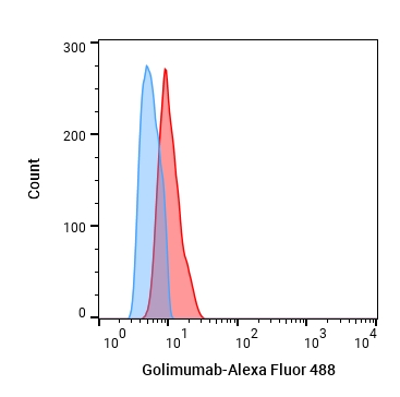 Flow cytometric analysis of 1.5X10⁶ RAW264.7 cells treated with 1µg/mL LPS for 24h labeling Golimumab (HY-P99111, red). Cells were fixed with 4% paraformaldehyde. Then stained with the primary antibody at 1/200 dilution for an hour at 4℃. Goat Anti-Human IgG H&L (AF488) (HY-P83776) was used as the secondary antibody at 1/1,000 dilution for 30 minutes at 4℃, Human IgG1 kappa Isotype Control (HY-P99001, blue) was used as the isotype control,cells without incubation with primary antibody were used as the unlabeled control (black).


Chemical Information
-
CAS No. 476181-74-5
-
Appearance Liquid
-
Molecular Weight 146.94 kDa
-
Color Colorless to light yellow
-
SMILES
[Golimumab]
-
Synonyms
CNTO-148
-
Formulation
Please refer to the lot-specific COA for specific buffer information.
-
Storage
Please store the product under the recommended conditions in the Certificate of Analysis.

Protocols
-
Collagen-Induced Arthritis
Collagen-induced arthritis (CIA) is an autoimmune murine model of rheumatoid arthritis in which immunization with type II collagen (CII) emulsified in an adjuvant induces a T cell- and autoantibody-driven inflammatory arthritis characterized by synovial hyperplasia, immune cell infiltration, and joint destruction. The model typically relies on genetically susceptible mouse strains (e. g. , DBA/1) and reproduces key features of human rheumatoid arthritis, including anti-collagen immune responses and progressive joint inflammation. Disease onset generally occurs within ~3-4 weeks after immunization, depending on antigen/adjuvant combinations and protocol variation. The immunopathology is driven by adaptive immune activation against CII, leading to systemic and local joint inflammation mediated by pro-inflammatory cytokines and effector immune cells, making CIA a standard preclinical platform for evaluating immunomodulatory and anti-arthritic interventions.
-
Apoptosis
Apoptosis, also called programmed cell death, is generally characterized by distinct morphological characteristics.
-
TUNEL staining for apoptotic DNA fragmentation
TUNEL staining detects DNA strand breaks by using terminal deoxynucleotidyl transferase to add labeled nucleotides to exposed 3′-OH DNA termini, generating either microscopic staining in fixed cells or tissue sections, or fluorescence/cytometric signal in cell suspensions. TUNEL positivity reflects DNA fragmentation but should not be interpreted alone as definitive apoptosis, because TUNEL can also label necrotic, autolytic, mechanically damaged, or DNA-repair-associated DNA breaks.
-
Annexin V plus membrane-impermeant dye apoptosis staining
Annexin V-based apoptosis assays rely on the detection of phosphatidylserine (PS) externalization from the inner leaflet of the plasma membrane to the outer leaflet, an early biochemical hallmark of apoptosis. Fluorescently labeled Annexin V binds PS in a calcium-dependent manner, enabling identification of early apoptotic cells by flow cytometry or fluorescence microscopy. When combined with a membrane-impermeant DNA-binding dye (e. g. , propidium iodide), this approach allows discrimination between viable (Annexin V−/dye−), early apoptotic (Annexin V+/dye−), and late apoptotic or necrotic (Annexin V+/dye+) cell populations by assessing membrane integrity and PS exposure.
-
Apoptosis Solutions
Apoptosis is a regulated, generally non-lytic cell-death pathway that removes unwanted, damaged, infected, or abnormal cells through coordinated morphological changes, caspase activation, DNA fragmentation, and membrane remodeling. The intrinsic apoptosis pathway is controlled mainly by mitochondrial outer membrane permeabilization, BCL-2 family proteins, cytochrome c release, apoptosome formation, caspase-9 activation, and downstream executioner caspase-3/7 activation. The extrinsic apoptosis pathway is initiated by death receptors such as Fas, TNFR, and TRAIL receptors, which recruit adaptor proteins and activate caspase-8 before engaging executioner caspases or mitochondrial amplification through BID cleavage. Apoptosis is linked to many phenotypes, including cancer cell killing, tissue homeostasis, immune regulation, neurodegeneration, infection response, and treatment-induced cytotoxicity; unresolved questions include how apoptosis interacts with necroptosis, pyroptosis, ferroptos
-
Research Protocol for Inflammation-related Diseases
The NLRP3 inflammasome is a cytosolic innate immune signaling platform that integrates priming signals and danger-signal activation to promote caspase-1 activation, maturation of IL-1β and IL-18, and gasdermin D-mediated pyroptotic cell death. The core experimental logic is to determine whether inflammatory disease phenotypes are driven by increased NLRP3 expression, ASC-containing inflammasome assembly, caspase-1 cleavage, GSDMD cleavage, and extracellular release of IL-1β/IL-18 rather than by nonspecific cell injury alone. The pathway is strongly linked to inflammation-related disease phenotypes because monosodium urate crystals activate NALP3/NLRP3 inflammasome signaling in gout-like crystal inflammation, cholesterol crystals activate NLRP3 inflammasomes in atherogenesis models, and DSS-induced intestinal inflammation has been reported to involve NLRP3 inflammasome activity. However, experimental colitis studies also show context-dependent protective effects of NLRP3 inflammasome co
-
Pyroptosis Solutions
Pyroptosis is a lytic inflammatory cell-death pathway executed by gasdermin pores, most classically through inflammasome-mediated activation of caspase-1, cleavage of gasdermin D, membrane pore formation, LDH release, and secretion of IL-1β and IL-18. The canonical pathway is commonly modeled by priming cells with an inflammatory signal such as LPS to induce pro-IL-1β and inflammasome components, followed by an activation signal such as ATP or nigericin to activate NLRP3, ASC speck formation, caspase-1 cleavage, GSDMD cleavage, cytokine release, and pyroptotic membrane rupture. The non-canonical pathway is triggered when cytosolic LPS activates mouse caspase-11 or human caspase-4/5, leading to GSDMD cleavage and pyroptosis, and this can secondarily activate NLRP3-dependent IL-1β release. Pyroptosis is linked to inflammatory injury, infection, cancer, liver disease, ocular disease, placental inflammation, and other disease phenotypes, but unresolved questions include which gasdermin fam


Purity & Documentation
-
Data Sheet (266 KB)
-
SDS (251 KB)
- English - EN (251 KB)
- Français - FR (251 KB)
- Deutsch - DE (251 KB)
- Norwegian - NO (251 KB)
- Español - ES (251 KB)
- Swedish - SV (251 KB)
- Italian - IT (251 KB)
- Korean - KR (251 KB)
- Portuguese - PT (251 KB)
-
Inhibitory Antibodies User Guide (603 KB)
References
[1]. Ueda N, et, al. The cytotoxic effects of certolizumab pegol and golimumab mediated by transmembrane tumor necrosis factor α. Inflamm Bowel Dis. 2013 May;19(6):1224-31. [Content Brief]
[2]. Pavitrakar V, et, al. Amelioration of Cisplatin-induced Renal Inflammation by Recombinant Human Golimumab in Mice. Curr Pharm Biotechnol. 2022;23(7):970-977. [Content Brief]
[3]. Shealy DJ, et, al. Characterization of golimumab, a human monoclonal antibody specific for human tumor necrosis factor α. MAbs. 2010 Jul-Aug;2(4):428-39. [Content Brief]

Calculators
Concentration (start) × Volume (start) = Concentration (final) × Volume (final)


Golimumab Related Classifications
HY-P99111 Related Classifications
 Powered by Bioz