



Hydralazine
Based on 4 publication(s) in Google Scholar
Hydralazine is an orally active, blood-brain barrier-permeable DNA methyltransferase inhibitor with vasodilatory, arterial smooth muscle relaxant and hypotensive activities. Hydralazine reactivates silenced tumor suppressor genes via mediating DNA demethylation, while exerting neuroprotective and anti-inflammatory properties. Hydralazine inhibits NOS-2 (iNOS) and COX-2, and reduces the production of NO and PGEE2; meanwhile, Hydralazine scavenges reactive oxygen species and inhibits macrophage activation. Hydralazine alleviates motor dysfunction, neuropathic inflammatory pain, and formalin-induced somatic and emotional pain responses. In addition, Hydralazine directly induces DNA strand breaks and sister chromatid exchange, exhibiting certain mutagenic characteristics. Hydralazine has been widely used in studies on hypertension, various cancers (such as cervical cancer, leukemia), spinal cord injury and the mechanisms of inflammatory pain.
Para uso exclusivo en investigación. No vendemos a pacientes.
- Pureza : 99.94%
- No. CAS: 86-54-4
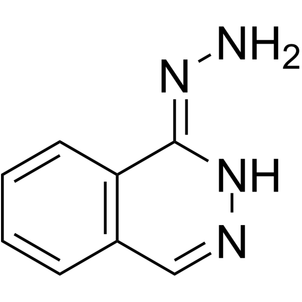
- Fòrmula: C8H8N4
- Peso molecular:160.18
-
Almacenamiento:Powder -20°C, 3 years ; In solvent -80°C, 6 months , -20°C, 1 month

To place orders, for customer services and technical support, please contact: MedChemExpress USA
Tel: 609-228-6898 E-mail: [email protected] [email protected]


-
 Actividad biológica
-
Chemical Information
-
Solvente y solubilidad
- Protocolo
- Pureza y Documentación
- Referencias
-
 Help & FAQs


Publications Citing Use of MedChemExpress (MCE) Hydralazine
More Customer Validation & Images

-
WB
Ver todos los productos específicos de isoformas DNA Methyltransferase
More

Actividad biológica
Descripciòn
IC50 & Target
|
COX-2 |
iNOS |
Cellular Effect
|
Cell Line

|
Type | Value | Description | References |
|---|---|---|---|---|
| NHDF | IC50 |
>50 μM
Compound: 36
|
Cytotoxicity against NHDF assessed as reduction in cell viability after 72 hrs by MTT assay
Cytotoxicity against NHDF assessed as reduction in cell viability after 72 hrs by MTT assay
|
[PMID: 28671460] |
In Vitro
Hydralazine (10 μM; 5 days) demethylates and reactivates expression of silenced tumor suppressor genes (ER, RARβ, p16) in MDA-231, MCF-7, and T24 cancer cell lines, respectively[1].
Hydralazine (10 μM; 24 hours) reduces DNMT1 and DNMT3a mRNA expression in MCF-7 breast cancer cells[1].
Hydralazine (0.1-10.0 mM) does not impair phagocytic function or viability of thioglycollate-prestimulated rat peritoneal macrophages[2].
Hydralazine (0.1-10.0 mM) significantly inhibits nitrite production by LPS/IFN-γ-stimulated thioglycollate-prestimulated rat peritoneal macrophages, with an IC50 of 0.43 mM[2].
Hydralazine (1.0-10.0 mM) inhibits both NOS-2 and COX-2 protein synthesis in LPS/IFNγ-stimulated thioglycollate-prestimulated rat peritoneal macrophages[2].
Hydralazine (3.1-50 nmol, plate incorporation test without S-9 mix; 1 mg per plate, spot test with/without S-9 mix) is a direct-acting, low-potency mutagen that induces mixed genetic mutation mechanisms in Salmonella typhimurium strains TA1535, TA100, TA1537, TA97, and TA98, with highest potency in strain TA97[3].
Hydralazine (5 mg per spot, spot test; serial twofold dilutions, micromethod assay with/without S-9 mix; 16 h incubation at 37°C) is a direct-acting genotoxin that is preferentially lethal to repair-deficient Escherichia coli strains WP67 and CM871, indicating involvement of recA recombination repair, lexA post-replication repair, and polA repair mechanisms, with no dependence on excision repair systems[3].
Hydralazine (≤25 μM) significantly mitigates acrolein-mediated cell death in PC12 cells[4].
MedChemExpress (MCE) has not independently confirmed the accuracy of these methods. They are for reference only.
-
Cell Line:MCF-7 breast cancer cells
-
Concentration:10 μM
-
Incubation Time:24 h
-
Result:Decreased the mRNA expression of DNMT1 and DNMT3a, as confirmed by reduced band intensity on PCR analysis relative to untreated controls.
Showed no change in DNMT3b mRNA expression.
Parmacokinetics
| Species | Dose | Route | Brain Concentration |
|---|---|---|---|
| Mice[4] | 5 mg/kg | i.p. | ~30 μM |
In Vivo
Hydralazine (used in combination with valproic acid (HY-10585)) eliminates tumor recurrence of HT1080 fibrosarcoma xenografts in nude mice[1].
Hydralazine (83 mg/kg; i.p.; once daily; 5 days) induces DNA fragmentation in the liver, kidney and spleen (but not the lung) of mice at 6 hours after the last administration, and the damage is completely repaired at 12 hours post-administration; meanwhile, it moderately increases the sister chromatid exchange rate of mouse bone marrow cells by 33%[3].
Hydralazine (5 mg/kg; i.p.; twice; 14 d) reduces acrolein levels by 50-70% in the injured spinal cord of rats, decreases post-injury cyst formation by 70%, improves motor function recovery, and alleviates mechanical hyperalgesia in rat models of spinal cord injury[4].
Hydralazine (0.1-10 mg/kg; i.p.; single dose) dose-dependently alleviates formalin-induced somatic and emotional inflammatory pain in male C57BL/6 mice, with a ED50 of 0.239-1.0160 mg/kg, while inhibiting excessive acrolein production and neuronal activation in the spinal cord[5].
Hydralazine (10 mg/kg; i.p.; single dose) does not impair central nervous system function in male C57BL/6 mice[5].
MedChemExpress (MCE) has not independently confirmed the accuracy of these methods. They are for reference only.
-
Animal Model:Nude mice[1]
-
Dosage:equivalent to those used in hypertensive patients
-
Administration:i.p.; daily; 7 days
-
Result:Induced demethylation of the estrogen receptor (ER) gene promoter in xenografted tumors, which correlated with re-expression of ER mRNA and protein.
-
Animal Model:Noninbred Swiss mice (male, 2-3 months old)[3]
-
Dosage:83 mg/kg (single dose); 28 mg/kg (daily x5)
-
Administration:i.p.; single dose; daily for 5 consecutive days
-
Result:Induced a modest but statistically significant increase in DNA elution rate in liver 6 hours post single 83 mg/kg dose.
Induced a modest but statistically significant increase in DNA elution rate in kidney 6 hours post single 83 mg/kg dose.
Induced a modest but statistically significant increase in DNA elution rate in spleen 6 hours post single 83 mg/kg dose.
Showed minimal or absent DNA damage in liver, kidney, and spleen 1 hour and 12 hours post single 83 mg/kg dose.
Showed no statistically significant DNA damage in lung at 1, 6, or 12 hours post single 83 mg/kg dose.
Showed no statistically significant DNA damage in liver, lung, kidney, or spleen 6 hours after final daily 28 mg/kg dose (x5).
-
Animal Model:Noninbred Swiss mice (male, 2-3 months old)[3]
-
Dosage:83 mg/kg
-
Administration:i.p.; single dose
-
Result:Induced a 33% increase in SCE frequency per diploid bone marrow cell relative to control mice, with a mean of 3.6 ± 1.8 SCEs per metaphase.
-
Animal Model:Sprague-Dawley (male, 200-250 grams at surgery, spinal cord contusion injury model)[4]
-
Dosage:5 mg/kg (tissue distribution); 5 mg/kg (two doses, acrolein reduction); 5 mg/kg (daily for 14 days, cyst formation, locomotor function, mechanical allodynia)
-
Administration:i.p.; single dose; two doses (immediately post-injury, 24 hours post-injury); daily; 14 days
-
Result:Reached concentrations of 2.9 μg/g (≈20 μM) in spinal cord tissue and 4.4 μg/g (≈30 μM) in brain tissue at 2 hours post-single 5 mg/kg i.p.
dose.
Reduced acrolein levels from 31.1 arbitrary units to 8.7 arbitrary units in moderate spinal cord injury, and from 42.7 arbitrary units to 21.3 arbitrary units in severe spinal cord injury after two 5 mg/kg i.p.
doses.
Reduced spinal cord cyst area from 38.0% of total cord area to 11.4% at 28 days post-injury after daily 5 mg/kg i.p.
treatment for 14 days.
Improved BBB locomotor scores to 16.1 at 28 days post-injury (compared to 12.4 in untreated injured rats), with significant improvements detected at 7, 14, 21, and 28 days post-injury after daily 5 mg/kg i.p.
treatment for 14 days.
Mitigated mechanical allodynia: at 14 days post-injury, paw withdrawal threshold was 8.1 g; at 21 days post-injury, threshold was 8.1 g; at 28 days post-injury, threshold was 9.7 g (all significantly higher than thresholds in untreated injured rats) after daily 5 mg/kg i.p.
treatment for 14 days.
Ensayo clínico
| NCT Number | Sponsor | Condition | Start Date |
Phase
|
|---|---|---|---|---|
| NCT01329991 | Plexxikon| | 2011-05 | PHASE1 |


Chemical Information
-
No. CAS 86-54-4
-
Appearance Solid
-
Peso molecular 160.18
-
Fòrmula C8H8N4
-
Color White to off-white
-
SMILES
N/N=C1NN=CC2=C\1C=CC=C2
-
Envío
Room temperature in continental US; may vary elsewhere.
-
Almacenamiento
Powder -20°C 3 years In solvent -80°C 6 months -20°C 1 month


Publications (4)
-
Journal Impact Factor
-
Most Recent
-
J Pharm Anal
Hydralazine represses Fpn ubiquitination to rescue injured neurons via competitive binding to UBA52. [Abstract]2024 Jan;14(1):86-99. PMID: 38352945 -
Neurosci Bull
Acrolein Aggravates Secondary Brain Injury After Intracerebral Hemorrhage Through Drp1-Mediated Mitochondrial Oxidative Damage in Mice. [Abstract]2020 Oct;36(10):1158-1170. PMID: 32436179
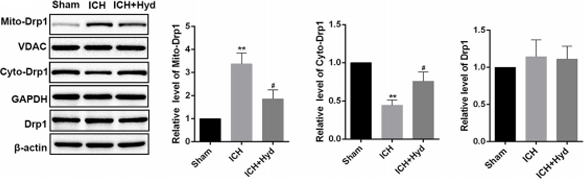
Hydralazine purchased from MedChemExpress. Usage Cited in: Neurosci Bull. 2020 Oct;36(10):1158-1170. [Abstract]
Western blots (left) and analysis (right) of cyto-Drp1, mito-Drp1, and T-Drp1 in different groups of mice. Hydralazine (acrolein scavenger) suppresses the translocation of Drp1 and alleviates the morphological disruption of mitochondria after ICH in mice.
-
-


Solvente y solubilidad
In Vitro:
DMSO : 20 mg/mL (124.86 mM; ultrasonic and warming and heat to 60°C; Hygroscopic DMSO has a significant impact on the solubility of product, please use newly opened DMSO)
Please refer to the solubility information to select the appropriate solvent. Once prepared, please aliquot and store the solution to prevent product inactivation from repeated freeze-thaw cycles.
Storage method and period of stock solution: -80°C, 6 months; -20°C, 1 month. When stored at -80°C, please use it within 6 months. When stored at -20°C, please use it within 1 month.
Please refer to the solubility information to select the appropriate solvent. Once prepared, please aliquot and store the solution to prevent product inactivation from repeated freeze-thaw cycles.
Storage method and period of stock solution: -80°C, 6 months; -20°C, 1 month. When stored at -80°C, please use it within 6 months. When stored at -20°C, please use it within 1 month.
Concentration (start) × Volume (start) = Concentration (final) × Volume (final)
In Vivo:
Select the appropriate dissolution method based on your experimental animal and administration route.
- For the following dissolution methods, please ensure to first prepare a clear stock solution using an In Vitro approach and then sequentially add co-solvents:
- To ensure reliable experimental results, the clarified stock solution can be appropriately stored based on storage conditions. As for the working solution for In Vivo experiments, it is recommended to prepare freshly and use it on the same day.
- The percentages shown for the solvents indicate their volumetric ratio in the final prepared solution. If precipitation or phase separation occurs during preparation, heat and/or sonication can be used to aid dissolution.
Add each solvent one by one: 10% DMSO 40% PEG300 5% Tween-80 45% Saline
Solubility: ≥ 2 mg/mL (12.49 mM); Clear solution
This protocol yields a clear solution of ≥ 2 mg/mL (saturation unknown).
Taking 1 mL working solution as an example, add 100 μL DMSO stock solution (20.0 mg/mL) to 400 μL PEG300, and mix evenly; then add 50 μL Tween-80 and mix evenly; then add 450 μL Saline to adjust the volume to 1 mL.
Preparation of Saline: Dissolve 0.9 g sodium chloride in ddH₂O and dilute to 100 mL to obtain a clear Saline solution.
Add each solvent one by one: 10% DMSO 90% (20% SBE-β-CD in Saline)
Solubility: ≥ 2 mg/mL (12.49 mM); Clear solution
This protocol yields a clear solution of ≥ 2 mg/mL (saturation unknown).
Taking 1 mL working solution as an example, add 100 μL DMSO stock solution (20.0 mg/mL) to 900 μL 20% SBE-β-CD in Saline, and mix evenly.
Preparation of 20% SBE-β-CD in Saline (4°C, storage for one week): 2 g SBE-β-CD powder is dissolved in 10 mL Saline, completely dissolve until clear.
In Vivo Dissolution Calculator
Please enter the basic information of animal experiments:
-
-
-
-
Recommended: Prepare an additional quantity of animals to account for potential losses during experiments.
Please enter your animal formula composition:
-
%DMSO +
Recommended: Keep the proportion of DMSO in working solution below 2% if your animal is weak.
-
%+
-
+%Tween-80 + +
-
%Saline +
The co-solvents required include: DMSO, . All of co-solvents are available by MedChemExpress (MCE). , Tween 80. All of co-solvents are available by MedChemExpress (MCE).
Working solution concentration: 0.22 mg/mL
Method for preparing stock solution: mg drug dissolved in μL DMSO. Stock solution concentration: mg/mL.
1. Take μL DMSO stock solution;
2. Add μL .
μL , mix evenly;
3. Then add μL Tween 80, mix evenly;
4. Then add μL
Please ensure that the stock solution in the first step is dissolved to a clear state, and add co-solvents in sequence. You can use ultrasonic heating (ultrasonic cleaner, recommended frequency 20-40 kHz), vortexing, etc. to assist dissolution.


Pureza y Documentación
-
Ficha de datos (289 KB)
-
SDS (394 KB)
- English - EN (394 KB)
- Français - FR (394 KB)
- Deutsch - DE (394 KB)
- Norwegian - NO (394 KB)
- Español - ES (394 KB)
- Swedish - SV (394 KB)
- Italian - IT (394 KB)
- Korean - KR (394 KB)
- Portuguese - PT (394 KB)
-
Instrucciones de manejo (2659 KB)
Referencias
[1]. Arce C, et al. Hydralazine target: from blood vessels to the epigenome. J Transl Med. 2006;4:10. Published 2006 Feb 28. [Content Brief]
[2]. Leiro JM, et al. Antioxidant activity and inhibitory effects of hydralazine on inducible NOS/COX-2 gene and protein expression in rat peritoneal macrophages. Int Immunopharmacol. 2004;4(2):163-177. [Content Brief]
[3]. de Flora S, et al. In vivo and in vitro genotoxicity of three antihypertensive hydrazine derivatives (hydralazine, dihydralazine, and endralazine). Environ Mutagen. 1982;4(5):605-619. [Content Brief]
[4]. Park J, et al. Neuroprotective role of hydralazine in rat spinal cord injury-attenuation of acrolein-mediated damage. J Neurochem. 2014;129(2):339-349. [Content Brief]
[5]. Bai L, et al. Attenuation of mouse somatic and emotional inflammatory pain by hydralazine through scavenging acrolein and inhibiting neuronal activation. Pain Physician. 2012;15(4):311-326. [Content Brief]


Complete Stock Solution Preparation Table
Please refer to the solubility information to select the appropriate solvent. Once prepared, please aliquot and store the solution to prevent product inactivation from repeated freeze-thaw cycles.
Storage method and period of stock solution: -80°C, 6 months; -20°C, 1 month. When stored at -80°C, please use it within 6 months. When stored at -20°C, please use it within 1 month.
| Optional Solvent | Concentration Solvent Mass | 1 mg | 5 mg | 10 mg | 25 mg |
|---|---|---|---|---|---|
| DMSO | 1 mM | 6.2430 mL | 31.2149 mL | 62.4298 mL | 156.0744 mL |
| 5 mM | 1.2486 mL | 6.2430 mL | 12.4860 mL | 31.2149 mL | |
| 10 mM | 0.6243 mL | 3.1215 mL | 6.2430 mL | 15.6074 mL | |
| 15 mM | 0.4162 mL | 2.0810 mL | 4.1620 mL | 10.4050 mL | |
| 20 mM | 0.3121 mL | 1.5607 mL | 3.1215 mL | 7.8037 mL | |
| 25 mM | 0.2497 mL | 1.2486 mL | 2.4972 mL | 6.2430 mL | |
| 30 mM | 0.2081 mL | 1.0405 mL | 2.0810 mL | 5.2025 mL | |
| 40 mM | 0.1561 mL | 0.7804 mL | 1.5607 mL | 3.9019 mL | |
| 50 mM | 0.1249 mL | 0.6243 mL | 1.2486 mL | 3.1215 mL | |
| 60 mM | 0.1040 mL | 0.5202 mL | 1.0405 mL | 2.6012 mL | |
| 80 mM | 0.0780 mL | 0.3902 mL | 0.7804 mL | 1.9509 mL | |
| 100 mM | 0.0624 mL | 0.3121 mL | 0.6243 mL | 1.5607 mL |


Hydralazine Related Classifications
HY-B0464A Related Classifications
 Powered by Bioz