



Flubendazole
Based on 7 publication(s) in Google Scholar
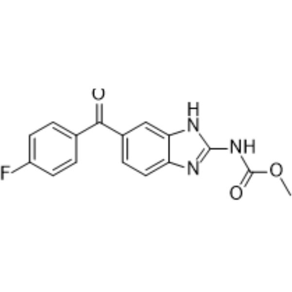
Flubendazole is an anthelmintic drug based on altering microtubule structure, inhibition of tubulin polymerization and disruption of microtubule function. Flubendazole induces apoptosis in human colorectal cancer (CRC) by blocking the STAT3 signaling axis and activation of autophagy. Flubendazole induces P53 expression and reduced Cyclin B1 and p-cdc2 expression. Flubendazole is an antitumor agent. Flubendazole can be used for worm and intestinal parasites.
For research use only. We do not sell to patients.
- Purity : 99.05%
- CAS No.: 31430-15-6
- Formula: C16H12FN3O3
- Molecular Weight:313.28
-
Storage:Powder -20°C, 3 years , 4°C, 2 years ; In solvent -80°C, 2 years , -20°C, 1 year

To place orders, for customer services and technical support, please contact: MedChemExpress USA
Tel: 609-228-6898 E-mail: [email protected] [email protected]


-
 Biological Activity
-
Chemical Information
-
Solvent & Solubility
- Protocols
- Purity & Documentation
- References
-
 Help & FAQs


-
HY-17595
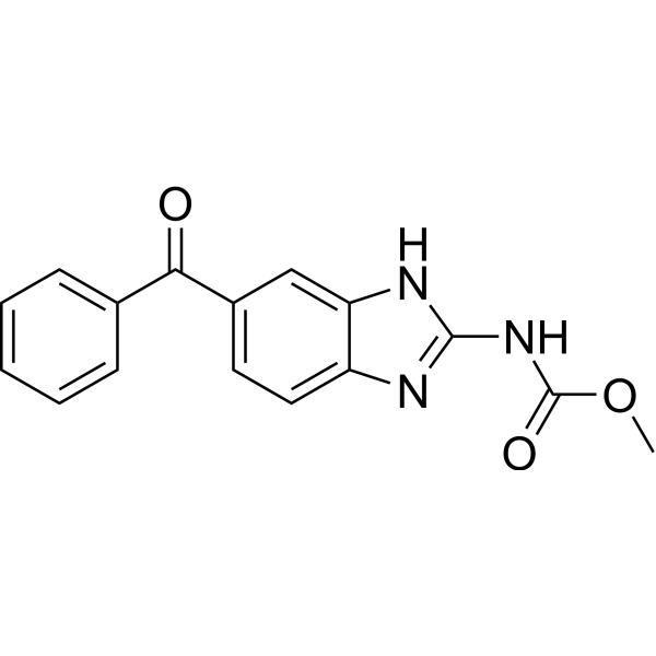
MebendazoleApoptosis Inducer -
HY-B0223
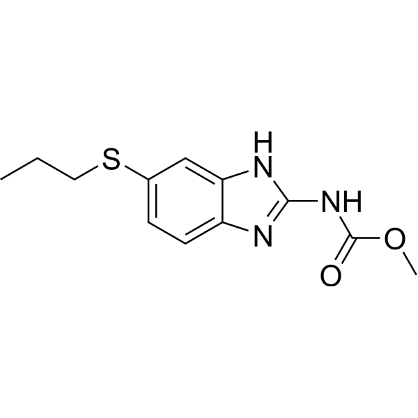
AlbendazoleParasiticide -
HY-B0299
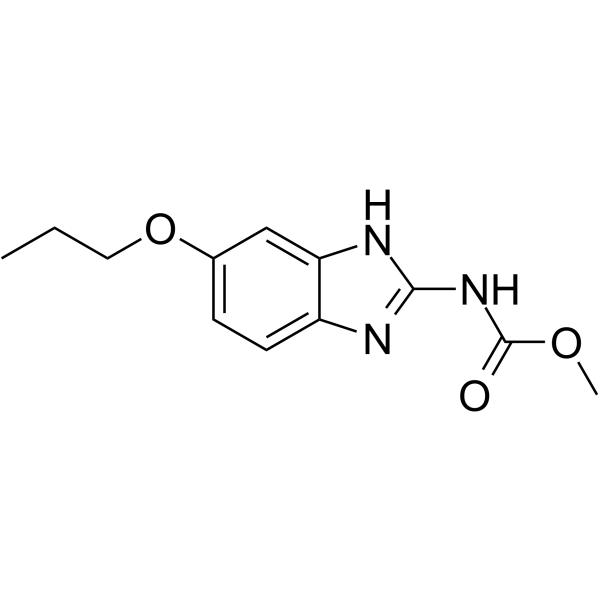
OxibendazolePolymerase Inhibitor -
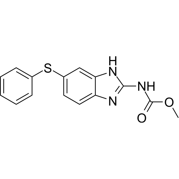
HY-B0413
FenbendazoleAntibiotic -

HY-B0294R
Flubendazole (Standard)Tubulin Polymerization Inhibitor -
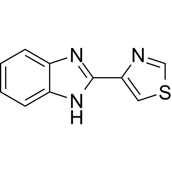
HY-B0263
Thiabendazolefungicide -
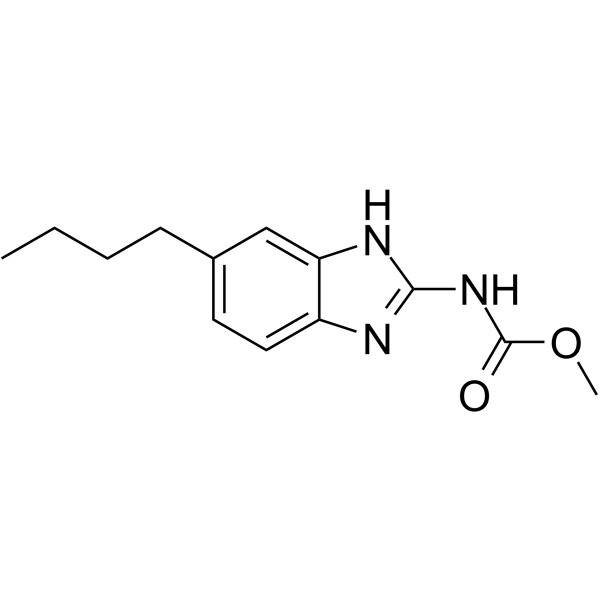
HY-115364
ParbendazoleMicrotubule Polymerization Inhibitor -
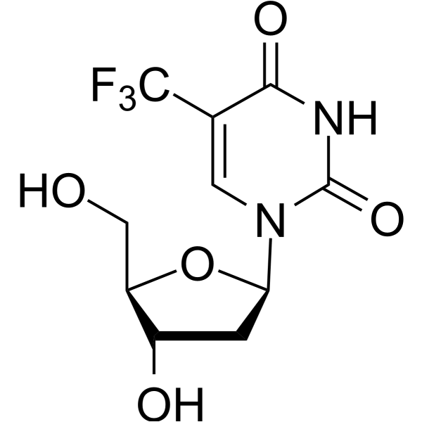
HY-A0061
TrifluridineAnti-Cancer Agent/Anti-Viral Agent -
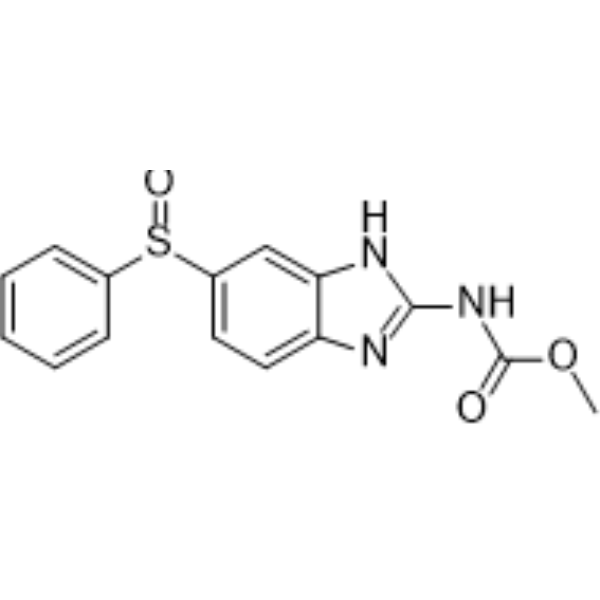
HY-B0291
OxfendazoleParasite Inhibitor -
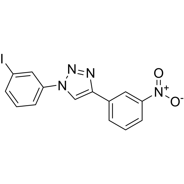
HY-115576
P62-mediated mitophagy inducerMitochondrial Autophagy Regulator -
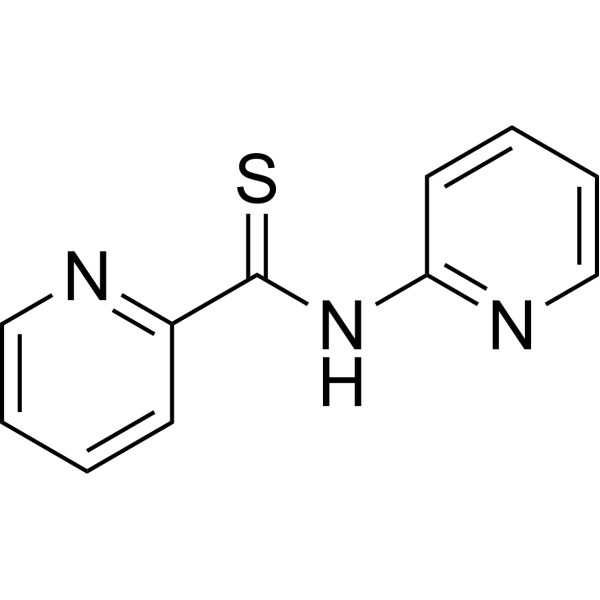
HY-125169
NSC 185058ATG4B Inhibitor -
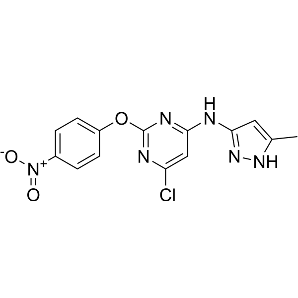
HY-101920
AutophinibVPS34 Inhibitor
Publications Citing Use of MedChemExpress (MCE) Flubendazole
More- Theranostics. 2021 Jun 1;11(15):7491-7506. [Abstract]
- Int J Biol Sci. 2023 Apr 23;19(7):2270-2288. [Abstract]
- Biochem Pharmacol. 2025 Aug 28;242(Pt 1):117287. [Abstract]
- Commun Biol. 2024 Jan 24;7(1):123. [Abstract]
- ACS Omega. 2020 Nov 15;5(46):29935-29942. [Abstract]
- J Cell Mol Med. 2024 Nov;28(22):e70188. [Abstract]
- Research Square Preprint. 2021 Jul.
All Parasite Isoforms
More

Biological Activity
Description
|
STAT3 |
Cellular Effect
|
Cell Line

|
Type | Value | Description | References |
|---|---|---|---|---|
| BT-549 | IC50 |
0.125 μM
Compound: 28; 65
|
Anticancer activity against human BT-549 cells assessed as cell growth inhibition by MTS assay
Anticancer activity against human BT-549 cells assessed as cell growth inhibition by MTS assay
|
[PMID: 33650861] |
| Hs-578T | IC50 |
0.125 μM
Compound: 28; 65
|
Anticancer activity against human Hs-578T cells assessed as cell growth inhibition by MTS assay
Anticancer activity against human Hs-578T cells assessed as cell growth inhibition by MTS assay
|
[PMID: 33650861] |
| MDA-MB-231 | IC50 |
0.25 μM
Compound: 28; 65
|
Anticancer activity against human MDA-MB-231 cells assessed as cell growth inhibition by MTS assay
Anticancer activity against human MDA-MB-231 cells assessed as cell growth inhibition by MTS assay
|
[PMID: 33650861] |
| MDA-MB-231 | IC50 |
0.5 μM
Compound: 28; 65
|
Anticancer activity against human MDA-MB-231 cells assessed as cell growth inhibition incubated for 24 hrs by MTT assay
Anticancer activity against human MDA-MB-231 cells assessed as cell growth inhibition incubated for 24 hrs by MTT assay
|
[PMID: 33650861] |
| MDA-MB-231 | IC50 |
0.75 μM
Compound: 14
|
Antiproliferative activity against human MDA-MB-231 cells assessed reduction in cell viability incubated for 24 hrs by MTT assay
Antiproliferative activity against human MDA-MB-231 cells assessed reduction in cell viability incubated for 24 hrs by MTT assay
|
[PMID: 37595546] |
| PC-3M | EC50 |
1.43 μM
Compound: Flubendazole
|
Cytotoxicity against human PC3M cells assessed as reduction in cell viability after 48 hrs by cyquant reagent based fluorescence spectrometric assay
Cytotoxicity against human PC3M cells assessed as reduction in cell viability after 48 hrs by cyquant reagent based fluorescence spectrometric assay
|
[PMID: 29288939] |
In Vitro
Flubendazole (0-400 μM; 48 h) inhibits human colorectal cancer (CRC) cells proliferation[1].
Flubendazole (0.3-1.2 μM; 48 h) induces apoptosis in CRC cells[1].
Flubendazole (0.3-1.2 μM; 24 h) induces autophagy initiation by inactivating mTOR and P62, and upregulating LC3-I/II in CRC cells[1].
Flubendazole (0.3-1.2 μM; 24 h) strongly reduces the expression of P-STAT3 in a dose and time-dependent manner[1].
MedChemExpress (MCE) has not independently confirmed the accuracy of these methods. They are for reference only.
-
Cell Line:CRC cells (HCT116, RKO and SW480)
-
Concentration:0-400 μM
-
Incubation Time:48 h
-
Result:Effectively reduces the viability of CRC cells (HCT116, RKO and SW480) in a concentration-dependent manner, with an IC50 of 2-5 μM.
-
Cell Line:CRC cells (HCT116, RKO and SW480)
-
Concentration:0.3, 0.6, 1.2 μM
-
Incubation Time:48 h
-
Result:Increased the proportion of apoptotic cells in a dose-dependent manner.
Dose-dependently effectively increases caspase-3 activity.
-
Cell Line:CRC cells (HCT116, RKO and SW480)
-
Concentration:0.3, 0.6, 1.2 μM
-
Incubation Time:24 h
-
Result:Induced autophagy initiation by inactivating mTOR and P62, and upregulating LC3-I/II, which are classical marker of autophagy.
-
Cell Line:CRC cells (HCT116, RKO and SW480)
-
Concentration:0.3, 0.6, 1.2 μM
-
Incubation Time:24 h
-
Result:Strongly reduced the expression of phosphorylated STAT3 (P-STAT3) in a dose and time-dependent manner.
No obvious change in total STAT3 expression.
Decreased expression of MCL1 and survivin in a dose-dependent manner.
In Vivo
MedChemExpress (MCE) has not independently confirmed the accuracy of these methods. They are for reference only.
-
Animal Model:Female BALB/c athymic nude mice (6-8 weeks) with HCT116 cells[1]
-
Dosage:10 or 30 mg/kg
-
Administration:Intraperitoneal injection; every other day; 14 days
-
Result:Markedly reduced the tumor volume.
Significantly reduces the protein level of P-STAT3, promotes autophagy and induces apoptosis in vivo.


Chemical Information
-
CAS No. 31430-15-6
-
Appearance Solid
-
Molecular Weight 313.28
-
Formula C16H12FN3O3
-
Color White to off-white
-
SMILES
O=C(OC)NC1=NC2=CC=C(C(C3=CC=C(F)C=C3)=O)C=C2N1
-
Shipping
Room temperature in continental US; may vary elsewhere.
-
Storage
Powder -20°C 3 years 4°C 2 years In solvent -80°C 2 years -20°C 1 year


Publications (7)
-
Journal Impact Factor
-
Most Recent
-
Theranostics
Microbial and genetic-based framework identifies drug targets in inflammatory bowel disease. [Abstract]2021 Jun 1;11(15):7491-7506. PMID: 34158863 -
Int J Biol Sci
Drug Repurposing Flubendazole to Suppress Tumorigenicity via PCSK9-dependent Inhibition and Potentiate Lenvatinib Therapy for Hepatocellular Carcinoma. [Abstract]2023 Apr 23;19(7):2270-2288. PMID: 37151886 -
Biochem Pharmacol
Flubendazole inhibits cervical carcinoma by targeting DHODH to induce ferroptosis and mitophagy. [Abstract]2025 Aug 28;242(Pt 1):117287. PMID: 40885318 -
Commun Biol
Parbendazole as a promising drug for inducing differentiation of acute myeloid leukemia cells with various subtypes. [Abstract]2024 Jan 24;7(1):123. PMID: 38267545 -
ACS Omega
Computational Approaches to Identify Molecules Binding to Mycobacterium tuberculosis KasA. [Abstract]2020 Nov 15;5(46):29935-29942. PMID: 33251429 -
J Cell Mol Med
Repurposing flubendazole for glioblastoma ferroptosis by affecting xCT and TFRC proteins. [Abstract]2024 Nov;28(22):e70188. PMID: 39543084 -


Solvent & Solubility
In Vitro:
DMSO : 4.17 mg/mL (13.31 mM; Need ultrasonic; Hygroscopic DMSO has a significant impact on the solubility of product, please use newly opened DMSO)
Please refer to the solubility information to select the appropriate solvent. Once prepared, please aliquot and store the solution to prevent product inactivation from repeated freeze-thaw cycles.
Storage method and period of stock solution: -80°C, 2 years; -20°C, 1 year. When stored at -80°C, please use it within 2 years. When stored at -20°C, please use it within 1 year.
Please refer to the solubility information to select the appropriate solvent. Once prepared, please aliquot and store the solution to prevent product inactivation from repeated freeze-thaw cycles.
Storage method and period of stock solution: -80°C, 2 years; -20°C, 1 year. When stored at -80°C, please use it within 2 years. When stored at -20°C, please use it within 1 year.
Concentration (start) × Volume (start) = Concentration (final) × Volume (final)
In Vivo:
For the following dissolution methods, please prepare the working solution directly:
It is recommended to prepare fresh solutions and use them promptly within a short period of time.
The percentages shown for the solvents indicate their volumetric ratio in the final prepared solution. If precipitation or phase separation occurs during preparation, heat and/or sonication can be used to aid dissolution.
Add each solvent one by one: 50% PEG300 50% Saline
Solubility: 5 mg/mL (15.96 mM); Suspended solution; Need ultrasonic


Purity & Documentation
-
Data Sheet (277 KB)
-
SDS (393 KB)
- English - EN (393 KB)
- Français - FR (393 KB)
- Deutsch - DE (393 KB)
- Norwegian - NO (393 KB)
- Español - ES (393 KB)
- Swedish - SV (393 KB)
- Italian - IT (393 KB)
- Korean - KR (393 KB)
- Portuguese - PT (393 KB)
-
Handling Instructions (2659 KB)
References
[1]. Shichong Lin, et al. Flubendazole demonstrates valid antitumor effects by inhibiting STAT3 and activating autophagy. J Exp Clin Cancer Res. 2019 Jul 8;38(1):293. [Content Brief]
[2]. Zhou X, et al. Flubendazole inhibits glioma proliferation by G2/M cell cycle arrest and pro-apoptosis. Cell Death Discov. 2018 Feb 14;4:18. [Content Brief]


Complete Stock Solution Preparation Table
Please refer to the solubility information to select the appropriate solvent. Once prepared, please aliquot and store the solution to prevent product inactivation from repeated freeze-thaw cycles.
Storage method and period of stock solution: -80°C, 2 years; -20°C, 1 year. When stored at -80°C, please use it within 2 years. When stored at -20°C, please use it within 1 year.
| Optional Solvent | Concentration Solvent Mass | 1 mg | 5 mg | 10 mg | 25 mg |
|---|---|---|---|---|---|
| DMSO | 1 mM | 3.1920 mL | 15.9602 mL | 31.9203 mL | 79.8008 mL |
| 5 mM | 0.6384 mL | 3.1920 mL | 6.3841 mL | 15.9602 mL | |
| 10 mM | 0.3192 mL | 1.5960 mL | 3.1920 mL | 7.9801 mL |


Flubendazole Related Classifications
HY-B0294 Related Classifications
 Powered by Bioz