



Dynorphin A TFA
Based on 2 publication(s) in Google Scholar
Dynorphin A TFA is an endogenous opioid peptide involved in inhibitory neurotransmission in the central nervous system (CNS). Dynorphin A TFA is a highy potent kappa opioid receptor (KOR) agonist, and is also an agonist for other opioid receptors, such as mu (MOR) and delta (DOR). Dynorphin A TFA can induce neuronal death, and can be used in the research of neurological disease.
商品は「研究用試薬」です。人や動物の医療用・臨床診断用・食品用の製品ではありません。
研究用途以外に使用した場合、当社は一切の責任を負いかねます。
- 分子式: C101H156F3N31O25
- 分子量:2261.50
-
保管条件:
Please store the product under the recommended conditions in the Certificate of Analysis.

To place orders, for customer services and technical support, please contact: MedChemExpress USA
Tel: 609-228-6898 E-mail: [email protected] [email protected]


-
 生物活性
生物活性
-
化学情報
- プロトコル
- 純度とドキュメンテーション
- 参考文献
-
 Help & FAQs


MedChemExpress(MCE)の使用を引用している文献 Dynorphin A TFA
MoreOpioid Receptor アイソフォーム固有の製品をすべて表示
MoreCaspase アイソフォーム固有の製品をすべて表示
MoreEndogenous Metabolite アイソフォーム固有の製品をすべて表示
More

生物活性
製品説明
IC50 & Target
[3]|
Human Endogenous Metabolite |
Caspase-3 |
κ Opioid Receptor/KOR |
体外実験
Dynorphin A TFA (10 μM, 4 h/72 h) increases caspase-3 activity and the level of cytochrome c released from mitochondria in mouse striatal neurons, and induces neuronal death[3].
dynorphin A TFA (33 μM, 4 h) elevates [Ca2+]i and causes a significant loss of neurons[4].
dynorphin A TFA (1 μM) inhibits the release of vasopressin (VP) from the isolated neural lobe[5].
MedChemExpress (MCE) has not independently confirmed the accuracy of these methods. They are for reference only.
-
Cell Line:Mouse striatal neurons
-
Concentration:10 μM
-
Incubation Time:0, 24, 48, 72 h
-
Result:Induced neuronal death (identified by the fragmentation and destruction of the cell body and neurites).
体内実験
Dynorphin A TFA (intracerebroventricular injection, 500 pmol/5 μL per day for 4 d) alleviates stress-induced behavioral impairments in ddY mice accompanied by regulation of the 5-HTergic system in the brain[6].
MedChemExpress (MCE) has not independently confirmed the accuracy of these methods. They are for reference only.
-
Animal Model:24 h water-deprived male rats[5]
-
Dosage:1 μg of 2 μL
-
Administration:Intracerebroventricular injection
-
Result:Inhibited vasopressin (VP) release 30 min upon injection.
-
Animal Model:Male ddY mice[6]
-
Dosage:15, 150, 1500 pmol/5 μL per day for 4 days
-
Administration:Intracerebroventricular injection
-
Result:Attenuated the repeated stress-induced escape failures from the shock.


化学情報
-
分子量 2261.50
-
分子式 C101H156F3N31O25
-
配列
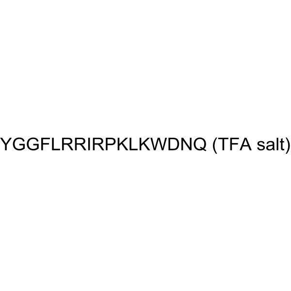
Tyr-Gly-Gly-Phe-Leu-Arg-Arg-Ile-Arg-Pro-Lys-Leu-Lys-Trp-Asp-Asn-Gln
-
シーケンスの短縮
YGGFLRRIRPKLKWDNQ
-
輸送条件
Room temperature in continental US; may vary elsewhere.
-
保管条件
Please store the product under the recommended conditions in the Certificate of Analysis.


Publications (2)
-
Journal Impact Factor
-
Most Recent
-
Neuron
Astrocyte-mediated central amygdala microcircuit gates comorbid anxiety symptoms in chronic pain. [Abstract]2025 Oct 2:S0896-6273(25)00670-1. PMID: 41043420 -


純度とドキュメンテーション
参考文献
[1]. Zhang, et al.Dynorphin A as a Potential Endogenous Ligand for Four Members of the Opioid Receptor Gene Family. J Pharmacol Exp Ther. 1998 Jul;286(1):136-41. [Content Brief]
[2]. Aruna Sharma, et al. Monoclonal antibodies as novel neurotherapeutic agents in CNS injury and repair. Int Rev Neurobiol. 2012;102:23-45. [Content Brief]
[3]. I. N. SINGH, et al. Dynorphin A (1–17) induces apoptosis in striatal neurons in vitro through AMPA/kainate receptor-mediated cytochrome c release and caspase-3 activation. Neuroscience. 2003;122(4):1013-23. [Content Brief]
[4]. B J Van de Heijning, et al. Dynorphin-A and vasopressin release in the rat: a structure-activity study. Neuropeptides. 1994 Jun;26(6):371-8. [Content Brief]
[5]. Takayoshi Mamiya, et al. Dynorphin a (1-13) alleviated stress-induced behavioral impairments in mice. Biol Pharm Bull. 2014;37(8):1269-73. [Content Brief]
[6]. K F Hauser, et al. Dynorphin A (1-13) neurotoxicity in vitro: opioid and non-opioid mechanisms in mouse spinal cord neurons. Exp Neurol. 1999 Dec;160(2):361-75. [Content Brief]

Calculators
濃度 (開始) × 体積 (開始) = 濃度 (終了) × 体積 (終了)


Dynorphin A TFA 関連分類
HY-P1333A 関連分類
 Powered by Bioz