



Mirificin

Based on 1 Customer Validation
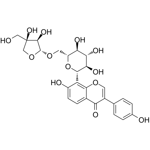
Mirificin (Puerarin apioside) is a blood-brain barrier-permeable tyrosinase inhibitor, with an IC50 value of 12.66 μM against mushroom tyrosinase. Mirificin reduces the expression of ALOX12 in cardiomyocytes and alleviates hypoxia/reoxygenation-induced apoptosis. The neuroprotective effect of Mirificin depends on the activation of VEGFR2 and HSP1A1, which reduces cerebral infarct size and improves neurological function. Mirificin can be used in the research of hyperpigmentation, acute myocardial infarction and ischemic stroke.
For research use only. We do not sell to patients.
- Purity : 99.66%
- CAS No.: 103654-50-8
- Formula: C26H28O13
- Molecular Weight:548.49
-
Storage:
4°C, protect from light
* In solvent : -80°C, 6 months; -20°C, 1 month (protect from light)

To place orders, for customer services and technical support, please contact: MedChemExpress USA
Tel: 609-228-6898 E-mail: [email protected] [email protected]


-
 Biological Activity
-
Chemical Information
- Protocols
- Purity & Documentation
- References
-
 Help & FAQs


-
HY-N0192
ArbutinEndogenous Metabolite -
HY-N0616
TrifolirhizinPterocarpan Flavonoid -
HY-107369
4-ButylresorcinolTyrosinase Inhibitor -

HY-N0192R
Arbutin (Standard)Endogenous Metabolite -
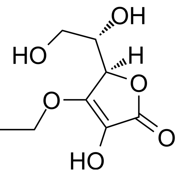
HY-W003607
3-O-Ethyl-L-ascorbic acidVitamin C Derivative -
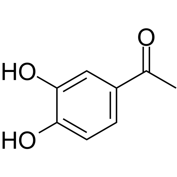
HY-N1775
3',4'-DihydroxyacetophenonePhenolic Compound -
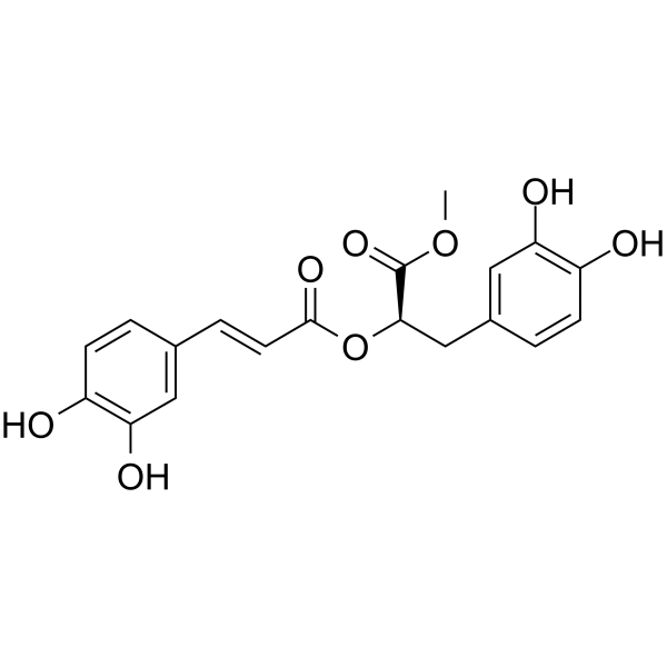
HY-N3266
Methyl rosmarinateHydroxycinnamic Acid -
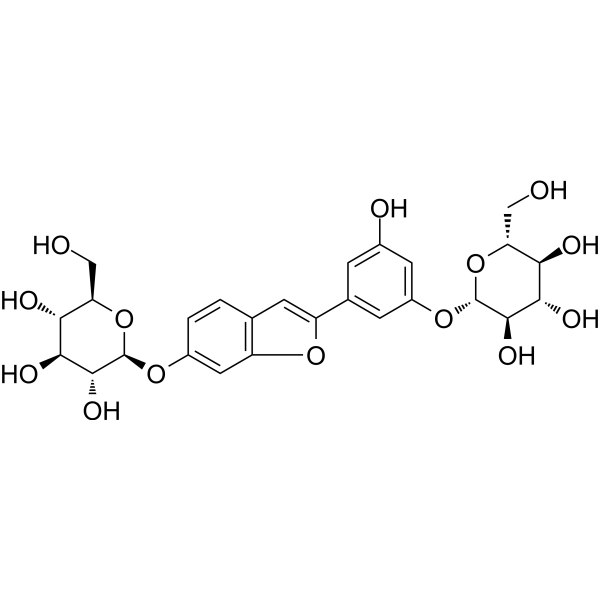
HY-N3518
Mulberroside FTyrosinase Inhibitor -
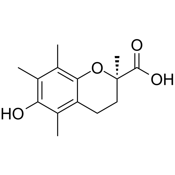
HY-101445A
(R)-TroloxTyrosinase Inhibitor -
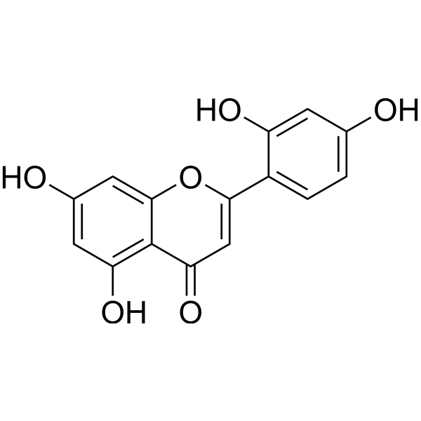
HY-N10503
NorartocarpetinTyrosinase Inhibitor -
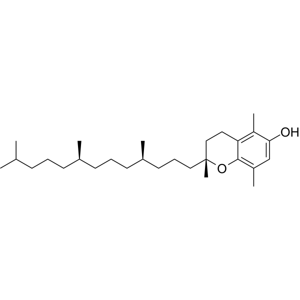
HY-113068
(rel)-β-Tocopherolβ-Tocopherol Isomer -
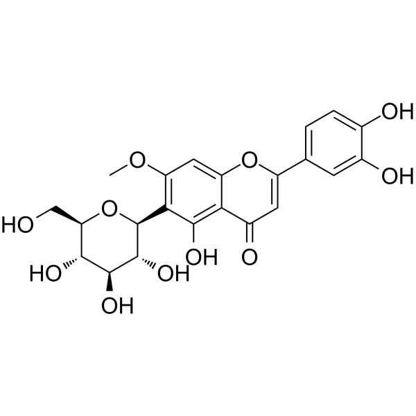
HY-N2204
SwertiajaponinTyrosinase Inhibitor
All VEGFR Isoforms
More

Biological Activity
Description
|
12-LOX |
VEGFR-2 |
HSP1A1 |
In Vitro
Mirificin (20.0 μM) shows no toxicity to H9c2 rat cardiomyocytes, even at the highest concentration tested[2].
Mirificin (5-50 μM; 24 h) dose-dependently increases the viability and reduces the cytotoxicity of differentiated PC12 cells with oxygen-glucose deprivation/reperfusion (OGD/R) injury[3].
Mirificin (0.1-10.0 μM) significantly reduces the mRNA expression level of Alox12 in H9c2 rat cardiomyocytes[2].
Mirificin (0.1 μM; 3 h) protects H9c2 rat cardiomyocytes against hypoxia/reoxygenation (H/R)-induced injury by inhibiting ALOX12 protein expression, restoring the BCL-2/BAX ratio, and reducing apoptosis[2].
MedChemExpress (MCE) has not independently confirmed the accuracy of these methods. They are for reference only. Further protocols information, click here.
-
Cell Line:Alox12-silenced H9c2 rat cardiomyocytes
-
Concentration:0.1 μM
-
Incubation Time:3 h (pretreatment prior to H/R)
-
Result:Produced no significant change in the apoptotic cell ratio compared to the H/R + siAlox12 control group.
-
Cell Line:OGD/R-injured differentiated PC12 cells
-
Concentration:5; 20; 50 μM
-
Incubation Time:24 h (pre-incubated prior to OGD/R)
-
Result:Increased cell viability to 65.8% at 5 μmol/L.
Increased cell viability to 67.4% at 20 μmol/L.
Increased cell viability to 69.5% at 50 μmol/L.
Reduced LDH leakage to 83.3% at 5 μmol/L.
Reduced LDH leakage to 84.8% at 20 μmol/L.
Reduced LDH leakage to 70.5% at 50 μmol/L.
In Vivo
Mirificin (12.5-25 mg/kg; i.p.; two administrations) reduces the cerebral infarction volume and improves neurological function in mice with ischemic stroke induced by middle cerebral artery occlusion (MCAO)[3].
MedChemExpress (MCE) has not independently confirmed the accuracy of these methods. They are for reference only.
-
Animal Model:C57BL/6 (male, 6 weeks old, 18-22g, intraluminal middle cerebral artery occlusion with 2 hours of occlusion followed by reperfusion)[3]
-
Dosage:12.5 mg/kg; 25 mg/kg
-
Administration:i.p.; two time points (1 hour pre-MCAO and 1 hour post-MCAO)
-
Result:Showed a significant reduction in Longa's Neurological Severity Score and a significant decrease in cerebral infarct area at 12.5 mg/kg.
Showed significant reductions in both neurological deficit score and cerebral infarct area, with a larger reduction in infarct area than the 12.5 mg/kg group at 25 mg/kg.


Chemical Information
-
CAS No. 103654-50-8
-
Appearance Solid
-
Molecular Weight 548.49
-
Formula C26H28O13
-
Color White to light yellow
-
SMILES
OC1=CC=C2C(OC=C(C3=CC=C(O)C=C3)C2=O)=C1[C@@H]([C@@H]([C@@H](O)[C@@H]4O)O)O[C@@H]4CO[C@H](OC[C@]5(O)CO)[C@@H]5O
-
Synonyms
Puerarin apioside
-
Structure Classification
-
Initial Source
-
Shipping
Room temperature in continental US; may vary elsewhere.
-
Storage
4°C, protect from light
* In solvent : -80°C, 6 months; -20°C, 1 month (protect from light)

Protocols
-
RNA extraction experimental
By lysing cells, releasing RNA, and removing impurities such as proteins and DNA, high-purity RNA products are finally obtained. The commonly used traditional method is the guanidine isothiocyanate/phenol/chloroform method (Trizol), which is suitable for a variety of animal materials including animal tissues, microorganisms, cultured cells, etc., and most plant materials.
-
Apoptosis
Apoptosis, also called programmed cell death, is generally characterized by distinct morphological characteristics.
-
TUNEL staining for apoptotic DNA fragmentation
TUNEL staining detects DNA strand breaks by using terminal deoxynucleotidyl transferase to add labeled nucleotides to exposed 3′-OH DNA termini, generating either microscopic staining in fixed cells or tissue sections, or fluorescence/cytometric signal in cell suspensions. TUNEL positivity reflects DNA fragmentation but should not be interpreted alone as definitive apoptosis, because TUNEL can also label necrotic, autolytic, mechanically damaged, or DNA-repair-associated DNA breaks.
-
Cell Cytotoxicity Assay
Cytotoxicity assays are usually based on the assessment of cell membrane damage, which can also be indirectly detected by measuring cell viability. Detection methods include MTT assay, CKK-8 assay, LDH assay and ATP assay, etc.
-
Annexin V plus membrane-impermeant dye apoptosis staining
Annexin V-based apoptosis assays rely on the detection of phosphatidylserine (PS) externalization from the inner leaflet of the plasma membrane to the outer leaflet, an early biochemical hallmark of apoptosis. Fluorescently labeled Annexin V binds PS in a calcium-dependent manner, enabling identification of early apoptotic cells by flow cytometry or fluorescence microscopy. When combined with a membrane-impermeant DNA-binding dye (e. g. , propidium iodide), this approach allows discrimination between viable (Annexin V−/dye−), early apoptotic (Annexin V+/dye−), and late apoptotic or necrotic (Annexin V+/dye+) cell populations by assessing membrane integrity and PS exposure.
-
Apoptosis Solutions
Apoptosis is a regulated, generally non-lytic cell-death pathway that removes unwanted, damaged, infected, or abnormal cells through coordinated morphological changes, caspase activation, DNA fragmentation, and membrane remodeling. The intrinsic apoptosis pathway is controlled mainly by mitochondrial outer membrane permeabilization, BCL-2 family proteins, cytochrome c release, apoptosome formation, caspase-9 activation, and downstream executioner caspase-3/7 activation. The extrinsic apoptosis pathway is initiated by death receptors such as Fas, TNFR, and TRAIL receptors, which recruit adaptor proteins and activate caspase-8 before engaging executioner caspases or mitochondrial amplification through BID cleavage. Apoptosis is linked to many phenotypes, including cancer cell killing, tissue homeostasis, immune regulation, neurodegeneration, infection response, and treatment-induced cytotoxicity; unresolved questions include how apoptosis interacts with necroptosis, pyroptosis, ferroptos
-
Research Protocol for Cardiovascular Diseases
Cardiovascular disease can be modeled as maladaptive cardiac remodeling, where ischemic injury or pressure overload activates inflammatory signaling, fibroblast activation, extracellular-matrix deposition, cardiomyocyte hypertrophy, vascular remodeling, and progressive ventricular dysfunction. The TGF-β/SMAD axis is a central profibrotic pathway after myocardial injury and pressure overload, while innate immune and cytokine pathways regulate leukocyte recruitment, scar formation, and adverse remodeling. Key unresolved questions include which inflammatory signals are reparative versus harmful, when fibrosis is protective versus maladaptive, and whether pathway inhibition improves function without weakening necessary infarct healing or compensatory remodeling.


Purity & Documentation
-
Data Sheet (279 KB)
-
SDS (252 KB)
- English - EN (252 KB)
- Français - FR (252 KB)
- Deutsch - DE (252 KB)
- Norwegian - NO (252 KB)
- Español - ES (252 KB)
- Swedish - SV (252 KB)
- Italian - IT (252 KB)
- Korean - KR (252 KB)
- Portuguese - PT (252 KB)
-
Handling Instructions (2659 KB)
References
[1]. Liu H, et al. Enzyme-Site Blocking Combined with Optimization of Molecular Docking for Efficient Discovery of Potential Tyrosinase Specific Inhibitors from Puerariae lobatae Radix. Molecules. 2018 Oct 11;23(10):2612. [Content Brief]
[2]. Xing H, et al. The potential effective components from Danlou tablet attenuates acute myocardial infarction by restoring ALOX12-mediated perturbed oxylipins. Journal of ethnopharmacology. 2025 Apr 09;345:119617. [Content Brief]
[3]. Zhou Z, et al. Integrating UHPLC-MS, Network Pharmacology, and Molecular Docking techniques to explore the neuroprotective effect of Mirificin. Naunyn-Schmiedeberg's archives of pharmacology. 2026 Feb;399(4):5447-5462. [Content Brief]

Calculators
Concentration (start) × Volume (start) = Concentration (final) × Volume (final)


Mirificin Related Classifications
HY-N2134 Related Classifications
 Powered by Bioz