



Moracin N
Based on 2 publication(s) in Google Scholar
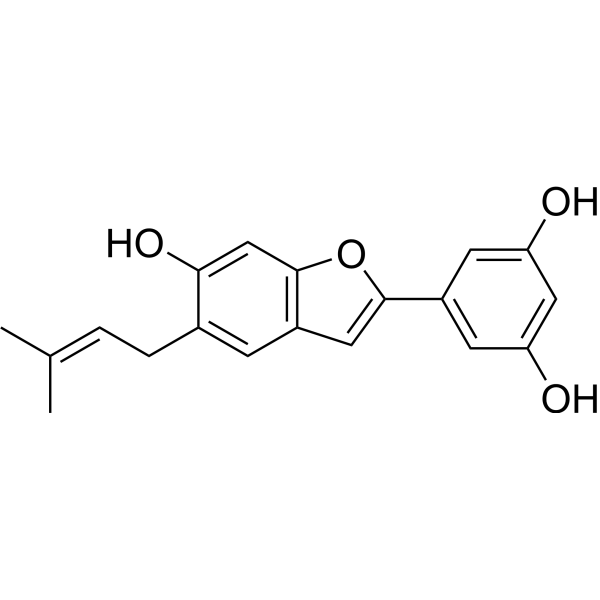
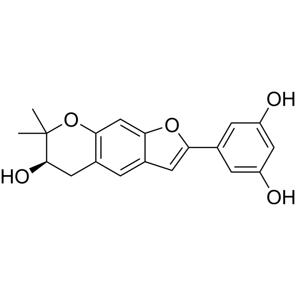
Moracin N is a benzofuran compound. At low concentrations, Moracin N activates the Keap1/Nrf2 pathway, downregulates the expression of FTH1 and ACSL4, enhances glutathione synthesis, and inhibits neuronal ferroptosis. At high concentrations, Moracin N induces ROS accumulation, activates mitochondrial apoptosis, and triggers autophagy-mediated cell death by inhibiting the AKT/mTOR pathway. Moracin N can be used in research related to non-small cell lung cancer, hepatocellular carcinoma and ischemic brain injury.
For research use only. We do not sell to patients.
- Purity : 98.02%
- CAS No.: 135248-05-4
- Formula: C19H18O4
- Molecular Weight:310.34
-
Storage:
-20°C, protect from light
* In solvent : -80°C, 6 months; -20°C, 1 month (protect from light)

To place orders, for customer services and technical support, please contact: MedChemExpress USA
Tel: 609-228-6898 E-mail: [email protected] [email protected]


-
 Biological Activity
-
Chemical Information
-
Solvent & Solubility
- Protocols
- Purity & Documentation
- References
-
 Help & FAQs


-
HY-122942
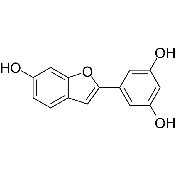
Moracin MEndogenous Metabolite -
HY-12364
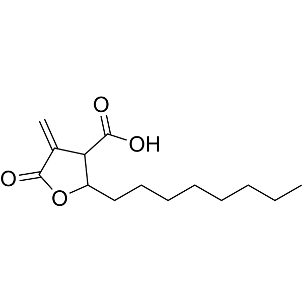
C75Fatty Acid Synthase Inhibitor -
HY-N0716B
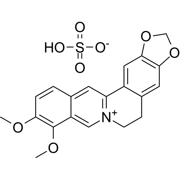
Berberine sulfateAntibiotic -
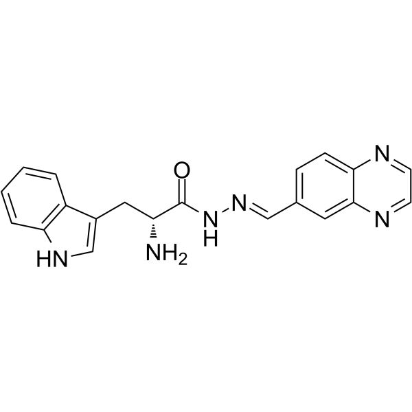
HY-12646A
RhosinRho Inhibitor -
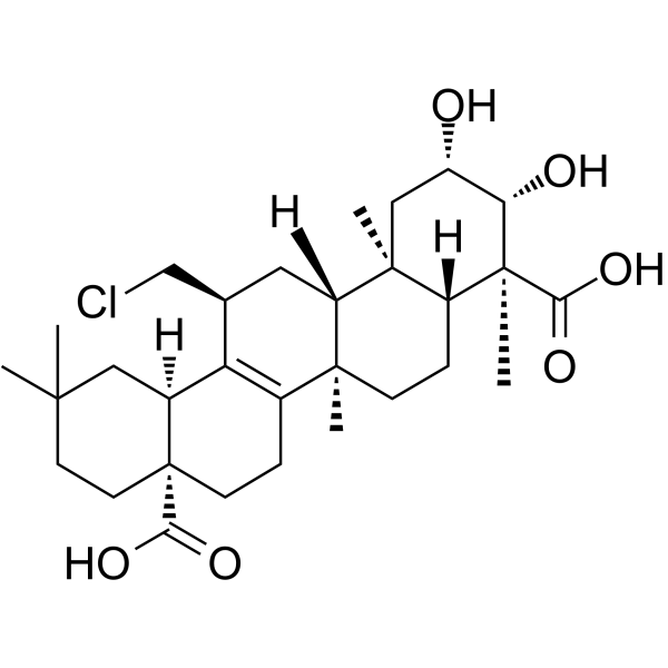
HY-N0802
TenuigeninNF-κB Inhibitor -
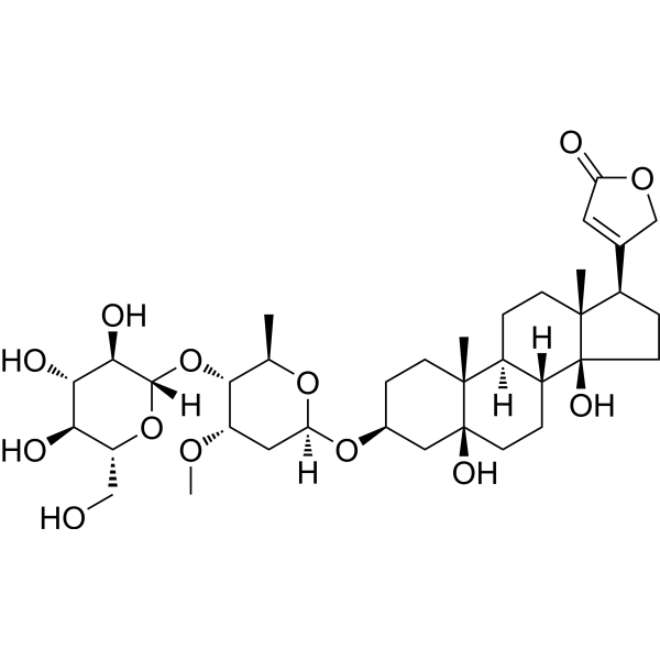
HY-N1381
PeriplocinAkt Inhibitor -
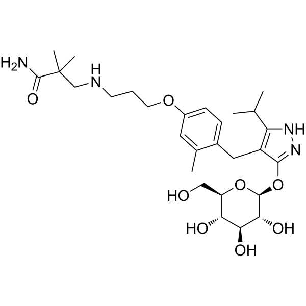
HY-17638
MizagliflozinSGLT1 Inhibitor -
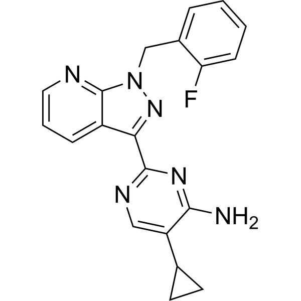
HY-12376
BAY 41-2272Guanylate Cyclase Activator -
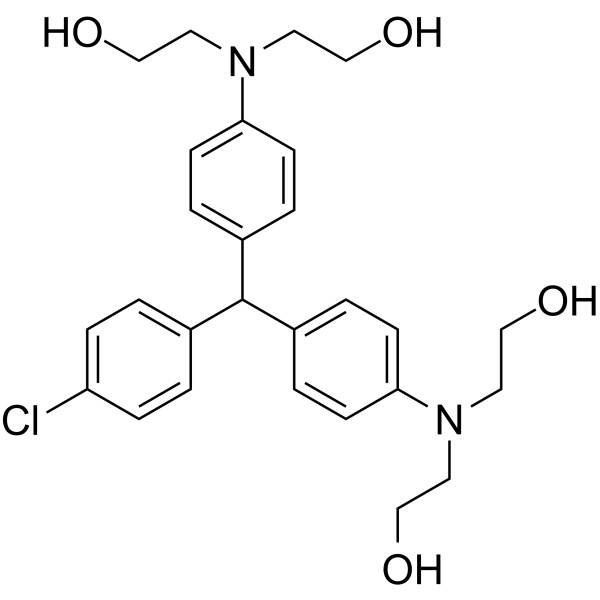
HY-104047
LM22B-10TrkB/TrkC Activator -
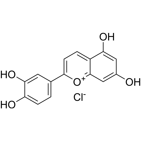
HY-129997
Luteolinidin chlorideCD38 Inhibitor -
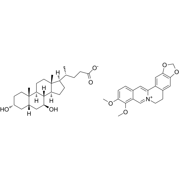
HY-147246
Berberine ursodeoxycholateHypolipidemic Agent -
HY-N3243
Moracin PReactive Oxygen Species Inhibitor
Publications Citing Use of MedChemExpress (MCE) Moracin N
More

Biological Activity
Description
Cellular Effect
|
Cell Line

|
Type | Value | Description | References |
|---|---|---|---|---|
| A2780 | IC50 |
>10 μg/mL
Compound: 14, Moracin N
|
Cytotoxicity against human A2780 cells after 96 hrs by MTT assay
Cytotoxicity against human A2780 cells after 96 hrs by MTT assay
|
[PMID: 25461329] |
| A549 | IC50 |
>10 μg/mL
Compound: 14, Moracin N
|
Cytotoxicity against human A549 cells after 96 hrs by MTT assay
Cytotoxicity against human A549 cells after 96 hrs by MTT assay
|
[PMID: 25461329] |
| Bel-7402 | IC50 |
>10 μg/mL
Compound: 14, Moracin N
|
Cytotoxicity against human Bel7402 cells after 96 hrs by MTT assay
Cytotoxicity against human Bel7402 cells after 96 hrs by MTT assay
|
[PMID: 25461329] |
| BGC-823 | IC50 |
>10 μg/mL
Compound: 14, Moracin N
|
Cytotoxicity against human BGC823 cells after 96 hrs by MTT assay
Cytotoxicity against human BGC823 cells after 96 hrs by MTT assay
|
[PMID: 25461329] |
| HCT-8 | IC50 |
>10 μg/mL
Compound: 14, Moracin N
|
Cytotoxicity against human HCT8 cells after 96 hrs by MTT assay
Cytotoxicity against human HCT8 cells after 96 hrs by MTT assay
|
[PMID: 25461329] |
In Vitro
Moracin N (0-100 μM; 24-72 h) inhibits the viability of human non-small cell lung cancer cells A549 and PC9 in a time- and dose-dependent manner, with IC50 values of 48.4 μM and 6.6 μM, respectively[1].
Moracin N (8-30 μM; 48 h) inhibits colony formation in human non-small cell lung cancer A549 and PC9 cells[1].
Moracin N (6-30 μM; 48 h) inhibits the migration of human non-small cell lung cancer A549 and PC9 cells in a dose-dependent manner[1].
Moracin N (0-100 μM; 72 h) exerts dose-dependent antiproliferative activity against HepG2 cells, with an IC50 of 50 μM after 72 h of incubation[3].
Moracin N (10-45 μM; 48 h) induces G0/G1 phase arrest in human non-small cell lung cancer PC9 cells (but exerts no such effect on A549 cells)[1].
Moracin N (10-45 μM; 48 h) induces dose-dependent apoptosis in human non-small cell lung cancer A549 and PC9 cells, with maximum apoptosis rates of 17% and 40%, respectively[1].
Moracin N (10-45 μM; 48 h) activates the mitochondrial apoptotic pathway in human non-small cell lung cancer A549 and PC9 cells by increasing the Bax/Bcl-2 ratio, inducing cytochrome c release, and activating the caspase cascade[1].
Moracin N (10-30 μM; 48 h) induces mitochondrial dysfunction, including mitochondrial fragmentation and reduced membrane potential, in human non-small cell lung cancer A549 and PC9 cells[1].
Moracin N (15-45 μM; 6-48 h) induces complete autophagic flux in A549, PC9 and HeLa-GFP-LC3 cells by promoting LC3-II conversion, inducing autophagosome formation and enhancing autophagosome-lysosome fusion[1].
Moracin N (15-45 μM; 6-48 h) inhibits the AKT/mTOR signaling pathway in a time- and dose-dependent manner, thereby inducing autophagy in human non-small cell lung cancer A549 cells[1].
Moracin N (10-45 μM; 6-24 h) activates lysosomal function in A549, PC9, HeLa-GFP-LC3 and L929-tfLC3 cells, promotes autophagosome-lysosome fusion, thereby enhancing complete autophagic flux[1].
Moracin N (10-45 μM; 24-48 h) induces dose-dependent reactive oxygen species (ROS) accumulation in human non-small cell lung cancer A549 and PC9 cells[1].
Moracin N (5 μM) inhibits ferroptosis in primary mouse embryonic neurons treated with oxygen-glucose deprivation/reoxygenation (OGD/R) and enhances their cell viability by regulating ferroptosis-related proteins, alleviating oxidative stress and iron overload, and increasing the activity of antioxidant enzymes[2].
Moracin N exhibits peroxyl radical scavenging activity in cell-free ORAC assays, with an ORAC value of 1.70 μmol TE/μmol[3].
Moracin N (10-60 μM; 30 min) potently scavenges DPPH free radicals in cell-free assays, with an IC50 of 40.00 μM[3].
Moracin N (10-40 μM; 1 h) exhibits potent cellular antioxidant activity in HepG2 cells, with EC50 values of 24.92 μM (without PBS washing) and 21.82 μM (with PBS washing), and shows efficient cellular uptake capacity[3].
Moracin N (0-100 μM; 24 h) shows no significant cytotoxicity against HepG2 cells even at concentrations up to 100 μM after 24 h of incubation[3].
MedChemExpress (MCE) has not independently confirmed the accuracy of these methods. They are for reference only.
-
Cell Line:human NSCLC A549 and PC9 cells
-
Concentration:20 μM, 30 μM (A549); 6 μM, 8 μM (PC9)
-
Incubation Time:48 h
-
Result:Inhibited cell migration in a dose-dependent manner.
Reduced A549 cell wound closure to ~25% at 20 μM and to ~10% at 30 μM relative to control.
Reduced PC9 cell wound closure to ~25% at 6 μM and ~15% at 8 μM, with statistically significant decreases relative to control groups.
-
Cell Line:human NSCLC A549 and PC9 cells
-
Concentration:15 μM, 30 μM, 45 μM (A549); 10 μM, 20 μM, 30 μM (PC9)
-
Incubation Time:48 h
-
Result:Increased the percentage of PC9 cells in the G0/G1 phase in a dose-dependent manner.
Had no effect on the cell cycle distribution of A549 cells.
-
Cell Line:human NSCLC A549 and PC9 cells
-
Concentration:15 μM, 30 μM, 45 μM (A549); 10 μM, 20 μM, 30 μM (PC9)
-
Incubation Time:48 h
-
Result:Increased the percentage of apoptotic (Annexin V+) cells in a dose-dependent manner.
Increased A549 cell apoptotic rates to ~5% at 15 , ~9% at 30 μM, and ~17% at 45 μM.
Increased PC9 cell apoptotic rates to ~15% at 10 μM, ~35% at 20 μM, and ~40% at 30 μM.
-
Cell Line:human NSCLC A549 cells
-
Concentration:15 μM, 30 μM, 45 μM
-
Incubation Time:6 h, 12 h, 24 h, 48 h
-
Result:Decreased the phosphorylation levels of AKT, mTOR, and S6 in a time- and dose-dependent manner.
Left total protein levels of AKT, mTOR, and S6 unchanged.
In Vivo
MedChemExpress (MCE) has not independently confirmed the accuracy of these methods. They are for reference only.
-
Animal Model:C57BL/6JNifdc (male, 20-22 g, ischaemic brain injury induced via 60-minute middle cerebral artery occlusion followed by reperfusion)[2]
-
Dosage:10 μg/kg; 20 μg/kg; 50 μg/kg
-
Administration:i.c.v.; administered at 1 h, 6 h, 12 h, 24 h post-reperfusion then daily for 2 additional days
-
Result:Significantly reduced brain water content, cerebral infarct volume, and modified neurological severity score (mNSS) at 20 μg/kg and 50 μg/kg compared to vehicle-treated mice.
Showed no significant difference from vehicle for brain water content, cerebral infarct volume, and mNSS at 10 μg/kg.
Improved long-term sensorimotor function at 20 μg/kg: reduced time to complete adhesive touch, adhesive removal, and pole tests; lower asymmetric rate in the cylinder test; longer latency to fall in the rotarod test; and fewer foot faults over 28 days post-MCAO.
Improved spatial learning and memory at 20 μg/kg: shorter escape latencies in Morris water maze (MWM) training trials, more platform crossings, and longer time spent in the target quadrant during MWM probe trials; increased novel object exploration time and higher discrimination index in the novel object recognition test.
Reversed MCAO-induced changes in ferroptosis-related proteins at 20 μg/kg: upregulated SLC7A11, FTH1, and GPX4; downregulated ACSL4 and 15-LOX2.
Reduced ROS production at 20 μg/kg: DHE fluorescence intensity fold change reduced from ~5 to ~2.
Increased GSH/GSSG ratio, GSH-Px, CAT, and SOD activity, and decreased MDA levels and free iron content in brain tissue at 20 μg/kg.
Activated the Keap1/Nrf2 pathway at 20 μg/kg: decreased Keap1 protein expression, increased Nrf2 protein expression, reduced Keap1-Nrf2 binding, repressed Nrf2 ubiquitination, increased nuclear Nrf2 translocation, enhanced Nrf2 DNA binding activity, and upregulated Nrf2 target genes NQO1, HO-1, GCLC, GCLM, and SOD2.
Abolished all neuroprotective and ferroptosis-inhibiting effects of 20 μg/kg moracin N when co-administered with Nrf2 inhibitor ML385, including reversing reductions in infarct volume, brain water content, and mNSS; restoring ROS, MDA, and free iron levels; and reducing GSH/GSSG ratio and antioxidant enzyme activity.
- RNA extraction experimental
- Mammalian live/dead viability and cytotoxicity staining
- Annexin V plus membrane-impermeant dye apoptosis staining
- ROS/oxidative-stress fluorescent staining
- Liver Cancer Modeling
- Detection of Apoptosis
- TUNEL staining for apoptotic DNA fragmentation
- Cell Cytotoxicity Assay
- Autophagy
- Apoptosis Solutions
- Ferroptosis Solutions
- Lysosome and acidic-vesicle live-cell staining
- Macroautophagy Solutions


Chemical Information
-
CAS No. 135248-05-4
-
Appearance Solid
-
Molecular Weight 310.34
-
Formula C19H18O4
-
Color White to off-white
-
SMILES
OC1=CC(C2=CC3=C(C=C(C(C/C=C(C)\C)=C3)O)O2)=CC(O)=C1
-
Structure Classification
-
Initial Source
-
Shipping
Room temperature in continental US; may vary elsewhere.
-
Storage
-20°C, protect from light
* In solvent : -80°C, 6 months; -20°C, 1 month (protect from light)


Publications (2)
-
Journal Impact Factor
-
Most Recent
-
Phytomedicine
Moracin N alleviates ischaemic brain injury in mice by suppressing neuronal ferroptosis via the activation of the Keap1/Nrf2 signalling pathway. [Abstract]2025 Sep 12:148:157253. PMID: 40972263 -
Environ Microbiol
Unravelling Wood Extractive Resistance in Phanerochaete chrysosporium Through Random Mutagenesis. [Abstract]2025 Dec;27(12):e70205. PMID: 41342371


Solvent & Solubility
In Vitro:
DMSO : 100 mg/mL (322.23 mM; Need ultrasonic; Hygroscopic DMSO has a significant impact on the solubility of product, please use newly opened DMSO)
Please refer to the solubility information to select the appropriate solvent. Once prepared, please aliquot and store the solution to prevent product inactivation from repeated freeze-thaw cycles.
Storage method and period of stock solution: -80°C, 6 months; -20°C, 1 month (protect from light). When stored at -80°C, please use it within 6 months. When stored at -20°C, please use it within 1 month.
Please refer to the solubility information to select the appropriate solvent. Once prepared, please aliquot and store the solution to prevent product inactivation from repeated freeze-thaw cycles.
Storage method and period of stock solution: -80°C, 6 months; -20°C, 1 month (protect from light). When stored at -80°C, please use it within 6 months. When stored at -20°C, please use it within 1 month.
Concentration (start) × Volume (start) = Concentration (final) × Volume (final)
In Vivo:
Select the appropriate dissolution method based on your experimental animal and administration route.
- For the following dissolution methods, please ensure to first prepare a clear stock solution using an In Vitro approach and then sequentially add co-solvents:
- To ensure reliable experimental results, the clarified stock solution can be appropriately stored based on storage conditions. As for the working solution for In Vivo experiments, it is recommended to prepare freshly and use it on the same day.
- The percentages shown for the solvents indicate their volumetric ratio in the final prepared solution. If precipitation or phase separation occurs during preparation, heat and/or sonication can be used to aid dissolution.
Add each solvent one by one: 10% DMSO 90% (20% SBE-β-CD in Saline)
Solubility: ≥ 2.5 mg/mL (8.06 mM); Clear solution
This protocol yields a clear solution of ≥ 2.5 mg/mL (saturation unknown).
Taking 1 mL working solution as an example, add 100 μL DMSO stock solution (25.0 mg/mL) to 900 μL 20% SBE-β-CD in Saline, and mix evenly.
Preparation of 20% SBE-β-CD in Saline (4°C, storage for one week): 2 g SBE-β-CD powder is dissolved in 10 mL Saline, completely dissolve until clear.
In Vivo Dissolution Calculator
Please enter the basic information of animal experiments:
-
-
-
-
Recommended: Prepare an additional quantity of animals to account for potential losses during experiments.
Please enter your animal formula composition:
-
%DMSO +
Recommended: Keep the proportion of DMSO in working solution below 2% if your animal is weak.
-
%+
-
+%Tween-80 + +
-
%Saline +
The co-solvents required include: DMSO, . All of co-solvents are available by MedChemExpress (MCE). , Tween 80. All of co-solvents are available by MedChemExpress (MCE).
Working solution concentration: 0.22 mg/mL
Method for preparing stock solution: mg drug dissolved in μL DMSO. Stock solution concentration: mg/mL. * In solvent : -80°C, 6 months; -20°C, 1 month (protect from light)
1. Take μL DMSO stock solution;
2. Add μL .
μL , mix evenly;
3. Then add μL Tween 80, mix evenly;
4. Then add μL
Please ensure that the stock solution in the first step is dissolved to a clear state, and add co-solvents in sequence. You can use ultrasonic heating (ultrasonic cleaner, recommended frequency 20-40 kHz), vortexing, etc. to assist dissolution.


Purity & Documentation
-
Data Sheet (294 KB)
-
SDS (252 KB)
- English - EN (252 KB)
- Français - FR (252 KB)
- Deutsch - DE (252 KB)
- Norwegian - NO (252 KB)
- Español - ES (252 KB)
- Swedish - SV (252 KB)
- Italian - IT (252 KB)
- Korean - KR (252 KB)
- Portuguese - PT (252 KB)
-
Handling Instructions (2659 KB)
References
[1]. Gao C, et al. A Novel Benzofuran Derivative Moracin N Induces Autophagy and Apoptosis Through ROS Generation in Lung Cancer. Frontiers in pharmacology. 2020;11:391. [Content Brief]
[2]. Zhang J, et al. Moracin N alleviates ischaemic brain injury in mice by suppressing neuronal ferroptosis via the activation of the Keap1/Nrf2 signalling pathway. Phytomedicine : international journal of phytotherapy and phytopharmacology. 2025 Nov 25;148:157253. [Content Brief]
[3]. Tu J, et al. Identification of moracin N in mulberry leaf and evaluation of antioxidant activity. Food and chemical toxicology : an international journal published for the British Industrial Biological Research Association. 2019 Oct;132:110730. [Content Brief]


Complete Stock Solution Preparation Table
Please refer to the solubility information to select the appropriate solvent. Once prepared, please aliquot and store the solution to prevent product inactivation from repeated freeze-thaw cycles.
Storage method and period of stock solution: -80°C, 6 months; -20°C, 1 month (protect from light). When stored at -80°C, please use it within 6 months. When stored at -20°C, please use it within 1 month.
| Optional Solvent | Concentration Solvent Mass | 1 mg | 5 mg | 10 mg | 25 mg |
|---|---|---|---|---|---|
| DMSO | 1 mM | 3.2223 mL | 16.1114 mL | 32.2227 mL | 80.5568 mL |
| 5 mM | 0.6445 mL | 3.2223 mL | 6.4445 mL | 16.1114 mL | |
| 10 mM | 0.3222 mL | 1.6111 mL | 3.2223 mL | 8.0557 mL | |
| 15 mM | 0.2148 mL | 1.0741 mL | 2.1482 mL | 5.3705 mL | |
| 20 mM | 0.1611 mL | 0.8056 mL | 1.6111 mL | 4.0278 mL | |
| 25 mM | 0.1289 mL | 0.6445 mL | 1.2889 mL | 3.2223 mL | |
| 30 mM | 0.1074 mL | 0.5370 mL | 1.0741 mL | 2.6852 mL | |
| 40 mM | 0.0806 mL | 0.4028 mL | 0.8056 mL | 2.0139 mL | |
| 50 mM | 0.0644 mL | 0.3222 mL | 0.6445 mL | 1.6111 mL | |
| 60 mM | 0.0537 mL | 0.2685 mL | 0.5370 mL | 1.3426 mL | |
| 80 mM | 0.0403 mL | 0.2014 mL | 0.4028 mL | 1.0070 mL | |
| 100 mM | 0.0322 mL | 0.1611 mL | 0.3222 mL | 0.8056 mL |


Moracin N Related Classifications
HY-N11849 Related Classifications
 Powered by Bioz
Keywords
- Moracin N
- 135248-05-4
- Tyrosinase
- Ferroptosis
- Mitochondrial Metabolism
- Apoptosis
- Autophagy
- Keap1-Nrf2
- ACSL Family
- Akt
- mTOR
- Reactive Oxygen Species (ROS)
- Keap1/Nrf2 signaling pathway
- ischaemic brain injury
- PC9 cells
- non-small-cell lung carcinoma
- Bcl-2
- hepatocellular carcinoma
- A549 cells
- HepG2 cells
- Bax
- Inhibitor
- inhibitor
- inhibit