



Niazirin

Based on 1 Customer Validation
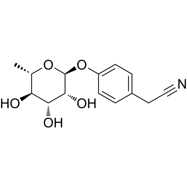
Niazirin is an orally active antioxidant. Niazirin can be isolated from Moringa oleifera Lam. Niazirin reduces the production levels of ROS and MDA, while increasing the levels of superoxide dismutase SOD and glutathione peroxidase GPx. Niazirin also abolishes high glucose-induced PKCζ activation and inhibits Nox4 protein expression. Niazirin exhibits excellent free radical scavenging activity. Niazirin significantly inhibits high glucose-induced proliferation of vascular smooth muscle cells. Niazirin can be used in the research of diabetic atherosclerosis.
For research use only. We do not sell to patients.
- Purity : 99.9%
- CAS No.: 122001-32-5
- Formula: C14H17NO5
- Molecular Weight:279.29
-
Storage:Powder -20°C, 3 years , 4°C, 2 years ; In solvent -80°C, 6 months , -20°C, 1 month

To place orders, for customer services and technical support, please contact: MedChemExpress USA
Tel: 609-228-6898 E-mail: [email protected] [email protected]


-
 Biological Activity
-
Chemical Information
- Protocols
- Purity & Documentation
- References
-
 Help & FAQs




Biological Activity
Description
IC50 & Target
[1]|
PKCζ |
NOX4 |
Cellular Effect
|
Cell Line

|
Type | Value | Description | References |
|---|---|---|---|---|
| RAW264.7 | IC50 |
>45 μM
Compound: 8
|
Antiinflammatory activity in mouse RAW264.7 cells assessed as inhibition of LPS-induced NO production incubated 30 mins prior to LPS challenge
Antiinflammatory activity in mouse RAW264.7 cells assessed as inhibition of LPS-induced NO production incubated 30 mins prior to LPS challenge
|
[PMID: 20685125] |
In Vitro
Niazirin (2-64 μM; 24 h) dose-dependently inhibits high glucose-induced abnormal proliferation of porcine aortic VSMCs, with no effect on VSMCs under normal glucose conditions[1].
Niazirin (5-25 μM; 24 h) significantly reduces high glucose-induced ROS production in porcine aortic VSMCs[1].
Niazirin (5-25 μM; 48 h) reverses high glucose-induced oxidant-antioxidant imbalance in porcine aortic VSMCs by reducing MDA production and increasing T-AOC, GPx, and SOD levels[1].
Niazirin (5-25 μM) inhibits high glucose-induced activation of the PKCζ/Nox4 pathway in porcine aortic VSMCs by reducing PKCζ Thr410 phosphorylation and Nox4 protein expression[1].
MedChemExpress (MCE) has not independently confirmed the accuracy of these methods. They are for reference only.
-
Cell Line:porcine aortic vascular smooth muscle cells (VSMCs) under normal glucose (NG) or high glucose (HG) conditions
-
Concentration:2 μM, 4 μM, 8 μM, 16 μM, 32 μM, 64 μM
-
Incubation Time:24 h
-
Result:Inhibited high glucose-induced VSMCs proliferation in a dose-dependent manner at all tested concentrations (p < 0.01).
Had no obvious effect on VSMCs growth under normal glucose conditions.
In Vivo
MedChemExpress (MCE) has not independently confirmed the accuracy of these methods. They are for reference only.
-
Animal Model:ICR mice (male, 20-25 g, diabetes induced by 8 weeks of high fat and carbohydrate diet followed by streptozotocin injection)[1]
-
Dosage:10 mg/kg; 40 mg/kg
-
Administration:i.g.; daily; 14 days
-
Result:Significantly increased serum total antioxidant capacity (T-AOC), superoxide dismutase (SOD), and glutathione peroxidase (GPx) levels relative to diabetic controls (p < 0.05 for T-AOC, GPx; p < 0.01 for SOD) at 10 mg/kg.
Significantly decreased serum malondialdehyde (MDA) production relative to diabetic controls (p < 0.01) at 10 mg/kg.
Significantly reduced thoracic aorta PKCζ Thr410 phosphorylation and Nox4 protein expression relative to diabetic controls (p < 0.05 for both) at 10 mg/kg.
Reduced aortic Ki67 positive area in diabetic controls.
Significantly increased serum T-AOC, SOD, and GPx levels relative to diabetic controls at 40 mg/kg.
Significantly decreased serum MDA production relative to diabetic controls (p < 0.01) at 40 mg/kg.
Significantly reduced thoracic aorta PKCζ Thr410 phosphorylation and Nox4 protein expression relative to diabetic controls (p < 0.01 for both) at 40 mg/kg.


Chemical Information
-
CAS No. 122001-32-5
-
Appearance Solid
-
Molecular Weight 279.29
-
Formula C14H17NO5
-
Color White to off-white
-
SMILES
O[C@H]([C@@H]([C@H]1O)O)[C@@H](O[C@H]1C)OC(C=C2)=CC=C2CC#N
-
Structure Classification
-
Initial Source
-
Shipping
Room temperature in continental US; may vary elsewhere.
-
Storage
Powder -20°C 3 years 4°C 2 years In solvent -80°C 6 months -20°C 1 month


Purity & Documentation
-
Data Sheet (273 KB)
-
SDS (251 KB)
- English - EN (251 KB)
- Français - FR (251 KB)
- Deutsch - DE (251 KB)
- Norwegian - NO (251 KB)
- Español - ES (251 KB)
- Swedish - SV (251 KB)
- Italian - IT (251 KB)
- Korean - KR (251 KB)
- Portuguese - PT (251 KB)
-
Handling Instructions (2659 KB)
References

Calculators
Concentration (start) × Volume (start) = Concentration (final) × Volume (final)


Niazirin Related Classifications
HY-N9097 Related Classifications
 Powered by Bioz