



Ophiopogonin D
Based on 2 publication(s) in Google Scholar
Ophiopogonin D can be isolated from the tubers of Ophiopogon japonicus, is a rare naturally occurring C29 steroidal glycoside. Ophiopogonin D is a CYP2J3 inducer that significantly inhibits Ang II induced NF-κB nuclear translocation, IκBα down-regulation, intracellular Ca2+ overload and activation of pro-inflammatory cytokines by increasing the expression of CYP2J2/EETs and PPARα in human umbilical vein endothelial cells (HUVECs). Ophiopogonin D can inhibit isteoclastic differentiation in RAW264.7 cells. Ophiopogonin D has protective effect as an antioxidant in H2O2-induced endothelial injury. Ophiopogonin D blocks ERK signaling cascades. Ophiopogonin D alleviates high-fat diet-induced metabolic syndrome and changes the structure of gut microbiota in mice. Ophiopogonin D has been used against inflammatory, metabolic and cardiovascular diseases.
For research use only. We do not sell to patients.
- Purity : 99.90%
- CAS No.: 945619-74-9
- Formula: C44H70O16
- Molecular Weight:855.02
-
Storage:
4°C, protect from light
* In solvent : -80°C, 6 months; -20°C, 1 month (protect from light)

To place orders, for customer services and technical support, please contact: MedChemExpress USA
Tel: 609-228-6898 E-mail: [email protected] [email protected]


-
 Biological Activity
-
Chemical Information
-
Solvent & Solubility
- Protocols
- Purity & Documentation
- References
-
 Help & FAQs


-
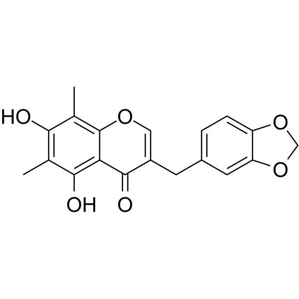
HY-N2441
Methylophiopogonone ANatural Product -
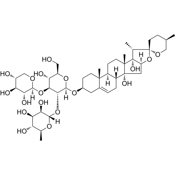
HY-N2175
Cixiophiopogon ANatural Product -

HY-N0515R
Ophiopogonin D (Standard)Calcium Channel Inhibitor -
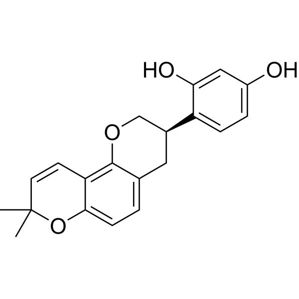
HY-N0393
GlabridinPPARγ Activator -
HY-N0292
OleuropeinApoptosis Inducer -
HY-N0441
NeferineAutophagy Modulator -
HY-N2065
Withaferin ANF-κB Inhibitor -
HY-N0604
Ginsenoside Rh1Endogenous Metabolite -
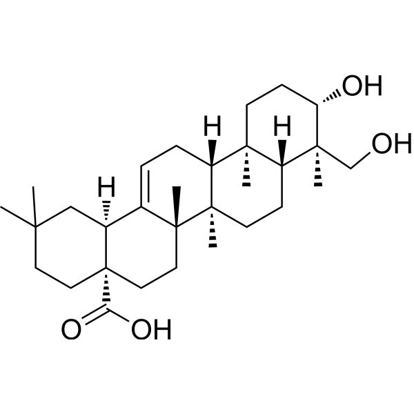
HY-N0256
HederageninCOX Inhibitor -
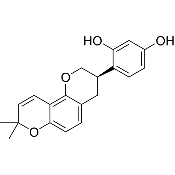
HY-N0393R
Glabridin (Standard) -
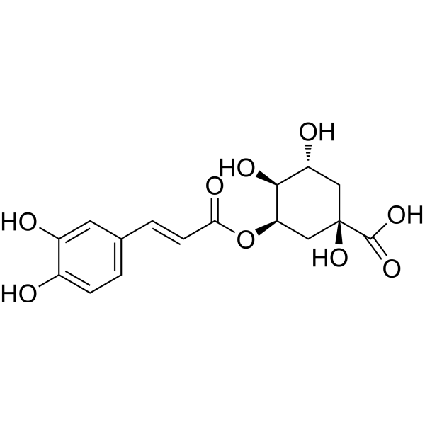
HY-N0722
Neochlorogenic acidCOX Inhibitor -
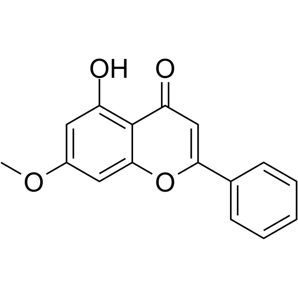
HY-14592
TectochrysinNF-κB Inhibitor
Publications Citing Use of MedChemExpress (MCE) Ophiopogonin D
MoreAll Calcium Channel Isoforms
More

Biological Activity
Description
IC50 & Target
|
PPARα |
NF-κB |
In Vitro
Ophiopogonin D (Compound OP-D) (0.1-200 μM, 24-48 h) only provokes cell viability at high concentrations above 40 μM in HUVECs[2].
Ophiopogonin D (5-20 μM, 24 h) significantly increases the expression of CYP2J2 and PPARα protein and mRNA expression dose-dependently[2].
Ophiopogonin D (1-100 μM, 24 h prior to H2O2) inhibits H2O2-induced cytotoxicity in MC3T3-E1 and RAW264.7 cells[3].
Ophiopogonin D (1-100 μM) inhibits osteoclastic differentiation in RAW264.7 cells[3].
Ophiopogonin D (0.6-60 μM, 2 h) dose-dependently prevents H2O2-induced oxidative stress in HUVECs[4].
MedChemExpress (MCE) has not independently confirmed the accuracy of these methods. They are for reference only. Further protocols information, click here.
-
Cell Line:HUVECs
-
Concentration:5, 10, 20 μM
-
Incubation Time:24 h
-
Result:Significantly increased 11,12-DHET levels.
-
Cell Line:HUVECs
-
Concentration:5, 10, 20 μM
-
Incubation Time:24 h
-
Result:Suppressed Ang II-induced inflammatory responses via CYP2J2-PPARα pathway.
In Vivo
MedChemExpress (MCE) has not independently confirmed the accuracy of these methods. They are for reference only.
-
Animal Model:BALB/c female OVX mice (19-21 g)[3]
-
Dosage:5, 25 mg/kg
-
Administration:Intraperitoneal injection (i.p.), daily for 12 w
-
Result:Inhibited serum osteoclastic markers in serum, such as CTX-1 and TRAP.
Partially inhibited osteoclastogenesis.


Chemical Information
-
CAS No. 945619-74-9
-
Appearance Solid
-
Molecular Weight 855.02
-
Formula C44H70O16
-
Color White to off-white
-
SMILES
C[C@@]12[C@]([C@@H]3C)([H])[C@](O[C@]34CC[C@@H](C)CO4)([H])C[C@@]1([H])[C@@](CC=C5[C@@]6([C@@H](C[C@H](O)C5)O[C@@](O[C@H](C)[C@H](O)[C@@H]7O[C@@](OC[C@@H](O)[C@@H]8O)([H])[C@@H]8O)([H])[C@@H]7O[C@@](O[C@@H](C)[C@H](O)[C@H]9O)([H])[C@@H]9O)C)([H])[C@]6([H])CC2
-
Structure Classification
-
Initial Source
-
Shipping
Room temperature in continental US; may vary elsewhere.
-
Storage
4°C, protect from light
* In solvent : -80°C, 6 months; -20°C, 1 month (protect from light)


Publications (2)
-
Journal Impact Factor
-
Most Recent
-
Part Fibre Toxicol
Exposure to polylactic acid microplastics during puberty increases the risk of skeletal dysplasia by disrupting arachidonic acid metabolism in osteoblasts. [Abstract]2026 Apr 22;23(1):26. PMID: 42021360 -
Int Immunopharmacol
Ophiopogonin D reprograms the polarization of macrophages through modulating PPM1K-mediated branched-chain amino acids catabolism to delay atherogenesis. [Abstract]2026 May 15:177:116551. PMID: 41871493


Solvent & Solubility
In Vitro:
DMSO : 100 mg/mL (116.96 mM; Need ultrasonic; Hygroscopic DMSO has a significant impact on the solubility of product, please use newly opened DMSO)
Please refer to the solubility information to select the appropriate solvent. Once prepared, please aliquot and store the solution to prevent product inactivation from repeated freeze-thaw cycles.
Storage method and period of stock solution: -80°C, 6 months; -20°C, 1 month (protect from light). When stored at -80°C, please use it within 6 months. When stored at -20°C, please use it within 1 month.
Please refer to the solubility information to select the appropriate solvent. Once prepared, please aliquot and store the solution to prevent product inactivation from repeated freeze-thaw cycles.
Storage method and period of stock solution: -80°C, 6 months; -20°C, 1 month (protect from light). When stored at -80°C, please use it within 6 months. When stored at -20°C, please use it within 1 month.
Concentration (start) × Volume (start) = Concentration (final) × Volume (final)
In Vivo:
Select the appropriate dissolution method based on your experimental animal and administration route.
- For the following dissolution methods, please ensure to first prepare a clear stock solution using an In Vitro approach and then sequentially add co-solvents:
- To ensure reliable experimental results, the clarified stock solution can be appropriately stored based on storage conditions. As for the working solution for In Vivo experiments, it is recommended to prepare freshly and use it on the same day.
- The percentages shown for the solvents indicate their volumetric ratio in the final prepared solution. If precipitation or phase separation occurs during preparation, heat and/or sonication can be used to aid dissolution.
In Vivo Dissolution Calculator
Please enter the basic information of animal experiments:
-
-
-
-
Recommended: Prepare an additional quantity of animals to account for potential losses during experiments.
Please enter your animal formula composition:
-
%DMSO +
Recommended: Keep the proportion of DMSO in working solution below 2% if your animal is weak.
-
%+
-
+%Tween-80 + +
-
%Saline +
The co-solvents required include: DMSO, . All of co-solvents are available by MedChemExpress (MCE). , Tween 80. All of co-solvents are available by MedChemExpress (MCE).
Working solution concentration: 0.22 mg/mL
Method for preparing stock solution: mg drug dissolved in μL DMSO. Stock solution concentration: mg/mL. * In solvent : -80°C, 6 months; -20°C, 1 month (protect from light)
1. Take μL DMSO stock solution;
2. Add μL .
μL , mix evenly;
3. Then add μL Tween 80, mix evenly;
4. Then add μL
Please ensure that the stock solution in the first step is dissolved to a clear state, and add co-solvents in sequence. You can use ultrasonic heating (ultrasonic cleaner, recommended frequency 20-40 kHz), vortexing, etc. to assist dissolution.

Protocols
-
Cytoplasmic-Nuclear Fractionated Protein Extraction
Cytoplasmic-nuclear fractionated protein extraction separates soluble cytoplasmic proteins from nuclear-enriched proteins by mild plasma-membrane permeabilization, differential centrifugation, washing of nuclei, and extraction of nuclear proteins for downstream immunoblotting or related molecular analysis. The readout is the relative abundance of a protein in cytoplasmic and nuclear fractions, commonly assessed by western blotting together with compartment markers such as tubulin or pyruvate kinase for cytoplasm and lamin, nucleoporin, hnRNP, H2AX, or Lamin B for nuclear fractions.
-
RNA extraction experimental
By lysing cells, releasing RNA, and removing impurities such as proteins and DNA, high-purity RNA products are finally obtained. The commonly used traditional method is the guanidine isothiocyanate/phenol/chloroform method (Trizol), which is suitable for a variety of animal materials including animal tissues, microorganisms, cultured cells, etc., and most plant materials.
-
LPS-Induced Endotoxemia/Systemic Inflammation
Lipopolysaccharide (LPS)-induced endotoxemia is a widely used in vivo model of acute systemic inflammation in which LPS, a Gram-negative bacterial endotoxin, activates innate immune signaling primarily through TLR4, leading to rapid and transient induction of pro-inflammatory cytokines such as TNF-α, IL-6, and IL-1β in circulation and tissues. This cytokine surge is commonly used as a measurable readout of systemic inflammatory activation and immune dysregulation, and is typically assessed within hours after intraperitoneal LPS administration in mouse models of endotoxemia. The model captures key features of systemic inflammatory response syndrome, including cytokine release, immune cell activation, and downstream tissue responses, and has been used to evaluate anti-inflammatory interventions such as cytokine modulation, lipid mediators, and immune cell-targeting therapies.
-
Endothelial Tube Formation Assay
Endothelial tube formation assay evaluates the ability of endothelial cells to attach, migrate, align, and organize into capillary-like networks when cultured on gelled basement membrane extract or Matrigel; the readout is the morphology and quantity of tube-like networks, which reflects an in vitro endothelial morphogenesis step related to angiogenesis. Basement membrane extract/Matrigel provides laminin-rich extracellular matrix cues that support endothelial differentiation into capillary-like structures, but it can contain biologically active growth factors, so growth-factor-reduced matrix is preferred when testing defined angiogenic stimulators or inhibitors.
-
Research Protocol for Microbiome Analysis
Microbiome analysis characterizes microbial communities in biological or environmental samples by measuring community composition, diversity, taxonomic structure, functional potential, and associations with host or environmental phenotypes. 16S rRNA gene amplicon sequencing is commonly used for bacterial and archaeal taxonomic profiling, while shotgun metagenomics provides higher taxonomic resolution and direct functional information, including microbial genes, pathways, viruses, fungi, and antimicrobial-resistance genes when sequencing depth and host-DNA contamination are adequately controlled. Microbiome results are strongly affected by sample collection, storage, DNA extraction, contamination, sequencing method, reference database, and bioinformatic pipeline; therefore, standardized protocols, negative controls, mock communities, and transparent analysis workflows are required. Unresolved issues include low-biomass contamination, compositional-data bias, inconsistent species-level c
-
Research Protocol for Inflammation-related Diseases
The NLRP3 inflammasome is a cytosolic innate immune signaling platform that integrates priming signals and danger-signal activation to promote caspase-1 activation, maturation of IL-1β and IL-18, and gasdermin D-mediated pyroptotic cell death. The core experimental logic is to determine whether inflammatory disease phenotypes are driven by increased NLRP3 expression, ASC-containing inflammasome assembly, caspase-1 cleavage, GSDMD cleavage, and extracellular release of IL-1β/IL-18 rather than by nonspecific cell injury alone. The pathway is strongly linked to inflammation-related disease phenotypes because monosodium urate crystals activate NALP3/NLRP3 inflammasome signaling in gout-like crystal inflammation, cholesterol crystals activate NLRP3 inflammasomes in atherogenesis models, and DSS-induced intestinal inflammation has been reported to involve NLRP3 inflammasome activity. However, experimental colitis studies also show context-dependent protective effects of NLRP3 inflammasome co
-
Ca2+ Staining Technique
Ca2+ staining is an experimental technique that utilizes specific fluorescent probes (such as Fluo-4 AM, Fura-2, etc.) to qualitatively or quantitatively detect dynamic changes in intracellular Ca2+ concentrations; this is achieved by monitoring the changes in fluorescent signals generated when these probes bind to free intracellular calcium ions. The underlying principle relies primarily on the presence of chelating groups within the probe's molecular structure that possess high affinity for calcium ions.
-
Research Protocol for Metabolic Diseases
AMP-activated protein kinase, AMPK, is a conserved cellular energy sensor that responds to reduced cellular energy status and coordinates metabolism by increasing ATP-generating catabolic pathways while suppressing ATP-consuming anabolic processes. In metabolic disease research, the AMPK pathway is experimentally relevant because it regulates hepatic lipid synthesis, fatty acid oxidation, glucose production, skeletal-muscle glucose disposal, mTORC1-linked biosynthesis, autophagy, mitochondrial homeostasis, and whole-body energy balance. The central pathway logic is that energy stress, metformin, exercise-like stimulation, or direct AMPK activators increase AMPKα Thr172 phosphorylation and downstream substrate phosphorylation, including ACC and RAPTOR. Phosphorylation of ACC suppresses lipogenesis and supports fatty acid oxidation, whereas phosphorylation of RAPTOR suppresses mTORC1 signaling and links cellular energy status to growth and protein synthesis control. The pathway is linked


Purity & Documentation
-
Data Sheet (284 KB)
-
SDS (393 KB)
- English - EN (393 KB)
- Français - FR (393 KB)
- Deutsch - DE (393 KB)
- Norwegian - NO (393 KB)
- Español - ES (393 KB)
- Swedish - SV (393 KB)
- Italian - IT (393 KB)
- Korean - KR (393 KB)
- Portuguese - PT (393 KB)
-
Handling Instructions (2659 KB)
References
[1]. Wang L , et al. Homo-aro-cholestane, furostane and spirostane saponins from the tubers of Ophiopogonjaponicus. Phytochemistry. 2017 Apr;136:125-132. [Content Brief]
[2]. Huang X, et al. Ophiopogonin D and EETs ameliorate Ang II-induced inflammatory responses via activating PPARα in HUVECs. Biochem Biophys Res Commun. 2017 Aug 19;490(2):123-133. [Content Brief]
[3]. Huang, Q., et al., (2015). Ophiopogonin D: A new herbal agent against osteoporosis. Bone, 74, 18–28. [Content Brief]
[4]. Qian, J., et al., (2010). Ophiopogonin D prevents H2O2-induced injury in primary human umbilical vein endothelial cells. Journal of ethnopharmacology, 128(2), 438–445. [Content Brief]


Complete Stock Solution Preparation Table
Please refer to the solubility information to select the appropriate solvent. Once prepared, please aliquot and store the solution to prevent product inactivation from repeated freeze-thaw cycles.
Storage method and period of stock solution: -80°C, 6 months; -20°C, 1 month (protect from light). When stored at -80°C, please use it within 6 months. When stored at -20°C, please use it within 1 month.
| Optional Solvent | Concentration Solvent Mass | 1 mg | 5 mg | 10 mg | 25 mg |
|---|---|---|---|---|---|
| DMSO | 1 mM | 1.1696 mL | 5.8478 mL | 11.6956 mL | 29.2391 mL |
| 5 mM | 0.2339 mL | 1.1696 mL | 2.3391 mL | 5.8478 mL | |
| 10 mM | 0.1170 mL | 0.5848 mL | 1.1696 mL | 2.9239 mL | |
| 15 mM | 0.0780 mL | 0.3899 mL | 0.7797 mL | 1.9493 mL | |
| 20 mM | 0.0585 mL | 0.2924 mL | 0.5848 mL | 1.4620 mL | |
| 25 mM | 0.0468 mL | 0.2339 mL | 0.4678 mL | 1.1696 mL | |
| 30 mM | 0.0390 mL | 0.1949 mL | 0.3899 mL | 0.9746 mL | |
| 40 mM | 0.0292 mL | 0.1462 mL | 0.2924 mL | 0.7310 mL | |
| 50 mM | 0.0234 mL | 0.1170 mL | 0.2339 mL | 0.5848 mL | |
| 60 mM | 0.0195 mL | 0.0975 mL | 0.1949 mL | 0.4873 mL | |
| 80 mM | 0.0146 mL | 0.0731 mL | 0.1462 mL | 0.3655 mL | |
| 100 mM | 0.0117 mL | 0.0585 mL | 0.1170 mL | 0.2924 mL |


Ophiopogonin D Related Classifications
HY-N0515 Related Classifications
 Powered by Bioz