



PD-166793
Based on 1 publication(s) in Google Scholar
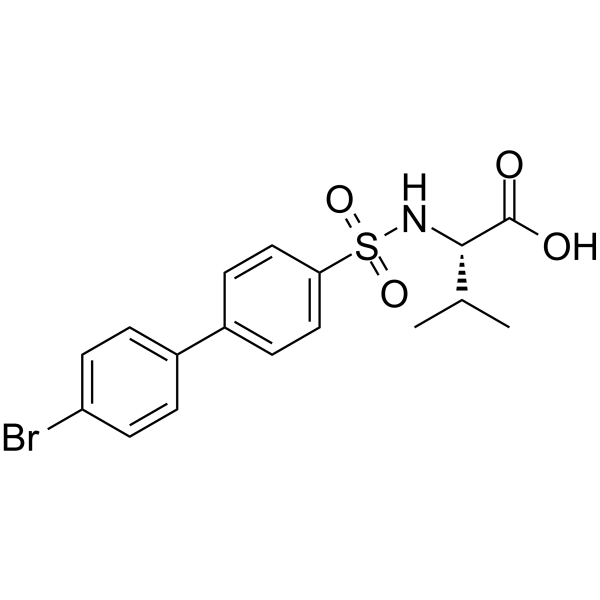
PD-166793 is a potent, selective, orally active and wide-broad spectrum inhibitor of MMP, exhibiting nanomolar potency against MMP-2, MMP-3 and MMP-13 (IC50=4, 7, and 8 nM, respectively) and micromolar potency vs MMP-1, -7 and -9 (IC50=6.0, 7.2, and 7.9 μM, respectively). PD-166793 can attenuate left ventricular remodeling and dysfunction in a rat model of progressive heart failure.
For research use only. We do not sell to patients.
- Purity : 99.21%
- CAS No.: 199850-67-4
- Formula: C17H18BrNO4S
- Molecular Weight:412.30
-
Storage:Powder -20°C, 3 years , 4°C, 2 years ; In solvent -80°C, 6 months , -20°C, 1 month

To place orders, for customer services and technical support, please contact: MedChemExpress USA
Tel: 609-228-6898 E-mail: [email protected] [email protected]


-
 Biological Activity
-
Chemical Information
-
Solvent & Solubility
- Protocols
- Purity & Documentation
- References
-
 Help & FAQs


-
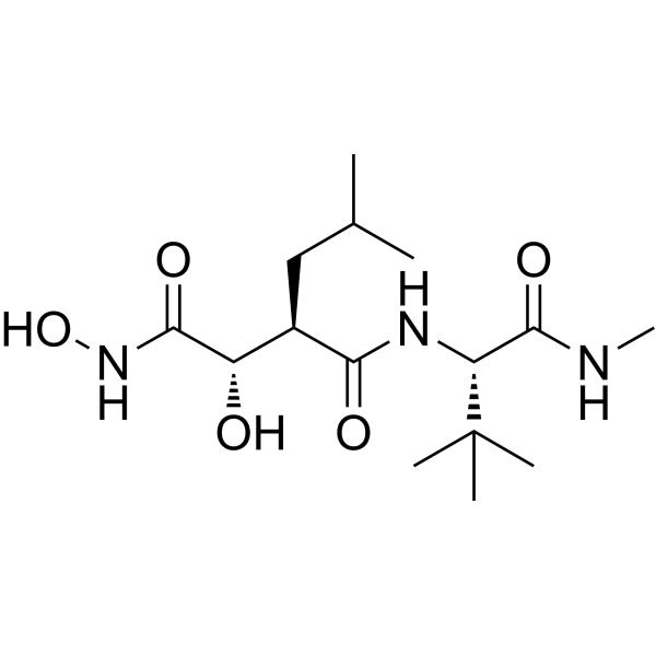
HY-12169
MarimastatMMP Inhibitor -
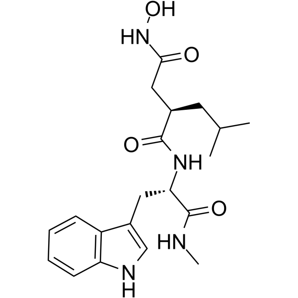
HY-15768
IlomastatMMP Inhibitor -
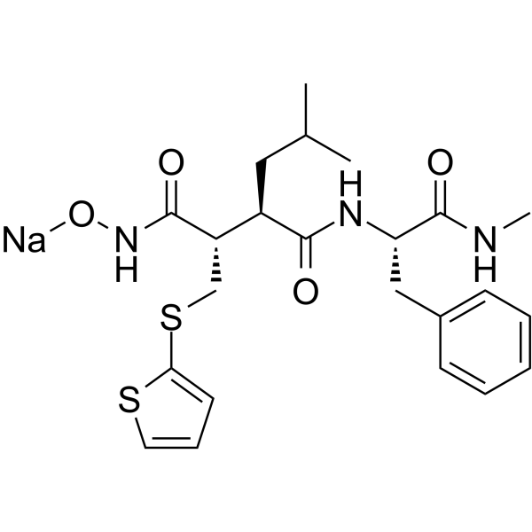
HY-13564A
Batimastat sodium salt -
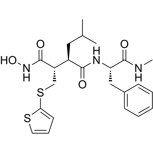
HY-13564
BatimastatMMP Inhibitor -
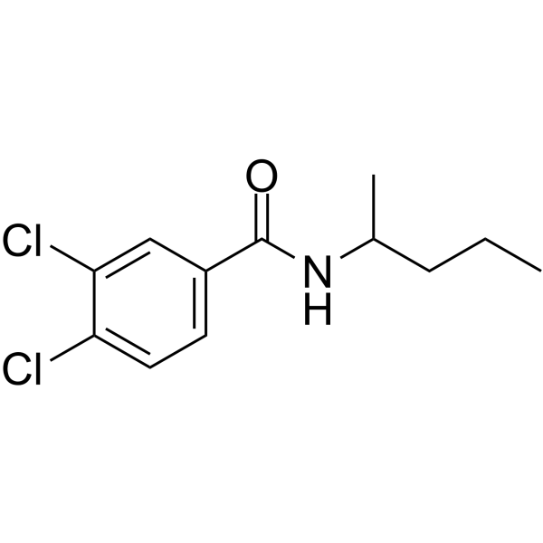
HY-15827
NSC 405020MMP14 Inhibitor -
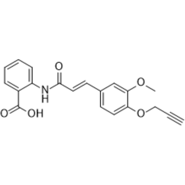
HY-100495
FT011Anti-fibrotic Agent -
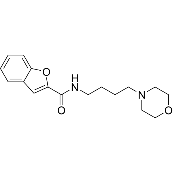
HY-100359
CL-82198MMP-13 Inhibitor -
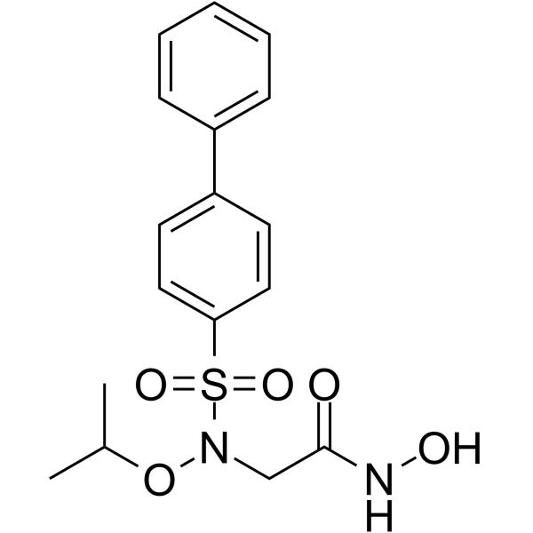
HY-103444
ARP-100MMP-2 Inhibitor -
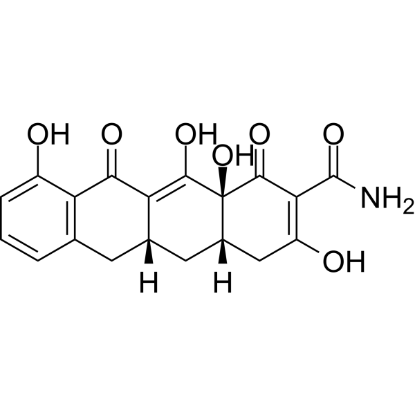
HY-13648
IncyclinideMMP Inhibitor -
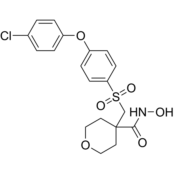
HY-10398
CTS-1027MMP Inhibitor -
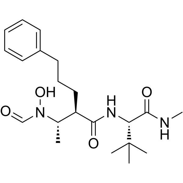
HY-19956
GI254023XADAM10/MMP9 Inhibitor -
HY-107394
UK 356618MMP-3 Inhibitor
Publications Citing Use of MedChemExpress (MCE) PD-166793
More

Biological Activity
Description
IC50 & Target
[1]|
MMP-2 4 nM (IC50) |
MMP-3 7 nM (IC50) |
MMP-13 8 nM (IC50) |
MMP-1 6.0 μM (IC50) |
MMP-7 7.2 μM (IC50) |
MMP-9 7.9 μM (IC50) |
In Vitro
PD-166793 (0.1 μM) leads to a 20% inhibition of AMP deaminase (AMPD) activity in rat heart homogenates[2].
PD-166793 (100 μM; 36 h) significantly reduces MMP-9 activity in normal human cardiac fibroblasts[2].
MedChemExpress (MCE) has not independently confirmed the accuracy of these methods. They are for reference only. Further protocols information, click here.
In Vivo
PD-166793 (5 mg/kg; oral gavage) exhibits superior pharmacokinetics (t1/2=43.6 h, Cmax=42.4 μg/mL, AUC0-∞=2822 μg h/mL) in rats[1].
MedChemExpress (MCE) has not independently confirmed the accuracy of these methods. They are for reference only.
-
Animal Model:Male Sprague-Dawley rats (6 weeks) were induced chronic biventricular volume overload[3]
-
Dosage:1 mg/kg
-
Administration:Daily gavage beginning 2 weeks before surgery and continued until 8 weeks after surgery
-
Result:Prevented ventricular dilatation and attenuated the hypertrophy typically induced by chronic volume overload.


Chemical Information
-
CAS No. 199850-67-4
-
Appearance Solid
-
Molecular Weight 412.30
-
Formula C17H18BrNO4S
-
Color White to pink
-
SMILES
CC(C)[C@@H](C(O)=O)NS(=O)(C1=CC=C(C2=CC=C(Br)C=C2)C=C1)=O
-
Shipping
Room temperature in continental US; may vary elsewhere.
-
Storage
Powder -20°C 3 years 4°C 2 years In solvent -80°C 6 months -20°C 1 month


Publications (1)
-
Journal Impact Factor
-
Most Recent
-
bioRxiv
ProNotch converts extracellular protease activity into programmable transcriptional outputs. [Abstract]2026 Jun 3:2026.06.01.728985. PMID: 42282718


Solvent & Solubility
In Vitro:
DMSO : 100 mg/mL (242.54 mM; Need ultrasonic; Hygroscopic DMSO has a significant impact on the solubility of product, please use newly opened DMSO)
Please refer to the solubility information to select the appropriate solvent. Once prepared, please aliquot and store the solution to prevent product inactivation from repeated freeze-thaw cycles.
Storage method and period of stock solution: -80°C, 6 months; -20°C, 1 month. When stored at -80°C, please use it within 6 months. When stored at -20°C, please use it within 1 month.
Please refer to the solubility information to select the appropriate solvent. Once prepared, please aliquot and store the solution to prevent product inactivation from repeated freeze-thaw cycles.
Storage method and period of stock solution: -80°C, 6 months; -20°C, 1 month. When stored at -80°C, please use it within 6 months. When stored at -20°C, please use it within 1 month.
Concentration (start) × Volume (start) = Concentration (final) × Volume (final)
In Vivo:
Select the appropriate dissolution method based on your experimental animal and administration route.
- For the following dissolution methods, please ensure to first prepare a clear stock solution using an In Vitro approach and then sequentially add co-solvents:
- To ensure reliable experimental results, the clarified stock solution can be appropriately stored based on storage conditions. As for the working solution for In Vivo experiments, it is recommended to prepare freshly and use it on the same day.
- The percentages shown for the solvents indicate their volumetric ratio in the final prepared solution. If precipitation or phase separation occurs during preparation, heat and/or sonication can be used to aid dissolution.
Add each solvent one by one: 10% DMSO 40% PEG300 5% Tween-80 45% Saline
Solubility: ≥ 2.5 mg/mL (6.06 mM); Clear solution
This protocol yields a clear solution of ≥ 2.5 mg/mL (saturation unknown).
Taking 1 mL working solution as an example, add 100 μL DMSO stock solution (25.0 mg/mL) to 400 μL PEG300, and mix evenly; then add 50 μL Tween-80 and mix evenly; then add 450 μL Saline to adjust the volume to 1 mL.
Preparation of Saline: Dissolve 0.9 g sodium chloride in ddH₂O and dilute to 100 mL to obtain a clear Saline solution.
Add each solvent one by one: 10% DMSO 90% (20% SBE-β-CD in Saline)
Solubility: ≥ 2.5 mg/mL (6.06 mM); Clear solution
This protocol yields a clear solution of ≥ 2.5 mg/mL (saturation unknown).
Taking 1 mL working solution as an example, add 100 μL DMSO stock solution (25.0 mg/mL) to 900 μL 20% SBE-β-CD in Saline, and mix evenly.
Preparation of 20% SBE-β-CD in Saline (4°C, storage for one week): 2 g SBE-β-CD powder is dissolved in 10 mL Saline, completely dissolve until clear.
In Vivo Dissolution Calculator
Please enter the basic information of animal experiments:
-
-
-
-
Recommended: Prepare an additional quantity of animals to account for potential losses during experiments.
Please enter your animal formula composition:
-
%DMSO +
Recommended: Keep the proportion of DMSO in working solution below 2% if your animal is weak.
-
%+
-
+%Tween-80 + +
-
%Saline +
The co-solvents required include: DMSO, . All of co-solvents are available by MedChemExpress (MCE). , Tween 80. All of co-solvents are available by MedChemExpress (MCE).
Working solution concentration: 0.22 mg/mL
Method for preparing stock solution: mg drug dissolved in μL DMSO. Stock solution concentration: mg/mL.
1. Take μL DMSO stock solution;
2. Add μL .
μL , mix evenly;
3. Then add μL Tween 80, mix evenly;
4. Then add μL
Please ensure that the stock solution in the first step is dissolved to a clear state, and add co-solvents in sequence. You can use ultrasonic heating (ultrasonic cleaner, recommended frequency 20-40 kHz), vortexing, etc. to assist dissolution.

Protocols
-
Mitochondrial membrane-potential fluorescent assay
Mitochondrial membrane potential fluorescent assays estimate ΔΨm in living cells using lipophilic cationic dyes such as TMRM, TMRE, rhodamine 123, and JC-1, which accumulate in mitochondria according to membrane polarization; loss of signal after FCCP or CCCP treatment is interpreted as mitochondrial depolarization. TMRM/TMRE and rhodamine 123 are commonly used for semi-quantitative live-cell microscopy or flow cytometry, while JC-1 can report a shift from red aggregate fluorescence to green monomer fluorescence during depolarization; interpretation requires controls because dye concentration, quenching mode, cell type, dye efflux, and mitochondrial mass can affect fluorescence independently of ΔΨm.
-
Cell Cytotoxicity Assay
Cytotoxicity assays are usually based on the assessment of cell membrane damage, which can also be indirectly detected by measuring cell viability. Detection methods include MTT assay, CKK-8 assay, LDH assay and ATP assay, etc.
-
How to Select a Suitable Non-Mouse Animal Model
Selecting a suitable non-mouse animal model is a structured decision based on the research question, required anatomy or physiology, disease mechanism, endpoint feasibility, translational relevance, and ethical justification. Non-mouse models are preferred when mice cannot reproduce key human-relevant features, such as organ size, surgical anatomy, cardiovascular physiology, neuroanatomy, immune features, pharmacology, toxicology, or long-term clinical procedures. Candidate species may include rats, rabbits, guinea pigs, ferrets, zebrafish, pigs, sheep, goats, dogs, cats, horses, and non-human primates, but each species must be justified by its specific scientific advantage rather than convenience or tradition. Unresolved questions include how to quantify translational superiority across species, how to balance increased biological relevance against higher ethical burden, and when human-derived systems or new approach methodologies should replace animal use.
-
Mitochondrial membrane-potential and mitochondrial mass staining
Mitochondrial membrane potential staining measures the electrochemical polarization across the mitochondrial inner membrane in live cells using lipophilic cationic fluorescent probes; early rhodamine-based work showed that selective mitochondrial dye accumulation is lost when the mitochondrial transmembrane potential is dissipated. JC-1 reports mitochondrial polarization by shifting from green monomer fluorescence to red J-aggregate fluorescence as dye concentration increases within energized mitochondria; therefore, the red/green fluorescence ratio is used as a relative readout of mitochondrial membrane potential. TMRE or TMRM staining provides a single-channel relative readout because these cationic rhodamine esters accumulate in polarized mitochondria, and lower fluorescence indicates reduced mitochondrial polarization when acquisition and dye-loading conditions are controlled. Mitochondrial mass staining is commonly performed with MitoTracker Green FM or related MitoTracker dyes as
-
Research Protocol for Cardiovascular Diseases
Cardiovascular disease can be modeled as maladaptive cardiac remodeling, where ischemic injury or pressure overload activates inflammatory signaling, fibroblast activation, extracellular-matrix deposition, cardiomyocyte hypertrophy, vascular remodeling, and progressive ventricular dysfunction. The TGF-β/SMAD axis is a central profibrotic pathway after myocardial injury and pressure overload, while innate immune and cytokine pathways regulate leukocyte recruitment, scar formation, and adverse remodeling. Key unresolved questions include which inflammatory signals are reparative versus harmful, when fibrosis is protective versus maladaptive, and whether pathway inhibition improves function without weakening necessary infarct healing or compensatory remodeling.
-
How to Choose the Right Model Animal
Choosing the right model animal is a validity-driven decision in which the species, strain, sex, age, genetic background, disease-induction method, outcome measures, and welfare burden must match the scientific question rather than laboratory tradition or convenience. A model should be selected by judging face validity, construct validity, and predictive validity: whether it resembles the human phenotype, whether it reproduces relevant mechanisms, and whether results are likely to predict human biology or treatment response. Animal studies often fail to translate because of species differences, weak disease resemblance, poor experimental design, inadequate reporting, publication bias, and underuse of randomization, blinding, and sample-size justification. Unresolved questions include how to rank competing models objectively, how much human-disease complexity must be reproduced for a given objective, and when non-animal systems such as organoids, ex vivo tissue, or computational models


Purity & Documentation
-
Data Sheet (274 KB)
-
SDS (251 KB)
- English - EN (251 KB)
- Français - FR (251 KB)
- Deutsch - DE (251 KB)
- Norwegian - NO (251 KB)
- Español - ES (251 KB)
- Swedish - SV (251 KB)
- Italian - IT (251 KB)
- Korean - KR (251 KB)
- Portuguese - PT (251 KB)
-
Handling Instructions (2659 KB)
References
[1]. O'Brien PM, et, al. Structure-activity relationships and pharmacokinetic analysis for a series of potent, systemically available biphenylsulfonamide matrix metalloproteinase inhibitors. J Med Chem. 2000 Jan 27;43(2):156-66. [Content Brief]
[2]. Kaludercic N, et, al. Inhibiting metalloproteases with PD 166793 in heart failure: impact on cardiac remodeling and beyond. Cardiovasc Ther. Spring 2008;26(1):24-37. [Content Brief]
[3]. Chancey AL, et, al. Effects of matrix metalloproteinase inhibition on ventricular remodeling due to volume overload. Circulation. 2002 Apr 23;105(16):1983-8. [Content Brief]


Complete Stock Solution Preparation Table
Please refer to the solubility information to select the appropriate solvent. Once prepared, please aliquot and store the solution to prevent product inactivation from repeated freeze-thaw cycles.
Storage method and period of stock solution: -80°C, 6 months; -20°C, 1 month. When stored at -80°C, please use it within 6 months. When stored at -20°C, please use it within 1 month.
| Optional Solvent | Concentration Solvent Mass | 1 mg | 5 mg | 10 mg | 25 mg |
|---|---|---|---|---|---|
| DMSO | 1 mM | 2.4254 mL | 12.1271 mL | 24.2542 mL | 60.6355 mL |
| 5 mM | 0.4851 mL | 2.4254 mL | 4.8508 mL | 12.1271 mL | |
| 10 mM | 0.2425 mL | 1.2127 mL | 2.4254 mL | 6.0635 mL | |
| 15 mM | 0.1617 mL | 0.8085 mL | 1.6169 mL | 4.0424 mL | |
| 20 mM | 0.1213 mL | 0.6064 mL | 1.2127 mL | 3.0318 mL | |
| 25 mM | 0.0970 mL | 0.4851 mL | 0.9702 mL | 2.4254 mL | |
| 30 mM | 0.0808 mL | 0.4042 mL | 0.8085 mL | 2.0212 mL | |
| 40 mM | 0.0606 mL | 0.3032 mL | 0.6064 mL | 1.5159 mL | |
| 50 mM | 0.0485 mL | 0.2425 mL | 0.4851 mL | 1.2127 mL | |
| 60 mM | 0.0404 mL | 0.2021 mL | 0.4042 mL | 1.0106 mL | |
| 80 mM | 0.0303 mL | 0.1516 mL | 0.3032 mL | 0.7579 mL | |
| 100 mM | 0.0243 mL | 0.1213 mL | 0.2425 mL | 0.6064 mL |


PD-166793 Related Classifications
HY-107428 Related Classifications
 Powered by Bioz