



Pterosin A

Based on 1 Customer Validation
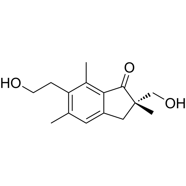
Pterosin A ((2S)-Pterosin A) is a sesquiterpene compound. Pterosin A is an orally active AMPK activator with anti-diabetic effect. Pterosin A can promote glucose uptake, increase serum insulin, and improve hyperglycemia and glucose intolerance. Pterosin A can prevent insulin-secreting cells death and reduce ROS production. Pterosin A can be used for the research of metabolic disease, such as diabetes.
Nos produits utilisent uniquement pour la recherche. Nous ne vendons pas aux patients.
- Pureté : 94.07%
- CAS No.: 35910-16-8
- Formule: C15H20O3
- Masse moléculaire:248.32
-
Stockage:
4°C, protect from light
* In solvent : -80°C, 6 months; -20°C, 1 month (protect from light)

To place orders, for customer services and technical support, please contact: MedChemExpress USA
Tel: 609-228-6898 E-mail: [email protected] [email protected]


-
 Activité biologique
-
Chemical Information
- Protocole
- Pureté et documentation
- Références
-
 Help & FAQs


Voir tous les produits spécifiques à Isoform AMPK
More

Activité biologique
Description
IC50 & Target
[1]|
GLUT4 |
p-GSK-3 |
In Vitro
Pterosin A (50 μg/mL, 0.5-4 h) enhances glucose uptake and AMPK phosphorylation in muscle cells[1].
Pterosin A (50-150 μg/mL, 0.5-4 h) inhibits PEPCK expression, triggers the phosphorylations of AMPK, ACC, and GSK-3, decreases glycogen synthase phosphorylation, and increases the intracellular glycogen level in liver cells[1].
Pterosin A (10-100 μM, 18 h) inhibits H2O2-induced ROS production in RINm5f β-cells[2].
Pterosin A (10-100 μM, 24 h) reduces lipotoxicity-induced cell death in RINm5f β-cells[2].
MedChemExpress (MCE) has not independently confirmed the accuracy of these methods. They are for reference only.
-
Cell Line:Liver cells
-
Concentration:50 and 150 μg/mL
-
Incubation Time:0.5, 1, 2 and 4 h
-
Result:Increased p-AMPK and P-ACC and p-GSK3-α/β levels.
In Vivo
MedChemExpress (MCE) has not independently confirmed the accuracy of these methods. They are for reference only.
-
Animal Model:Streptozotocin (HY-13753), high-fat diet-fed and db/db diabetic mice models[1]
-
Dosage:10, 30 and 100 mg/kg
-
Administration:Orally administration, daily for 4 weeks
-
Result:Reversed the increased serum insulin and insulin resistance in dexamethasone-IR mice.
Reversed the reduced muscle GLUT-4 translocation and the increased liver phosphoenolpyruvate carboxyl kinase (PEPCK) expression.
Reversed the decreased phosphorylations of AMP-activated protein kinase (AMPK) and Akt in muscles.
Reversed increased p38 phosphorylation in livers.


Chemical Information
-
CAS No. 35910-16-8
-
Appearance Solid
-
Masse moléculaire 248.32
-
Formule C15H20O3
-
Color White to off-white
-
SMILES
O=C1C(C(C[C@](C)1CO)=CC(C)=C2CCO)=C2C
-
Synonyms
(2S)-Pterosin A
-
Structure Classification
-
Initial Source
-
Livraison
Room temperature in continental US; may vary elsewhere.
-
Stockage
4°C, protect from light
* In solvent : -80°C, 6 months; -20°C, 1 month (protect from light)


Pureté et documentation
-
Fiche technique (272 KB)
-
SDS (252 KB)
- English - EN (252 KB)
- Français - FR (252 KB)
- Deutsch - DE (252 KB)
- Norwegian - NO (252 KB)
- Español - ES (252 KB)
- Swedish - SV (252 KB)
- Italian - IT (252 KB)
- Korean - KR (252 KB)
- Portuguese - PT (252 KB)
-
Instruction de manipulation (2659 KB)
Références
[1]. Hsu FL, et al. Antidiabetic effects of pterosin A, a small-molecular-weight natural product, on diabetic mouse models. Diabetes. 2013 Feb;62(2):628-38. [Content Brief]
[2]. Chen CY, et al. Chemical constituents analysis and antidiabetic activity validation of four fern species from Taiwan. Int J Mol Sci. 2015 Jan 22;16(2):2497-516. [Content Brief]

Calculators
Concentration (start) × Volume (start) = Concentration (final) × Volume (final)


Pterosin A Related Classifications
HY-N7912 Related Classifications
 Powered by Bioz