



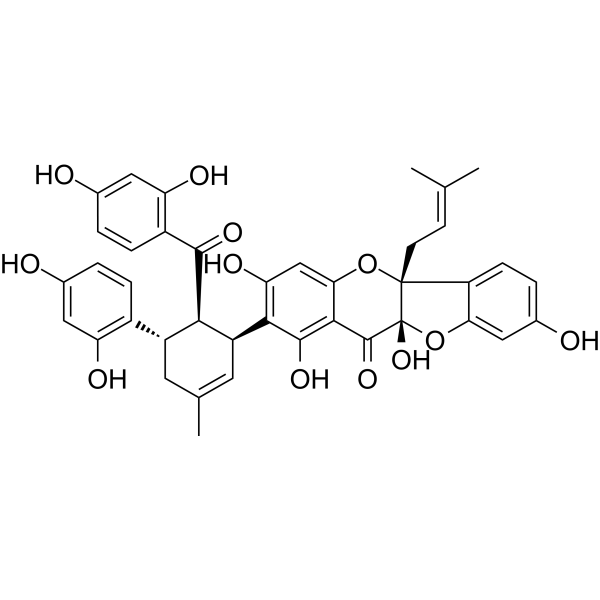
Sanggenon C
Based on 4 publication(s) in Google Scholar
Sanggenon C, a flavonoid, exerts protective effects against cardiac hypertrophy and fibrosis via suppression of the calcineurin/NFAT2 pathway. Sanggenon C inhibits mitochondrial fission to induce apoptosis by blocking the ERK signaling pathway. Sanggenon C inhibits inducible nitric oxide synthase expression in RAW264.7 cells, and TNF-α-stimulated cell adhesion and VCAM-1 expression, by suppressing NF-κB activity. Sanggenon C possesses antioxidant, anti-inflammatory and antitumor activities.
For research use only. We do not sell to patients.
- Purity : 98.46%
- CAS No.: 80651-76-9
- Formula: C40H36O12
- Molecular Weight:708.71
-
Storage:
-20°C, sealed storage, away from moisture and light
* In solvent : -80°C, 6 months; -20°C, 1 month (sealed storage, away from moisture and light)

To place orders, for customer services and technical support, please contact: MedChemExpress USA
Tel: 609-228-6898 E-mail: [email protected] [email protected]


-
 Biological Activity
-
Chemical Information
-
Solvent & Solubility
- Protocols
- Purity & Documentation
- References
-
 Help & FAQs


-
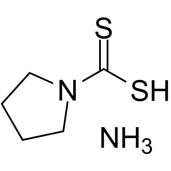
HY-18738
Pyrrolidinedithiocarbamate ammoniumSelective NF-κB Inhibitor -
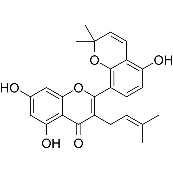
HY-N2300
Kuwanon ANO Synthase Inhibitor -
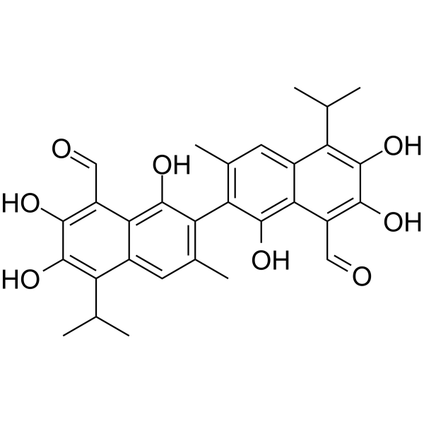
HY-13407
GossypolBcl-2 Family Antagonist -
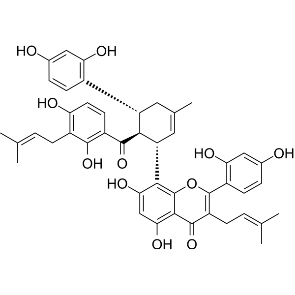
HY-N2600
Kuwanon HBombesin Receptor Antagonist/Platelet Activation Inhibitor/GRPR Antagonist -
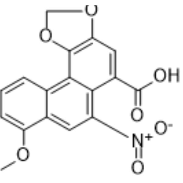
HY-N0510
Aristolochic acid ANF-κB Inhibitor -
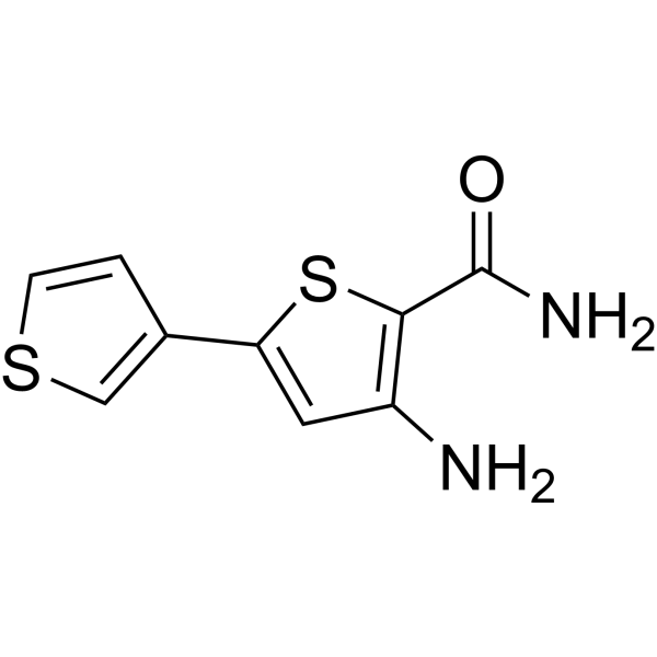
HY-13802
SC-514IKK Inhibitor -
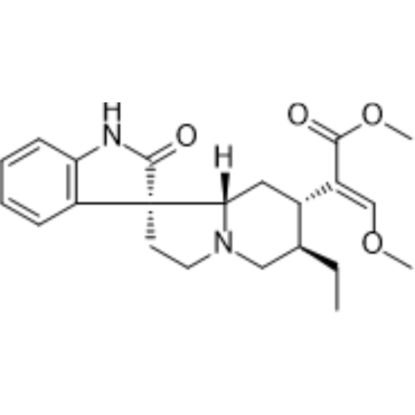
HY-N0387
RhynchophyllineNF-κB Inhibitor -
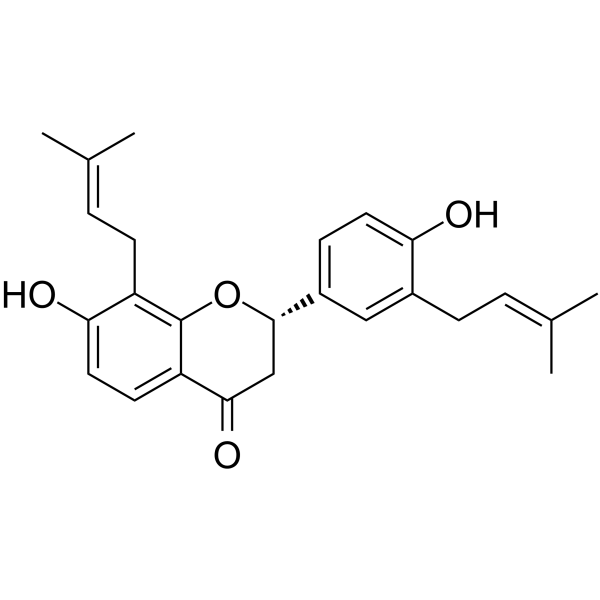
HY-N4193
GlabrolAcyltransferase Inhibitor -
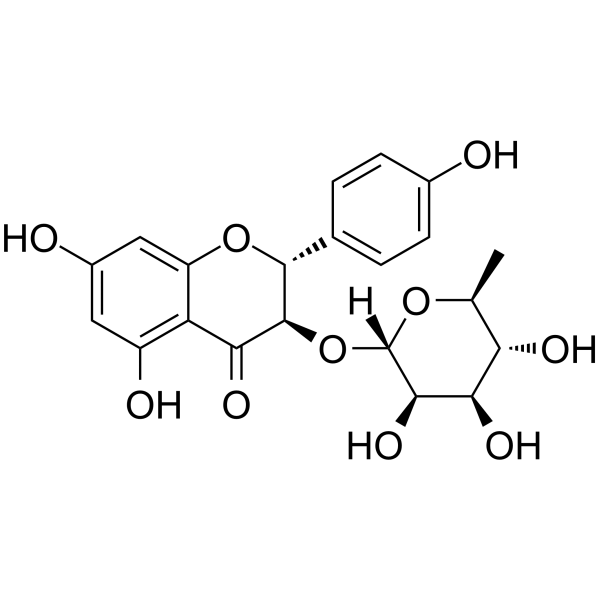
HY-N0436
EngeletinNF-κB Inhibitor -
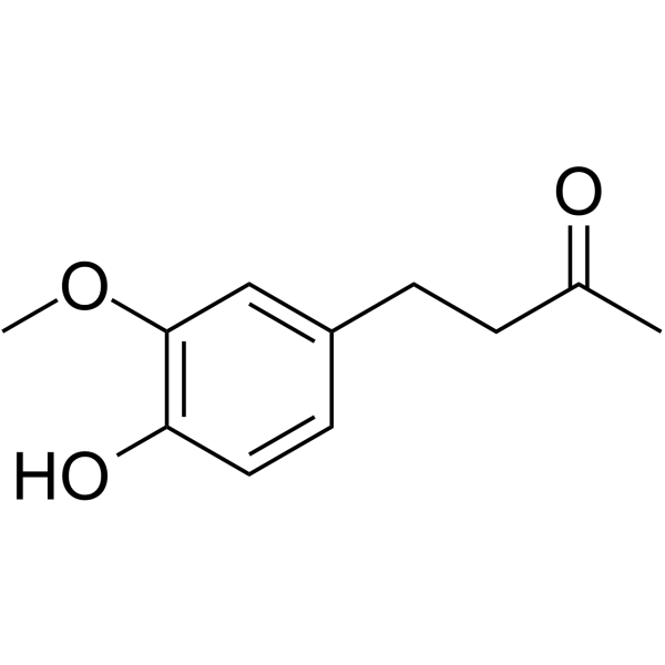
HY-14621
ZingeronePhenolic Ketone -
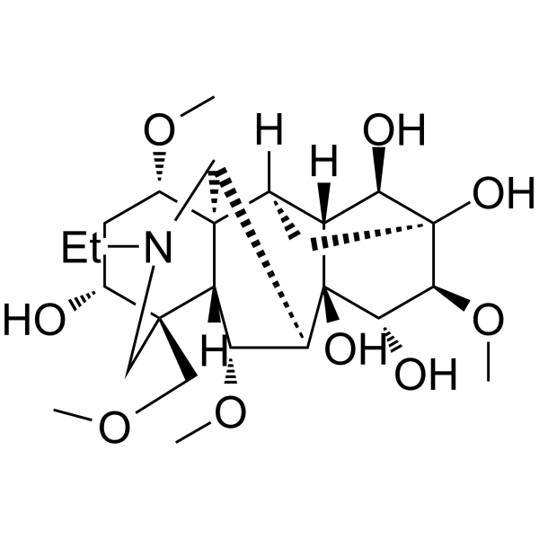
HY-N0277
AconineNF-κB Inhibitor -
HY-N0694
Schisantherin ANF-κB Inhibitor
Publications Citing Use of MedChemExpress (MCE) Sanggenon C
More Customer Validation & Images

-
WB
-
WB


Biological Activity
Description
|
NF-κB |
ERK |
Cellular Effect
|
Cell Line

|
Type | Value | Description | References |
|---|---|---|---|---|
| Calu-3 | CC50 |
23.6 μM
Compound: 12
|
Cytotoxicity against human Calu-3 cells assessed as reduction in cell viability
Cytotoxicity against human Calu-3 cells assessed as reduction in cell viability
|
[PMID: 36651644] |
| Hep 3B2 | IC50 |
1.26 μM
Compound: 8
|
Inhibition of hypoxia-induced HIF1alpha protein accumulation in human Hep3B cells treated for 30 mins measured after 12 hrs by Western blot analysis
Inhibition of hypoxia-induced HIF1alpha protein accumulation in human Hep3B cells treated for 30 mins measured after 12 hrs by Western blot analysis
|
[PMID: 19072214] |
| Hep 3B2 | IC50 |
3.2 μM
Compound: 8
|
Inhibition of hypoxia-induced VEGF protein secretion in human Hep3B cells after 16 hrs by ELISA
Inhibition of hypoxia-induced VEGF protein secretion in human Hep3B cells after 16 hrs by ELISA
|
[PMID: 19072214] |
| Hep 3B2 | IC50 |
8.26 μM
Compound: 8
|
Inhibition of hypoxia-induced HIF1 activation in human Hep3B cells by pGL3-HRE-luciferase reporter gene assay
Inhibition of hypoxia-induced HIF1 activation in human Hep3B cells by pGL3-HRE-luciferase reporter gene assay
|
[PMID: 19072214] |
| HSC-2 | CC50 |
18 μM
Compound: 7
|
Cytotoxicity against human HSC2 cells
Cytotoxicity against human HSC2 cells
|
[PMID: 11429996] |
| Platelet | IC50 |
11.2 x 10⁻⁵ M
Compound: sanggenon C
|
Inhibition of 12-hydroxy-5,8,10-heptadecatrienoic acid formation in Wistar King platelets
Inhibition of 12-hydroxy-5,8,10-heptadecatrienoic acid formation in Wistar King platelets
|
[PMID: 3097265] |
| Platelet | IC50 |
18 x 10-5 M
Compound: sanggenon C
|
Inhibition of thromboxane B2 formation in Wistar King platelets
Inhibition of thromboxane B2 formation in Wistar King platelets
|
[PMID: 3097265] |
In Vitro
Sanggenon C (4-12 μM; 24 h) inhibits the proliferation of human gastric cancer (GC) cells and greatly reduces the number of GC cell colonies formed[2].
Sanggenon C (6-10 μM; 24 h) inhibits cell cycle arrest and apoptosis of GC cells[2].
Sanggenon C (6-10 μM; 24 h) markedly downregulates the levels of p-ERK[2].
Sanggenon C (6-10 μM; 24 h) induces mitochondrial dysfunction of GC cells[2].
MedChemExpress (MCE) has not independently confirmed the accuracy of these methods. They are for reference only.
-
Cell Line:Human GC cell lines HGC-27 and AGS cells
-
Concentration:4-12 μM
-
Incubation Time:24 h
-
Result:Inhibited the proliferation of GC cells in a dose-dependent manner.
The IC50 values of were 9.129 μM for HGC-27 and 9.863 μM for AGS.
-
Cell Line:Human GC cell lines HGC-27 and AGS cells
-
Concentration:6, 8, 10 μM
-
Incubation Time:24 h
-
Result:The proportions of cells in the G0-G1 phase were increased and the levels of CDK4 and cyclin D1 were decreased.
-
Cell Line:Human GC cell lines HGC-27 and AGS cells
-
Concentration:6, 8, 10 μM
-
Incubation Time:24 h
-
Result:Exhibited a dose-dependent induction of apoptosis, with the percentage of apoptotic cells increasing from 7.3% to 24.8% and from 4.6% to 15.1% for HGC-27 and AGS cells, respectively.
-
Cell Line:Human GC cell lines HGC-27 and AGS cells
-
Concentration:6, 8, 10 μM
-
Incubation Time:24 h
-
Result:The levels of p-ERK were markedly downregulated.
In Vivo
Sanggenon C (10, 20 mg/kg/day; Intraperitoneally; for 21 days) suppresses the tumor burden in the nude mice bearing tumor xenografts derived from AGS. Sanggenon C downregulates levels of p-ERK expression[2].
MedChemExpress (MCE) has not independently confirmed the accuracy of these methods. They are for reference only.
-
Animal Model:Male C57/BL6 mice (weight 23.5-27.5 g; age, 8 weeks)[1]
-
Dosage:10, 20 mg/kg
-
Administration:Intraperitoneally; daily; for 3 weeks
-
Result:Prevented the development of ventricular dysfunction, as evidenced by decreased LV end-diastolic diameter, LV end-systolic diameter, and increased LVFS and LVEF.
- Fibrosis/Collagen Morphometry
- Collagen: Sirius Red Staining
- RNA extraction experimental
- Detection of Apoptosis
- TUNEL staining for apoptotic DNA fragmentation
- Annexin V plus membrane-impermeant dye apoptosis staining
- Connective Tissue: Masson's Trichrome/Collagen Trichrome Staining
- Apoptosis Solutions
- Research Protocol for Inflammation-related Diseases
- Cardiac Morphometry


Chemical Information
-
CAS No. 80651-76-9
-
Appearance Solid
-
Molecular Weight 708.71
-
Formula C40H36O12
-
Color Light yellow to green yellow
-
SMILES
C/C(C)=C/C[C@](C1=CC=C(O)C=C1O2)(OC3=CC(O)=C([C@@H](C=C(C)C[C@@H]4C(C=CC(O)=C5)=C5O)[C@@H]4C(C(C=CC(O)=C6)=C6O)=O)C(O)=C3C7=O)[C@]27O
-
Structure Classification
-
Initial Source
-
Shipping
Room temperature in continental US; may vary elsewhere.
-
Storage
-20°C, sealed storage, away from moisture and light
* In solvent : -80°C, 6 months; -20°C, 1 month (sealed storage, away from moisture and light)


Publications (4)
-
Journal Impact Factor
-
Most Recent
-
Talanta
A targeted strategy for screening bioactive inhibitors from Morus alba L.: Immobilized PTP1B affinity chromatography. [Abstract]2026 Sep 1:307:129772. PMID: 41955787 -
J Nat Prod
Sanggenon C Suppresses Tumorigenesis of Gastric Cancer by Blocking ERK-Drp1-Mediated Mitochondrial Fission. [Abstract]2022 Oct 28;85(10):2351-2362. PMID: 36256535
Sanggenon C purchased from MedChemExpress. Usage Cited in: J Nat Prod. 2022 Oct 28;85(10):2351-2362. [Abstract]
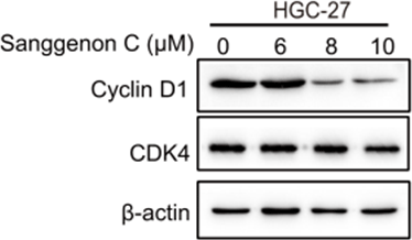
Sanggenon C (0, 6, 8, 10 µM) decreases the levels of CDK4 and cyclin D1 in HGC-27 cells.
Sanggenon C purchased from MedChemExpress. Usage Cited in: J Nat Prod. 2022 Oct 28;85(10):2351-2362. [Abstract]
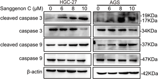
Sanggenon C (6, 8, 10 µM) induces an increase in apoptosis markers in HGC-27 and AGS cells.
-
-


Solvent & Solubility
In Vitro:
DMSO : 100 mg/mL (141.10 mM; Need ultrasonic; Hygroscopic DMSO has a significant impact on the solubility of product, please use newly opened DMSO)
Please refer to the solubility information to select the appropriate solvent. Once prepared, please aliquot and store the solution to prevent product inactivation from repeated freeze-thaw cycles.
Storage method and period of stock solution: -80°C, 6 months; -20°C, 1 month (sealed storage, away from moisture and light). When stored at -80°C, please use it within 6 months. When stored at -20°C, please use it within 1 month.
Please refer to the solubility information to select the appropriate solvent. Once prepared, please aliquot and store the solution to prevent product inactivation from repeated freeze-thaw cycles.
Storage method and period of stock solution: -80°C, 6 months; -20°C, 1 month (sealed storage, away from moisture and light). When stored at -80°C, please use it within 6 months. When stored at -20°C, please use it within 1 month.
Concentration (start) × Volume (start) = Concentration (final) × Volume (final)
In Vivo:
Select the appropriate dissolution method based on your experimental animal and administration route.
- For the following dissolution methods, please ensure to first prepare a clear stock solution using an In Vitro approach and then sequentially add co-solvents:
- To ensure reliable experimental results, the clarified stock solution can be appropriately stored based on storage conditions. As for the working solution for In Vivo experiments, it is recommended to prepare freshly and use it on the same day.
- The percentages shown for the solvents indicate their volumetric ratio in the final prepared solution. If precipitation or phase separation occurs during preparation, heat and/or sonication can be used to aid dissolution.
Add each solvent one by one: 10% DMSO 90% (20% SBE-β-CD in Saline)
Solubility: ≥ 2.5 mg/mL (3.53 mM); Clear solution
This protocol yields a clear solution of ≥ 2.5 mg/mL (saturation unknown).
Taking 1 mL working solution as an example, add 100 μL DMSO stock solution (25.0 mg/mL) to 900 μL 20% SBE-β-CD in Saline, and mix evenly.
Preparation of 20% SBE-β-CD in Saline (4°C, storage for one week): 2 g SBE-β-CD powder is dissolved in 10 mL Saline, completely dissolve until clear.
In Vivo Dissolution Calculator
Please enter the basic information of animal experiments:
-
-
-
-
Recommended: Prepare an additional quantity of animals to account for potential losses during experiments.
Please enter your animal formula composition:
-
%DMSO +
Recommended: Keep the proportion of DMSO in working solution below 2% if your animal is weak.
-
%+
-
+%Tween-80 + +
-
%Saline +
The co-solvents required include: DMSO, . All of co-solvents are available by MedChemExpress (MCE). , Tween 80. All of co-solvents are available by MedChemExpress (MCE).
Working solution concentration: 0.22 mg/mL
Method for preparing stock solution: mg drug dissolved in μL DMSO. Stock solution concentration: mg/mL. * In solvent : -80°C, 6 months; -20°C, 1 month (sealed storage, away from moisture and light)
1. Take μL DMSO stock solution;
2. Add μL .
μL , mix evenly;
3. Then add μL Tween 80, mix evenly;
4. Then add μL
Please ensure that the stock solution in the first step is dissolved to a clear state, and add co-solvents in sequence. You can use ultrasonic heating (ultrasonic cleaner, recommended frequency 20-40 kHz), vortexing, etc. to assist dissolution.


Purity & Documentation
-
Data Sheet (282 KB)
-
SDS (252 KB)
- English - EN (252 KB)
- Français - FR (252 KB)
- Deutsch - DE (252 KB)
- Norwegian - NO (252 KB)
- Español - ES (252 KB)
- Swedish - SV (252 KB)
- Italian - IT (252 KB)
- Korean - KR (252 KB)
- Portuguese - PT (252 KB)
-
Handling Instructions (2659 KB)
References
[1]. Xiao L, et al. Sanggenon C protects against pressure overload induced cardiac hypertrophy via the calcineurin/NFAT2 pathway. Mol Med Rep. 2017 Oct;16(4):5338-5346. [Content Brief]
[2]. Xiao-Jie Chen, et al. Sanggenon C Suppresses Tumorigenesis of Gastric Cancer by Blocking ERK-Drp1-Mediated Mitochondrial Fission. J Nat Prod. 2022 Oct 28;85(10):2351-2362. [Content Brief]


Complete Stock Solution Preparation Table
Please refer to the solubility information to select the appropriate solvent. Once prepared, please aliquot and store the solution to prevent product inactivation from repeated freeze-thaw cycles.
Storage method and period of stock solution: -80°C, 6 months; -20°C, 1 month (sealed storage, away from moisture and light). When stored at -80°C, please use it within 6 months. When stored at -20°C, please use it within 1 month.
| Optional Solvent | Concentration Solvent Mass | 1 mg | 5 mg | 10 mg | 25 mg |
|---|---|---|---|---|---|
| DMSO | 1 mM | 1.4110 mL | 7.0551 mL | 14.1101 mL | 35.2754 mL |
| 5 mM | 0.2822 mL | 1.4110 mL | 2.8220 mL | 7.0551 mL | |
| 10 mM | 0.1411 mL | 0.7055 mL | 1.4110 mL | 3.5275 mL | |
| 15 mM | 0.0941 mL | 0.4703 mL | 0.9407 mL | 2.3517 mL | |
| 20 mM | 0.0706 mL | 0.3528 mL | 0.7055 mL | 1.7638 mL | |
| 25 mM | 0.0564 mL | 0.2822 mL | 0.5644 mL | 1.4110 mL | |
| 30 mM | 0.0470 mL | 0.2352 mL | 0.4703 mL | 1.1758 mL | |
| 40 mM | 0.0353 mL | 0.1764 mL | 0.3528 mL | 0.8819 mL | |
| 50 mM | 0.0282 mL | 0.1411 mL | 0.2822 mL | 0.7055 mL | |
| 60 mM | 0.0235 mL | 0.1176 mL | 0.2352 mL | 0.5879 mL | |
| 80 mM | 0.0176 mL | 0.0882 mL | 0.1764 mL | 0.4409 mL | |
| 100 mM | 0.0141 mL | 0.0706 mL | 0.1411 mL | 0.3528 mL |


Sanggenon C Related Classifications
HY-N0617 Related Classifications
 Powered by Bioz