



SD-36
Based on 9 publication(s) in Google Scholar
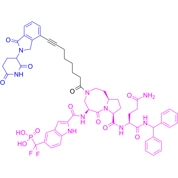
SD-36 is a potent STAT3 PROTAC degrader with a Kd of 44.4 nM and a Ki of 11 nM. SD-36 recruits the cereblon/culin 4A E3 ligase complex to mediate ubiquitination and proteasomal degradation of STAT3, inducing G1 phase cell cycle arrest and apoptosis. SD-36 is useful for research on acute myeloid leukemia, anaplastic large cell lymphoma, and hematopoietic and lymphoid malignancies.
(Pink: STAT3 ligand (HY-187011); Blue: Cereblon ligand (HY-43722); Black: linker (HY-69220)).
For research use only. We do not sell to patients.
- Purity : 99.73%
- CAS No.: 2429877-44-9
- Formula: C59H62F2N9O12P
- Molecular Weight:1158.15
-
Storage:
-80°C, stored under nitrogen

To place orders, for customer services and technical support, please contact: MedChemExpress USA
Tel: 609-228-6898 E-mail: [email protected] [email protected]


-
 Biological Activity
-
Chemical Information
-
Solvent & Solubility
- Protocols
- Purity & Documentation
- References
-
 Help & FAQs


-
HY-129603
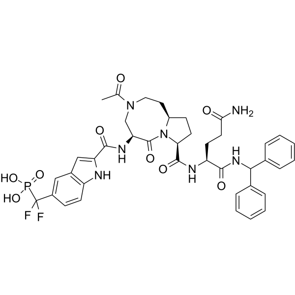
SI-109STAT3 Inhibitor -

HY-156730
KT-333STAT3 Degrader -
HY-112288
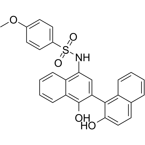
C188-9STAT3 Inhibitor -
HY-13818
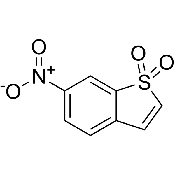
StatticSTAT3 Inhibitor -
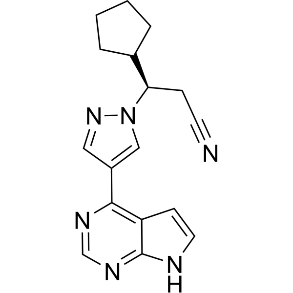
HY-50856
RuxolitinibJAK1/2 Inhibitor -
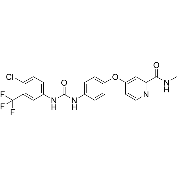
HY-10201
SorafenibMultikinase Inhibitor -

HY-100579
Ferrostatin-1Ferroptosis Inhibitor -
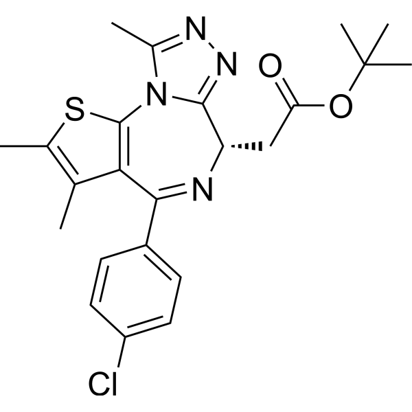
HY-13030
(+)-JQ-1BET Inhibitor -
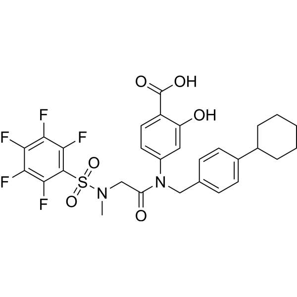
HY-100493
BP-1-102STAT3 Inhibitor -
HY-13919
NapabucasinSTAT3 Inhibitor -
HY-16658B
Z-VAD-FMKPan-Caspase Inhibitor -
HY-100716
EganelisibPI3Kγ Inhibitor
Publications Citing Use of MedChemExpress (MCE) SD-36
More- Sci Transl Med. 2023 Jun 28;15(702):eabo3826. [Abstract]
- J Med Chem. 2026 Feb 26;69(4):3932-3940.
- Stem Cell Reports. 2024 Jan 9;19(1):100-111. [Abstract]
- J Cell Mol Med. 2021 Sep;25(17):8261-8270. [Abstract]
- iScience. 2025 Apr 22;28(5):112502. [Abstract]
- bioRxiv. 2025 Dec 26:2025.12.25.695625. [Abstract]
- bioRxiv. 2025 Nov 10.
- Research Square Preprint. 2024 Feb 15.
- bioRxiv. 2023 Sep 28:2023.09.26.559434. [Abstract]
 Customer Validation & Images

-
WB
-
RT-PCR
-
WB
-
Cell Proliferation/Viability Assay
-
Cell Migration/Invasion Assay
All PROTACs Isoforms
More

Biological Activity
Description
IC50 & Target
[1]|
STAT3 44.4 nM (Kd) |
STAT3 11 nM (Ki) |
Cereblon |
Cellular Effect
|
Cell Line

|
Type | Value | Description | References |
|---|---|---|---|---|
| MOLM-16 | DC50 |
0.06 μM
|
Induction of total STAT3 and phosphorylated STAT3 (pSTAT3Y705) protein degradation in Molm-16 acute myeloid leukemia cells incubated for 4 hrs, measured via Western blotting.
Induction of total STAT3 and phosphorylated STAT3 (pSTAT3Y705) protein degradation in Molm-16 acute myeloid leukemia cells incubated for 4 hrs, measured via Western blotting.
|
31747516 |
| SU-DHL-1 | DC50 |
28 nM
|
Induction of total STAT3 and phosphorylated STAT3 (pSTAT3Y705) protein degradation in SU-DHL-1 anaplastic large cell lymphoma cells incubated for 16 hrs, measured via Western blotting.
Induction of total STAT3 and phosphorylated STAT3 (pSTAT3Y705) protein degradation in SU-DHL-1 anaplastic large cell lymphoma cells incubated for 16 hrs, measured via Western blotting.
|
31747516 |
| MOLM-16 | IC50 |
13 nM
|
Cell growth inhibition against Molm-16 acute myeloid leukemia cells assessed as reduction in cell viability incubated for 4 days.
Cell growth inhibition against Molm-16 acute myeloid leukemia cells assessed as reduction in cell viability incubated for 4 days.
|
31747516 |
| SU-DHL-1 | IC50 |
0.61 μM
|
Cell growth inhibition against SU-DHL-1 anaplastic large cell lymphoma cells assessed as reduction in cell viability incubated for 4 days.
Cell growth inhibition against SU-DHL-1 anaplastic large cell lymphoma cells assessed as reduction in cell viability incubated for 4 days.
|
31747516 |
| MOLM-16 | IC50 |
35 nM
|
Growth-inhibitory activity against human acute myeloid leukemia MOLM-16 cells assessed via luminescent cell viability assay after 4 days of treatment.
Growth-inhibitory activity against human acute myeloid leukemia MOLM-16 cells assessed via luminescent cell viability assay after 4 days of treatment.
|
31747516 |
In Vitro
SD-36 (10 nM-10 μM, 4 h) potently induces degradation of total STAT3 and pSTAT3Y705 in Molm-16 acute myeloid leukemia cells, with a DC50 of 0.06 μM after 4 h of treatment[2].
SD-36 (10 nM-10 μM; 18 h) potently induces degradation of total STAT3 and pSTAT3Y705 in anaplastic large cell lymphoma SU-DHL-1 cells, with a DC50 of 28 nM after 16 h of treatment[2].
SD-36 (4 days) potently inhibits the growth of Molm-16 acute myeloid leukemia cells and the anaplastic large cell lymphoma cell line SU-DHL-1, with IC50 values of 13 nM and 0.61 μM, respectively, after 4 days of incubation[2].
SD-36 (0.1-10 μM; 1-24 h) induces degradation of total STAT3 and phosphorylated STAT3 (Y705) in a time- and concentration-dependent manner in MOLM-16, SU-DHL-1, DEL, and KI-JK cells[1].
SD-36 (1 μM; 5 h) completely depletes both monomeric and dimeric STAT3 proteins in MOLM-16 cells[1].
SD-36 (1 μM; 8 h) abolishes the DNA-binding activity of STAT3 in MOLM-16 and SU-DHL-1 cells[1].
SD-36 (1 μM; 8 h) significantly downregulates STAT3 transcriptional target genes, including key oncogenic regulators in MOLM-16 cells[1].
SD-36 (up to 5 μM; 4 days) potently inhibits the proliferation of MOLM-16 acute myeloid leukemia cells (IC50 = 35 nM) and suppresses 5 of 9 tested lymphoma cell lines after 4 days of treatment[1].
SD-36 binds to the STAT3 SH2 domain with high affinity (Kd = 44.4 nM), while showing weaker affinity for other STAT family proteins[2].
SD-36 (1 h) binds to recombinant human STAT3 protein with high affinity, exhibiting a Ki of 11 nM, and shows weaker binding to other STAT family members[1].
SD-36 (up to 10 μM; 24 h) potently inhibits STAT3 transcriptional activity in a STAT3 luciferase reporter cell line, with an IC50 of 10 nM after 24 h of treatment[1].
SD-36 (1 μM; 48 h) induces G1 phase cell cycle arrest in MOLM-16 and SU-DHL-1 cells[1].
SD-36 (1 μM; 48 h) induces strong apoptotic cell death in SU-DHL-1 and SUP-M2 lymphoma cells[1].
MedChemExpress (MCE) has not independently confirmed the accuracy of these methods. They are for reference only. Further protocols information, click here.
-
Cell Line:MOLM-16, SU-DHL-1, DEL, KI-JK
-
Concentration:0.1, 1, 10 μM
-
Incubation Time:3, 24 h
-
Result:Depleted the total STAT3 protein in the cells.
-
Cell Line:MOLM-16 and SU-DHL-1 cells
-
Concentration:8, 24 h
-
Incubation Time:1 μM
-
Result:Significantly down-regulated the mRNA expression levels of typical STAT3 downstream target genes (such as BCL3, JAK3, MYC, PIM1, SOCS3, etc.
-
Cell Line:MOLM-16 and SU-DHL-1 cells
-
Concentration:48 h
-
Incubation Time:1 μM
-
Result:Caused profound G1 cell cycle arrest and effectively reduced the proportion of S phase cells.
-
Cell Line:SU-DHL-1 and SUP-M2 cells
-
Concentration:48 h
-
Incubation Time:1 μM
-
Result:Triggered strong apoptotic cell death in the tested lymphoma cells.
-
Cell Line:MOLM-16
-
Concentration:0.25 μM
-
Incubation Time:1, 2, 4, 7, 10 h
-
Result:Decreased the levels of total STAT3 and pSTAT3(Y705) proteins in a time-dependent manner.
-
Cell Line:SU-DHL-1
-
Concentration:0.01, 0.03, 0.1, 0.3, 1, 3, 10 μM
-
Incubation Time:18 h
-
Result:Highly selectively degraded the STAT3 protein, and had no obvious effects on other proteins such as STAT1, STAT2, STAT4, STAT5A/B, and STAT6.
In Vivo
SD-36 (25-100 mg/kg; i.v.; once weekly or twice weekly for 4 consecutive weeks; approximately 65 days) completely and durably induces tumor regression in the MOLM-16 subcutaneous xenograft NOD/SCID female mouse model[1].
SD-36 (25-100 mg/kg; i.v.; 3 times per week or once weekly for multiple consecutive weeks; up to 125 days) induces complete tumor regression in SU-DHL-1 and SUP-M2 subcutaneous xenograft NOD/SCID female mouse models[1].
SD-36 (25-50 mg/kg; i.v.; 3 times per week for 3 consecutive weeks or once weekly; approximately 40 days) induces complete tumor regression in the MOLM-16 xenograft SCID mouse model[2].
SD-36 (50 mg/kg; i.v.; single administration; 1-72 hr) rapidly and persistently degrades intratumoral STAT3 in the MOLM-16 xenograft SCID mouse model[2].
SD-36 (100 mg/kg; i.v.; single dose; 1-96 hr) deeply depletes STAT3 protein in healthy tissues including liver, spleen, heart, and kidney in wild-type immunocompetent CD-1 female mice[1].
SD-36 (50-100 mg/kg; i.v.; 3 times per week for 3 consecutive weeks; 27 days) is well tolerated with no obvious toxicity in wild-type immunocompetent CD-1 female mice[1].
MedChemExpress (MCE) has not independently confirmed the accuracy of these methods. They are for reference only.
-
Animal Model:NOD/SCID (female, acute myeloid leukemia xenograft via subcutaneous injection of 3×10⁶ MOLM-16 cells in 5 mg/mL Matrigel, tumors grown to average volume of 150 mm3)[1]
-
Dosage:25, 50, 100 mg/kg
-
Administration:i.v.; once or twice a week; 4 consecutive weeks
-
Result:Inhibited tumor growth.
Achieved complete and long-lasting tumor regression at higher doses.
-
Animal Model:CD-1 (female, immune competent, healthy)[1]
-
Dosage:50, 100 mg/kg
-
Administration:i.v.; single dose; three times per week for 3 weeks
-
Result:Did not cause weight loss or abnormal organ coefficients in mice.
Had no significant adverse effects on blood routine and liver/kidney function indexes, showing good tolerability.
-
Animal Model:CD-1 female mice[2]
-
Dosage:25 mg/kg
-
Administration:i.v.; single dose; Samples collected at 3, 24, 48, 72 hours post-treatment
-
Result:Effectively reduced STAT3 protein levels in spleen tissue, and the effect persisted for over 48 hours.
-
Animal Model:CB.17 SCID mice were subcutaneously injected of 3×10⁶ MOLM-16 cells[2]
-
Dosage:50 mg/kg
-
Administration:i.v.; single dose; samples collected at 1, 3, 24, 48, 72 hours post-treatment
-
Result:Decreased the levels of total STAT3 and pSTAT3(Y705) proteins in tumor tissues by over 95% within 1 hour after administration.
-
Animal Model:CB.17 SCID mice were subcutaneously injected of 3×10⁶ MOLM-16 cells[2]
-
Dosage:25, 50 mg/kg
-
Administration:i.v.; three times a week (25, 50 mg/kg) or once weekly (50 mg/kg); administered for 3 consecutive weeks
-
Result:Achieved complete tumor regression after 2 weeks of administration under the dosing schedule of 25 or 50 mg/kg three times a week.
Induced complete and durable tumor regression, with no recurrence observed for over 2 weeks after stopping the treatment.


Chemical Information
-
CAS No. 2429877-44-9
-
Appearance Solid
-
Molecular Weight 1158.15
-
Formula C59H62F2N9O12P
-
Color White to off-white
-
SMILES
O=C([C@@H](NC([C@@H]1CC[C@H](CCN(C(CCCCCC#CC2=CC=CC3=C2CN(C4CCC(NC4=O)=O)C3=O)=O)C[C@@H]5NC(C6=CC(C=C(C(F)(P(O)(O)=O)F)C=C7)=C7N6)=O)N1C5=O)=O)CCC(N)=O)NC(C8=CC=CC=C8)C9=CC=CC=C9
-
Shipping
Shipping with dry ice.
-
Storage
-80°C, stored under nitrogen


Publications (9)
-
Journal Impact Factor
-
Most Recent
-
Sci Transl Med
Targeting CCR7-PI3Kγ overcomes resistance to tyrosine kinase inhibitors in ALK-rearranged lymphoma. [Abstract]2023 Jun 28;15(702):eabo3826. PMID: 37379367
SD-36 purchased from MedChemExpress. Usage Cited in: Sci Transl Med. 2023 Jun 28;15(702):eabo3826. [Abstract]
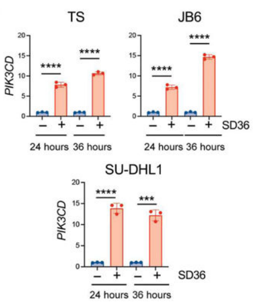
qRT-PCR analysis of PIK3CD mRNA expression performed on ALK+ ALCL cell lines treated with SD-36 (1 μM). n = 3 technical replicates.
SD-36 purchased from MedChemExpress. Usage Cited in: Sci Transl Med. 2023 Jun 28;15(702):eabo3826. [Abstract]
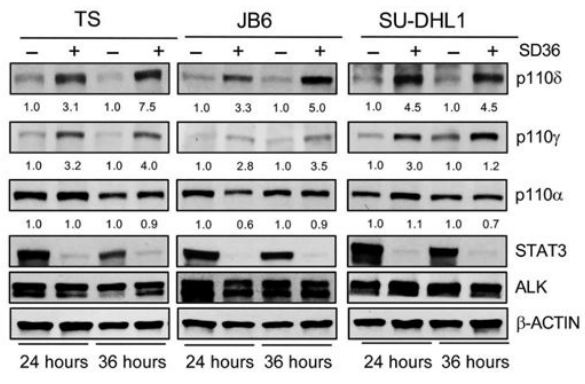
Western blot analysis on ALK+ ALCL human cell lines treated with SD-36 (1 μM).
-
-
Stem Cell Reports
Distinct roles of hematopoietic cytokines in the regulation of leukemia stem cells in murine MLL-AF9 leukemia. [Abstract]2024 Jan 9;19(1):100-111. PMID: 38101400
SD-36 purchased from MedChemExpress. Usage Cited in: Stem Cell Reports. 2024 Jan 9;19(1):100-111. [Abstract]
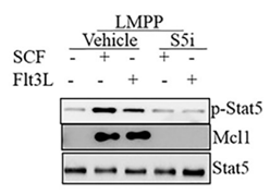
GMP-like LSCs isolated from 3-cytokine-medium-cultured AML cells. GMP-like LSCs were starved for 4 h followed by 1 μM SD-36 (Stat3 PROTAC). Cells were subsequently stimulated by IL-3 or IL-6 for 30 min and collected for western blotting.
-
J Cell Mol Med
SD-36 promotes growth inhibition and induces apoptosis via suppression of Mcl-1 in glioma. [Abstract]2021 Sep;25(17):8261-8270. PMID: 34291563
SD-36 purchased from MedChemExpress. Usage Cited in: J Cell Mol Med. 2021 Sep;25(17):8261-8270. [Abstract]
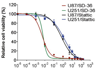
U87 and U251 cells were treated with increasing concentration of SD-36 or Stattic for 72 h. Cell viability was analysed by MTS assay.
SD-36 purchased from MedChemExpress. Usage Cited in: J Cell Mol Med. 2021 Sep;25(17):8261-8270. [Abstract]
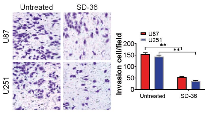
Invasion of U87 and U251 cells treated with SD-36 (100 nM; 24 h) was analysed by Transwell assay. The results sowed that SD-36 treatment decreases cell invasion in U87 and U251 cells, which was detected by Transwell assay.
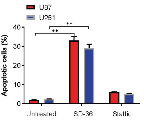
SD-36 purchased from MedChemExpress. Usage Cited in: J Cell Mol Med. 2021 Sep;25(17):8261-8270. [Abstract]
U87 and U251 cells were treated with 100 nmol/L SD-36 or 1 μmol/L Stattic for 24 h. Apoptosis was analysed by flow cytometry. The results sowed that SD-36 effectively enhanced apoptotic populations in the two cell lines.
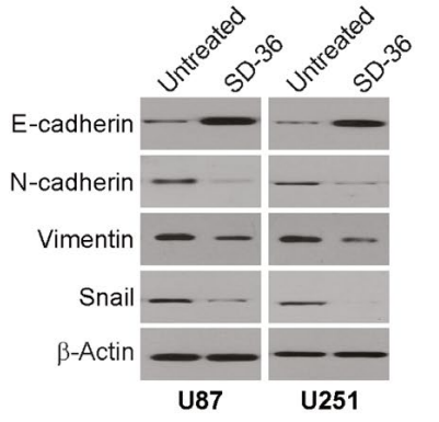
SD-36 purchased from MedChemExpress. Usage Cited in: J Cell Mol Med. 2021 Sep;25(17):8261-8270. [Abstract]
U87 and U251 cells were treated with 100 nmol/L SD-36 or 1 μmol/L Stattic for 24 h. Indicated protein levels were analysed by western blotting. The results sowed that SD-36 treatment resulted in increase in E-cadherin and decrease in N-cadherin, vimentin and snail in U87 and U251 cells.
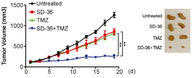
SD-36 purchased from MedChemExpress. Usage Cited in: J Cell Mol Med. 2021 Sep;25(17):8261-8270. [Abstract]
Tumour volume was quantified for established PDX tumours in NSG mice treated with vehicle, SD-36 (2 mg/kg/d; ip; twice a week), TMZ, or the combination of SD-36 and TMZ, and representative tumours were examined at the end of the experiment.
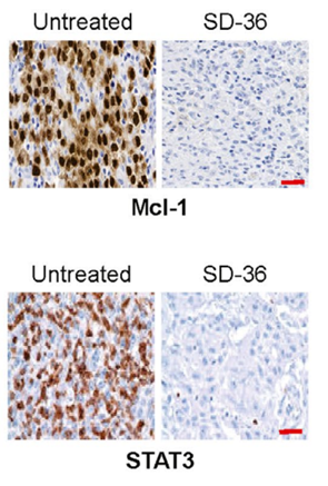
SD-36 purchased from MedChemExpress. Usage Cited in: J Cell Mol Med. 2021 Sep;25(17):8261-8270. [Abstract]
Mcl‐1 and STAT3 expression level was analysed in the indicated tumours by IHC. Scale bars: 25 μm. The results SD-36 (2 mg/kg/d; ip; twice a week) treatment decreased Mcl-1 and STAT3 expression in PDX mouse tumours.
-
iScience
Epigenomic landscapes define differential Janus kinases inhibitor sensitivity in IFN-γ-primed human macrophages. [Abstract]2025 Apr 22;28(5):112502. PMID: 40491958 -
bioRxiv
Combined antagonism of Oncostatin M (OSM) and Interleukin 6 (IL-6) provides both anti-fibrotic and anti-inflammatory benefit in pulmonary fibrosis. [Abstract]2025 Dec 26:2025.12.25.695625. PMID: 41497665 -
-
-
bioRxiv
2023 Sep 28:2023.09.26.559434. PMID: 37808649


Solvent & Solubility
In Vitro:
DMSO : 100 mg/mL (86.34 mM; Need ultrasonic; Hygroscopic DMSO has a significant impact on the solubility of product, please use newly opened DMSO)
Please refer to the solubility information to select the appropriate solvent. Once prepared, please aliquot and store the solution to prevent product inactivation from repeated freeze-thaw cycles.
Storage method and period of stock solution: -80°C, 6 months (stored under nitrogen). When stored at -80°C, please use it within 6 months.
Please refer to the solubility information to select the appropriate solvent. Once prepared, please aliquot and store the solution to prevent product inactivation from repeated freeze-thaw cycles.
Storage method and period of stock solution: -80°C, 6 months (stored under nitrogen). When stored at -80°C, please use it within 6 months.
Concentration (start) × Volume (start) = Concentration (final) × Volume (final)
In Vivo:
Select the appropriate dissolution method based on your experimental animal and administration route.
- For the following dissolution methods, please ensure to first prepare a clear stock solution using an In Vitro approach and then sequentially add co-solvents:
- To ensure reliable experimental results, the clarified stock solution can be appropriately stored based on storage conditions. As for the working solution for In Vivo experiments, it is recommended to prepare freshly and use it on the same day.
- The percentages shown for the solvents indicate their volumetric ratio in the final prepared solution. If precipitation or phase separation occurs during preparation, heat and/or sonication can be used to aid dissolution.
Add each solvent one by one: 10% DMSO 40% PEG300 5% Tween-80 45% Saline
Solubility: ≥ 1.25 mg/mL (1.08 mM); Clear solution
This protocol yields a clear solution of ≥ 1.25 mg/mL (saturation unknown).
Taking 1 mL working solution as an example, add 100 μL DMSO stock solution (12.5 mg/mL) to 400 μL PEG300, and mix evenly; then add 50 μL Tween-80 and mix evenly; then add 450 μL Saline to adjust the volume to 1 mL.
Preparation of Saline: Dissolve 0.9 g sodium chloride in ddH₂O and dilute to 100 mL to obtain a clear Saline solution.
Add each solvent one by one: 10% DMSO 90% (20% SBE-β-CD in Saline)
Solubility: ≥ 1.25 mg/mL (1.08 mM); Clear solution
This protocol yields a clear solution of ≥ 1.25 mg/mL (saturation unknown).
Taking 1 mL working solution as an example, add 100 μL DMSO stock solution (12.5 mg/mL) to 900 μL 20% SBE-β-CD in Saline, and mix evenly.
Preparation of 20% SBE-β-CD in Saline (4°C, storage for one week): 2 g SBE-β-CD powder is dissolved in 10 mL Saline, completely dissolve until clear.
In Vivo Dissolution Calculator
Please enter the basic information of animal experiments:
-
-
-
-
Recommended: Prepare an additional quantity of animals to account for potential losses during experiments.
Please enter your animal formula composition:
-
%DMSO +
Recommended: Keep the proportion of DMSO in working solution below 2% if your animal is weak.
-
%+
-
+%Tween-80 + +
-
%Saline +
The co-solvents required include: DMSO, . All of co-solvents are available by MedChemExpress (MCE). , Tween 80. All of co-solvents are available by MedChemExpress (MCE).
Working solution concentration: 0.22 mg/mL
Method for preparing stock solution: mg drug dissolved in μL DMSO. Stock solution concentration: mg/mL.
1. Take μL DMSO stock solution;
2. Add μL .
μL , mix evenly;
3. Then add μL Tween 80, mix evenly;
4. Then add μL
Please ensure that the stock solution in the first step is dissolved to a clear state, and add co-solvents in sequence. You can use ultrasonic heating (ultrasonic cleaner, recommended frequency 20-40 kHz), vortexing, etc. to assist dissolution.

Protocols
-
Apoptosis
Apoptosis, also called programmed cell death, is generally characterized by distinct morphological characteristics.
-
TUNEL staining for apoptotic DNA fragmentation
TUNEL staining detects DNA strand breaks by using terminal deoxynucleotidyl transferase to add labeled nucleotides to exposed 3′-OH DNA termini, generating either microscopic staining in fixed cells or tissue sections, or fluorescence/cytometric signal in cell suspensions. TUNEL positivity reflects DNA fragmentation but should not be interpreted alone as definitive apoptosis, because TUNEL can also label necrotic, autolytic, mechanically damaged, or DNA-repair-associated DNA breaks.
-
Flow cytometric DNA-content cell-cycle staining
Flow cytometric DNA-content cell-cycle staining measures the fluorescence intensity of DNA-bound fluorochromes in single cells or nuclei to estimate DNA content distributions, allowing assignment of populations to G0/G1, S, and G2/M phases by DNA histogram deconvolution. Propidium iodide (PI) intercalates into DNA, and PI fluorescence is proportional to cellular DNA content when staining is performed under conditions that make DNA accessible and minimize non-DNA signal. Cells with G2/M DNA content are expected to show approximately twice the fluorescence intensity of G0/G1 cells, while S-phase cells occupy intermediate fluorescence values. PI-based DNA-content analysis can also detect cells with fractional DNA content, often reported as sub-G1, when DNA fragmentation and extraction during staining reduce retained DNA signal in apoptotic cells. DAPI is an alternative DNA fluorochrome for univariate DNA-content analysis, while bivariate approaches combining DNA content with proliferation
-
Annexin V plus membrane-impermeant dye apoptosis staining
Annexin V-based apoptosis assays rely on the detection of phosphatidylserine (PS) externalization from the inner leaflet of the plasma membrane to the outer leaflet, an early biochemical hallmark of apoptosis. Fluorescently labeled Annexin V binds PS in a calcium-dependent manner, enabling identification of early apoptotic cells by flow cytometry or fluorescence microscopy. When combined with a membrane-impermeant DNA-binding dye (e. g. , propidium iodide), this approach allows discrimination between viable (Annexin V−/dye−), early apoptotic (Annexin V+/dye−), and late apoptotic or necrotic (Annexin V+/dye+) cell populations by assessing membrane integrity and PS exposure.
-
BrdU Incorporation Assay
Bromodeoxyuridine (BrdU) incorporation assay is based on the principle that BrdU, a thymidine analog, is incorporated into newly synthesized DNA during the S phase of the cell cycle, thereby serving as a marker of DNA replication and cellular proliferation. Incorporated BrdU can be detected using anti-BrdU antibodies following DNA denaturation, enabling visualization or quantification of proliferating cells through immunochemical detection methods such as immunofluorescence or immunohistochemistry.
-
Apoptosis Solutions
Apoptosis is a regulated, generally non-lytic cell-death pathway that removes unwanted, damaged, infected, or abnormal cells through coordinated morphological changes, caspase activation, DNA fragmentation, and membrane remodeling. The intrinsic apoptosis pathway is controlled mainly by mitochondrial outer membrane permeabilization, BCL-2 family proteins, cytochrome c release, apoptosome formation, caspase-9 activation, and downstream executioner caspase-3/7 activation. The extrinsic apoptosis pathway is initiated by death receptors such as Fas, TNFR, and TRAIL receptors, which recruit adaptor proteins and activate caspase-8 before engaging executioner caspases or mitochondrial amplification through BID cleavage. Apoptosis is linked to many phenotypes, including cancer cell killing, tissue homeostasis, immune regulation, neurodegeneration, infection response, and treatment-induced cytotoxicity; unresolved questions include how apoptosis interacts with necroptosis, pyroptosis, ferroptos
-
Protocol for Cell Cycle
Cell-cycle analysis by flow cytometry measures DNA content in single cells to estimate the fraction of cells in G0/G1, S, and G2/M phases. Propidium iodide intercalates into DNA, and after RNA removal with RNase, fluorescence intensity reflects cellular DNA content: 2N cells are assigned to G0/G1, cells between 2N and 4N to S phase, and 4N cells to G2/M. DNA-content analysis alone cannot reliably separate G0 from G1 or G2 from M. Ki-67 can distinguish quiescent G0 cells from cycling cells, EdU or BrdU incorporation marks active DNA synthesis in S phase, and phospho-histone H3 staining identifies mitotic cells within the 4N population.


Purity & Documentation
-
Data Sheet (304 KB)
-
SDS (252 KB)
- English - EN (252 KB)
- Français - FR (252 KB)
- Deutsch - DE (252 KB)
- Norwegian - NO (252 KB)
- Español - ES (252 KB)
- Swedish - SV (252 KB)
- Italian - IT (252 KB)
- Korean - KR (252 KB)
- Portuguese - PT (252 KB)
-
Handling Instructions (2659 KB)
References
[1]. Bai L, et al. A Potent and Selective Small-Molecule Degrader of STAT3 Achieves Complete Tumor Regression In Vivo. Cancer cell. 2019 Nov 11;36(5):498-511.e17. [Content Brief]
[2]. Zhou H, et al. Structure-Based Discovery of SD-36 as a Potent, Selective, and Efficacious PROTAC Degrader of STAT3 Protein. Journal of medicinal chemistry. 2019 Dec 26;62(24):11280-11300. [Content Brief]


Complete Stock Solution Preparation Table
Please refer to the solubility information to select the appropriate solvent. Once prepared, please aliquot and store the solution to prevent product inactivation from repeated freeze-thaw cycles.
Storage method and period of stock solution: -80°C, 6 months (stored under nitrogen). When stored at -80°C, please use it within 6 months.
| Optional Solvent | Concentration Solvent Mass | 1 mg | 5 mg | 10 mg | 25 mg |
|---|---|---|---|---|---|
| DMSO | 1 mM | 0.8634 mL | 4.3172 mL | 8.6345 mL | 21.5861 mL |
| 5 mM | 0.1727 mL | 0.8634 mL | 1.7269 mL | 4.3172 mL | |
| 10 mM | 0.0863 mL | 0.4317 mL | 0.8634 mL | 2.1586 mL | |
| 15 mM | 0.0576 mL | 0.2878 mL | 0.5756 mL | 1.4391 mL | |
| 20 mM | 0.0432 mL | 0.2159 mL | 0.4317 mL | 1.0793 mL | |
| 25 mM | 0.0345 mL | 0.1727 mL | 0.3454 mL | 0.8634 mL | |
| 30 mM | 0.0288 mL | 0.1439 mL | 0.2878 mL | 0.7195 mL | |
| 40 mM | 0.0216 mL | 0.1079 mL | 0.2159 mL | 0.5397 mL | |
| 50 mM | 0.0173 mL | 0.0863 mL | 0.1727 mL | 0.4317 mL | |
| 60 mM | 0.0144 mL | 0.0720 mL | 0.1439 mL | 0.3598 mL | |
| 80 mM | 0.0108 mL | 0.0540 mL | 0.1079 mL | 0.2698 mL |


SD-36 Related Classifications
HY-129602 Related Classifications
 Powered by Bioz