



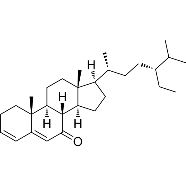
Stigmasta-3,5-dien-7-one

Based on 1 Customer Validation
Stigmasta-3,5-dien-7-one is a steroid compound that can be isolated from Harrisonia abyssinica. Stigmasta-3,5-dien-7-one blocks the NF-κB signaling pathway via down-regulation of phospho-p38 mitogen-activated protein kinase and phosphorylation and degradation of inhibitor of NF-κB α. Stigmasta-3,5-dien-7-one reduces LPS (HY-D1056)-induced nitric oxide, PGE2, and pro-inflammatory cytokine levels in macrophages. Stigmasta-3,5-dien-7-one can be used for inflammation diseases.
For research use only. We do not sell to patients.
- Purity : 99.84%
- CAS No.: 2034-72-2
- Formula: C29H46O
- Molecular Weight:410.67
-
Storage:Powder -20°C, 3 years , 4°C, 2 years ; In solvent -80°C, 6 months , -20°C, 1 month

To place orders, for customer services and technical support, please contact: MedChemExpress USA
Tel: 609-228-6898 E-mail: [email protected] [email protected]


-
 Biological Activity
-
Chemical Information
- Protocols
- Purity & Documentation
- References
-
 Help & FAQs




Biological Activity
Description
In Vitro
Stigmasta-3,5-dien-7-one (5-100 μg/mL; 1 h pre-incubation prior to LPS stimulation) dose-dependently inhibits LPS-induced NO production in RAW264.7 macrophage cells, with no cytotoxicity[1].
Stigmasta-3,5-dien-7-one (50 μg/mL; 1 h pre-incubation prior to LPS stimulation) significantly inhibits LPS-induced PGE2 production in RAW264.7 macrophage cells without altering basal PGE2 levels[1].
Stigmasta-3,5-dien-7-one (50 μg/mL; 1 h pre-incubation prior to LPS stimulation) significantly inhibits LPS-induced production of TNF-α, IL-1β, IL-6, iNOS, and COX-2 protein expression in RAW264.7 macrophage cells, without altering basal cytokine levels[1].
Stigmasta-3,5-dien-7-one (50 μg/mL; 1 h pre-incubation prior to LPS stimulation) inhibits LPS-induced NF-κB activation in RAW264.7 macrophage cells by blocking IκBα phosphorylation, IκBα degradation, and NF-κB nuclear translocation[1].
Stigmasta-3,5-dien-7-one (50 μg/mL; 1 h pre-incubation prior to LPS stimulation) inhibits LPS-induced p38 MAPK phosphorylation in RAW264.7 macrophage cells, while having no effect on ERK or SAPK/JNK phosphorylation[1].
MedChemExpress (MCE) has not independently confirmed the accuracy of these methods. They are for reference only. Further protocols information, click here.
-
Cell Line:RAW264.7 macrophage cells
-
Concentration:5; 10; 25; 50; 100 μg/mL
-
Incubation Time:1 h pre-incubation prior to LPS stimulation
-
Result:Showed no cytotoxicity at all tested concentrations.
Maintained cell viability at or near 100% across all doses, both in the presence and absence of LPS.
-
Cell Line:RAW264.7 macrophage cells
-
Concentration:50 μg/mL
-
Incubation Time:1 h pre-incubation prior to LPS stimulation
-
Result:Significantly inhibited LPS-induced PGE2 production compared to the LPS-only group.
Had no effect on basal PGE2 levels in the absence of LPS.\n
Significantly inhibited LPS-induced production of TNF-α, IL-1β, and IL-6 compared to the LPS-only group.
Had no effect on basal cytokine levels in the absence of LPS.
-
Cell Line:RAW264.7 macrophage cells
-
Concentration:50 μg/mL
-
Incubation Time:1 h pre-incubation prior to LPS stimulation, followed by co-incubation with LPS for experimental period
-
Result:Reduced LPS-induced iNOS and COX-2 protein expression compared to the LPS-only group.
Had no effect on basal iNOS or COX-2 expression in the absence of LPS.\nInhibited LPS-induced phosphorylation and degradation of IκBα compared to the LPS-only group.
Suppressed LPS-induced nuclear translocation of NF-κB compared to the LPS-only group.
-
Cell Line:RAW264.7 macrophage cells
-
Concentration:50 μg/mL
-
Incubation Time:1 h pre-incubation prior to 15-min LPS stimulation
-
Result:Suppressed LPS-induced phosphorylation of p38 MAPK compared to the LPS-only group.
Had no significant inhibitory effect on LPS-induced phosphorylation of ERK or SAPK/JNK MAPK compared to the LPS-only group.


Chemical Information
-
CAS No. 2034-72-2
-
Appearance Solid
-
Molecular Weight 410.67
-
Formula C29H46O
-
Color Off-white to light yellow
-
SMILES
O=C1[C@@]2([H])[C@@]3([H])[C@@]([C@@](CC3)([H])[C@H](C)CC[C@@H](CC)C(C)C)(CC[C@]2([H])[C@@]4(C(C=CCC4)=C1)C)C
-
Structure Classification
-
Initial Source
-
Shipping
Room temperature in continental US; may vary elsewhere.
-
Storage
Powder -20°C 3 years 4°C 2 years In solvent -80°C 6 months -20°C 1 month

Protocols
-
Kinase activity and phosphorylation assays
Kinase activity assays measure the ability of kinases to transfer phosphate groups from ATP to specific substrates, while phosphorylation assays detect the presence and levels of phosphorylated proteins. Common methods include radiolabeled ATP incorporation (e. g. ,), ADP release detection via bioluminescence (e. g. ,[3]), enzyme-linked immunosorbent assays (ELISA) for phospho-specific epitopes (e. g. ,[6]), and microtiter-based formats for high-throughput screening (e. g. ,[8]). The ADP-Glo assay quantifies kinase activity by measuring ADP produced during phosphorylation using a luciferase-based system. Radiometric assays involve autoradiography or scintillation counting after incorporation of 32P-labeled ATP into substrate proteins. ELISA-based approaches rely on phospho-specific antibodies to detect activated kinases in cell lysates or purified samples.
-
Western Blot
Western blotting (WB) is a commonly used experimental method in molecular biology, biochemistry, and immunogenetics for identifying and quantifying target proteins. It combines gel electrophoresis with immunoassay, enabling researchers to analyze protein expression, post-translational modifications, and molecular weight.
-
Research Protocol for Inflammation-related Diseases
The NLRP3 inflammasome is a cytosolic innate immune signaling platform that integrates priming signals and danger-signal activation to promote caspase-1 activation, maturation of IL-1β and IL-18, and gasdermin D-mediated pyroptotic cell death. The core experimental logic is to determine whether inflammatory disease phenotypes are driven by increased NLRP3 expression, ASC-containing inflammasome assembly, caspase-1 cleavage, GSDMD cleavage, and extracellular release of IL-1β/IL-18 rather than by nonspecific cell injury alone. The pathway is strongly linked to inflammation-related disease phenotypes because monosodium urate crystals activate NALP3/NLRP3 inflammasome signaling in gout-like crystal inflammation, cholesterol crystals activate NLRP3 inflammasomes in atherogenesis models, and DSS-induced intestinal inflammation has been reported to involve NLRP3 inflammasome activity. However, experimental colitis studies also show context-dependent protective effects of NLRP3 inflammasome co
-
Protocol for Kinase activity and phosphorylation assays
Kinase activity assays measure transfer of phosphate from ATP to a protein or peptide substrate, generating phosphorylated substrate, ADP, or incorporated radiolabeled phosphate as the readout; phosphorylation assays measure site-specific phosphorylation in cells or tissues as a proxy for kinase-pathway activation, inhibition, or substrate regulation. Phosphorylation can be detected by phospho-specific Western blot, immunoprecipitation kinase assay, phospho-immunofluorescence, phospho-flow cytometry, luminescent ADP detection, radiolabeled ATP incorporation, or reporter-based pathway assays, and these readouts can be applied to cancer cells, primary neurons, mouse tumors, organoids, inflammatory macrophages, ferroptosis studies, and mitophagy studies when the kinase target is biologically relevant.


Purity & Documentation
-
Data Sheet (276 KB)
-
SDS (251 KB)
- English - EN (251 KB)
- Français - FR (251 KB)
- Deutsch - DE (251 KB)
- Norwegian - NO (251 KB)
- Español - ES (251 KB)
- Swedish - SV (251 KB)
- Italian - IT (251 KB)
- Korean - KR (251 KB)
- Portuguese - PT (251 KB)
-
Handling Instructions (2659 KB)
References
[1]. Park SJ, et al. Anti-inflammatory Steroid from Phragmitis rhizoma Modulates LPS-Mediated Signaling Through Inhibition of NF-κB Pathway. Inflammation. 2016;39(2):727-734. [Content Brief]
[2]. Baldé AM, et al. Steroids from Harrisonia abyssinica. Planta Med. 2000;66(1):67-69. [Content Brief]
[3]. Szakiel A, et al. Triterpenoid content of berries and leaves of bilberry Vaccinium myrtillus from Finland and Poland. J Agric Food Chem. 2012;60(48):11839-11849. [Content Brief]

Calculators
Concentration (start) × Volume (start) = Concentration (final) × Volume (final)


Stigmasta-3,5-dien-7-one Related Classifications
HY-N12188 Related Classifications
 Powered by Bioz
Keywords
- Stigmasta-3,5-dien-7-one
- 2034-72-2
- NF-κB
- TNF Receptor
- Interleukin Related
- p38 MAPK
- phospho-p38 mitogen-activated protein kinase
- RAW264.7 macrophage cells
- inhibitor of NF-κB α
- NF-κB signaling pathway
- prostaglandin E2
- macrophages
- p38 mitogen-activated protein kinase
- LPS-induced inflammation
- nitric oxide
- Inhibitor
- inhibitor
- inhibit