



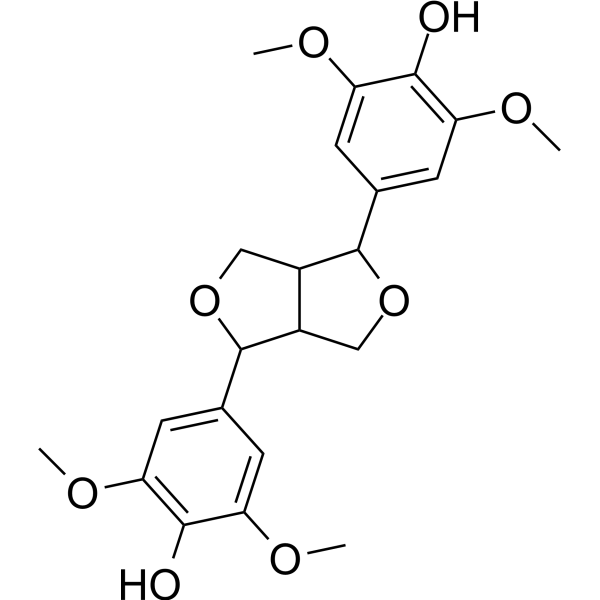
Syringaresinol

Based on 1 Customer Validation
Syringaresinol is a lignan-type phytochemical with anti-inflammatory activities. Syringaresinol inhibits the NF-κB and AKT signaling pathway in IL-1β-activated mouse chondrocytes. Syringaresinol increases phosphorylation of AMPK, eNOS, and intracellular Ca2+ levels in HUVECs. Syringaresinol attenuates osteoarthritis progression in mice with Destabilization of the Medial Meniscus (DMM)-induced osteoarthritis. Syringaresinol can be used for the study of osteoarthritis (OA).
For research use only. We do not sell to patients.
- Purity : 99.66%
- CAS No.: 487-35-4
- Formula: C22H26O8
- Molecular Weight:418.44
-
Storage:
-20°C, protect from light
* In solvent : -80°C, 6 months; -20°C, 1 month (protect from light)

To place orders, for customer services and technical support, please contact: MedChemExpress USA
Tel: 609-228-6898 E-mail: [email protected] [email protected]


-
 Biological Activity
-
Chemical Information
-
Solvent & Solubility
- Protocols
- Purity & Documentation
- References
-
 Help & FAQs




Biological Activity
Description
In Vitro
Syringaresinol (0-100 μM, 2 h pretreatment) dose-dependently decreases the production of inflammatory mediators (PGE2, IL-6, TNF-α, NO) and downregulates the protein expression of iNOS and COX-2 in IL-1β-activated mouse chondrocytes[1].
Syringaresinol (0-100 μM, 2 h pretreatment) upregulates the production of ECM components and downregulates the secretion of ECM-degrading enzymes in IL-1β-activated mouse chondrocytes[1].
Syringaresinol (0-100 μM, 2 h pretreatment) inhibits the activation of the NF-κB and AKT signaling pathway[1].
Syringaresinol (10-30 μM, 30 min) dose-dependently increases phosphorylation of Akt, AMPK, and eNOS, and increases intracellular Ca2+ levels in HUVECs[2].
Syringaresinol (30 μM, 30 min) significantly increases eNOS dimerization and decreases eNOS monomer in HUVECs[2].
MedChemExpress (MCE) has not independently confirmed the accuracy of these methods. They are for reference only.
-
Cell Line:IL-1β-activated mouse chondrocytes
-
Concentration:0, 25, 50, 100 μM
-
Incubation Time:2 h pretreatment
-
Result:Reduced the the production of inflammatory mediators (PGE2, IL-6, TNF-α).
Upregulated the production of ECM components (collagen II, aggrecan).
Downregulated the secretion of ECM-degrading enzymes (MMP-13, ADAMTS-5).
-
Cell Line:IL-1β-activated mouse chondrocytes
-
Concentration:0, 25, 50, 100 μM
-
Incubation Time:2 h pretreatment
-
Result:Downregulated the protein expression of iNOS and COX-2.
Suppressed IL-1β-induced degradation of IκB-α, phosphorylation of p65, and nuclear translocation of p65.
Decreased IL-1β-induced phosphorylation of AKT.
In Vivo
MedChemExpress (MCE) has not independently confirmed the accuracy of these methods. They are for reference only.
-
Animal Model:60-day-old male C57BL/6 mice with DMM-induced osteoarthritis[1]
-
Dosage:10 mg/kg
-
Administration:Intra-articular injection, once weekly for 4 consecutive weeks
-
Result:Reduced joint space narrowing, osteophyte formation and cartilage calcification (X-ray detection).
Alleviated cartilage erosion and proteoglycan depletion.
Reversed the elevation of Osteoarthritis Research Society International (OARSI) scores.
Decreased COX-2 protein expression in cartilage tissue.


Chemical Information
-
CAS No. 487-35-4
-
Appearance Solid
-
Molecular Weight 418.44
-
Formula C22H26O8
-
Color White to off-white
-
SMILES
OC1=C(OC)C=C(C2OCC3C2COC3C4=CC(OC)=C(O)C(OC)=C4)C=C1OC
-
Structure Classification
-
Shipping
Room temperature in continental US; may vary elsewhere.
-
Storage
-20°C, protect from light
* In solvent : -80°C, 6 months; -20°C, 1 month (protect from light)


Solvent & Solubility
In Vitro:
DMSO : 50 mg/mL (119.49 mM; Need ultrasonic; Hygroscopic DMSO has a significant impact on the solubility of product, please use newly opened DMSO)
Please refer to the solubility information to select the appropriate solvent. Once prepared, please aliquot and store the solution to prevent product inactivation from repeated freeze-thaw cycles.
Storage method and period of stock solution: -80°C, 6 months; -20°C, 1 month (protect from light). When stored at -80°C, please use it within 6 months. When stored at -20°C, please use it within 1 month.
Please refer to the solubility information to select the appropriate solvent. Once prepared, please aliquot and store the solution to prevent product inactivation from repeated freeze-thaw cycles.
Storage method and period of stock solution: -80°C, 6 months; -20°C, 1 month (protect from light). When stored at -80°C, please use it within 6 months. When stored at -20°C, please use it within 1 month.
Concentration (start) × Volume (start) = Concentration (final) × Volume (final)


Purity & Documentation
-
Data Sheet (280 KB)
-
SDS (393 KB)
- English - EN (393 KB)
- Français - FR (393 KB)
- Deutsch - DE (393 KB)
- Norwegian - NO (393 KB)
- Español - ES (393 KB)
- Swedish - SV (393 KB)
- Italian - IT (393 KB)
- Korean - KR (393 KB)
- Portuguese - PT (393 KB)
-
Handling Instructions (2659 KB)
References
[1]. Wang X, et al. Syringaresinol attenuates osteoarthritis via regulating the NF-κB pathway. Int Immunopharmacol. 2023 May;118:109982. [Content Brief]
[2]. Chung BH, et al. Syringaresinol causes vasorelaxation by elevating nitric oxide production through the phosphorylation and dimerization of endothelial nitric oxide synthase. Exp Mol Med. 2012 Mar 31;44(3):191-201. [Content Brief]


Complete Stock Solution Preparation Table
Please refer to the solubility information to select the appropriate solvent. Once prepared, please aliquot and store the solution to prevent product inactivation from repeated freeze-thaw cycles.
Storage method and period of stock solution: -80°C, 6 months; -20°C, 1 month (protect from light). When stored at -80°C, please use it within 6 months. When stored at -20°C, please use it within 1 month.
| Optional Solvent | Concentration Solvent Mass | 1 mg | 5 mg | 10 mg | 25 mg |
|---|---|---|---|---|---|
| DMSO | 1 mM | 2.3898 mL | 11.9491 mL | 23.8983 mL | 59.7457 mL |
| 5 mM | 0.4780 mL | 2.3898 mL | 4.7797 mL | 11.9491 mL | |
| 10 mM | 0.2390 mL | 1.1949 mL | 2.3898 mL | 5.9746 mL | |
| 15 mM | 0.1593 mL | 0.7966 mL | 1.5932 mL | 3.9830 mL | |
| 20 mM | 0.1195 mL | 0.5975 mL | 1.1949 mL | 2.9873 mL | |
| 25 mM | 0.0956 mL | 0.4780 mL | 0.9559 mL | 2.3898 mL | |
| 30 mM | 0.0797 mL | 0.3983 mL | 0.7966 mL | 1.9915 mL | |
| 40 mM | 0.0597 mL | 0.2987 mL | 0.5975 mL | 1.4936 mL | |
| 50 mM | 0.0478 mL | 0.2390 mL | 0.4780 mL | 1.1949 mL | |
| 60 mM | 0.0398 mL | 0.1992 mL | 0.3983 mL | 0.9958 mL | |
| 80 mM | 0.0299 mL | 0.1494 mL | 0.2987 mL | 0.7468 mL | |
| 100 mM | 0.0239 mL | 0.1195 mL | 0.2390 mL | 0.5975 mL |


Syringaresinol Related Classifications
HY-N8307 Related Classifications
 Powered by Bioz