



Teleocidin A1
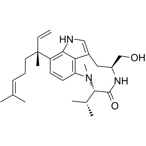
Teleocidin A1 (Lyngbyatoxin A) is a PKC activator with a Ki value of 0.11 nM for binding to the PKCδ-C1B peptide. Teleocidin A1 exhibits anticancer activity against cervical cancer and leukemia. Teleocidin A1 can induce seaweed dermatitis, food poisoning and local skin toxicity. Teleocidin A1 can be used in studies related to cervical cancer, seaweed dermatitis and food poisoning.
For research use only. We do not sell to patients.
- CAS No.: 70497-14-2
- Formula: C27H39N3O2
- Molecular Weight:437.62
-
Storage:
Please store the product under the recommended conditions in the Certificate of Analysis.

To place orders, for customer services and technical support, please contact: MedChemExpress USA
Tel: 609-228-6898 E-mail: [email protected] [email protected]


-
 Biological Activity
-
Chemical Information
- Protocols
- Purity & Documentation
- References
-
 Help & FAQs





Biological Activity
Description
IC50 & Target
[2]|
PKCδ-C1B 0.11 nM (Ki) |
Cellular Effect
|
Cell Line

|
Type | Value | Description | References |
|---|---|---|---|---|
| L1210 | IC50 |
8.1 μM
Compound: 25
|
Cytotoxicity against mouse L1210 cells assessed as inhibition of cell growth
Cytotoxicity against mouse L1210 cells assessed as inhibition of cell growth
|
[PMID: 32652435] |
In Vitro
Teleocidin A1 (incubated for 48 h) potently inhibits the proliferation of HeLa cervical cancer cells, with an IC50 value of 9.2 nM[1].
Teleocidin A1 (incubated for 24 h) inhibits the proliferation of L1210 mouse leukemia cells, with an IC50 value of 8.1 μM[2].
Teleocidin A1 binds to the PKCδ-C1B peptide segment with high affinity, with a Ki value of 0.11 nM for the inhibition of [3H]PDBu, a result that confirms the aforementioned binding characteristics[2].
Lyngbyatoxin A (26 μg/cm2; 1-48 h) rapidly penetrates the abdominal skin of ex vivo male Hartley guinea pigs, reaching a peak of 23% of the administered dose within 1 hour, with no significant changes in the penetration amount at subsequent time points[3].
MedChemExpress (MCE) has not independently confirmed the accuracy of these methods. They are for reference only. Further protocols information, click here.
In Vivo
MedChemExpress (MCE) has not independently confirmed the accuracy of these methods. They are for reference only.
-
Animal Model:Palaemon paucidens (average weight 0.5 g)[2]
-
Dosage:0.6 mg/kg
-
Administration:injection
-
Result:Reached an LD33 value of 0.6 mg/kg.
Was approximately 150-times more potent than compound 1 (LD33 89 mg/kg).
Was 40-times more potent than compound 2 (LD33 25 mg/kg).


Chemical Information
-
CAS No. 70497-14-2
-
Molecular Weight 437.62
-
Formula C27H39N3O2
-
SMILES
C/C(C)=C\CC[C@@](C)(C1=C2C3=C(C=C1)N([C@H](C(N[C@@H](CC3=CN2)CO)=O)C(C)C)C)C=C
-
Synonyms
Lyngbyatoxin A
-
Structure Classification
-
Initial Source
Streptomyces
-
Shipping
Room temperature in continental US; may vary elsewhere.
-
Storage
Please store the product under the recommended conditions in the Certificate of Analysis.

Protocols
-
TPA/Croton Oil Ear Edema and Dermatitis
The TPA (12-O-tetradecanoylphorbol-13-acetate) and croton oil-induced mouse ear edema model is a well-established acute cutaneous inflammation system used to evaluate topical anti-inflammatory activity by measuring edema formation, neutrophil infiltration, vascular permeability, and cytokine-mediated skin responses in vivo. The inflammatory response is triggered by topical application of phorbol esters (TPA) or croton oil constituents, leading to rapid activation of protein kinase C signaling, leukocyte recruitment, and increased vascular permeability, which can be quantified by ear thickness, weight, dye extravasation, and biochemical markers such as myeloperoxidase (MPO) activity and pro-inflammatory mediators in ear tissue homogenates. This model is widely used for screening anti-inflammatory agents, where reductions in edema and inflammatory biomarkers reflect suppression of acute dermal inflammation and immune cell infiltration. Histological evaluation typically confirms epidermal
-
Dermal Irritation/Dermal Toxicity Study
This protocol assesses dermal irritation using reconstructed human epidermis (RhE) models such as EpiDerm, EPISKIN, and SkinEthic RHE, in which a test substance is applied topically and tissue viability is measured after exposure; reduced viability reflects cytotoxic injury associated with skin irritation potential. The primary readout is MTT reduction, where viable cells convert tetrazolium salt into colored formazan measured by spectrophotometry; this signal is used as a quantitative viability endpoint for classifying irritant versus non-irritant responses. The historical in vivo comparator is the Draize rabbit skin irritation method, which scores erythema and edema after topical exposure, but validated RhE assays were developed to replace or reduce reliance on this animal-based endpoint.


Purity & Documentation
References
[1]. Youssef DT, et al. 2,3-seco-2,3-dioxo-lyngbyatoxin A from a Red Sea strain of the marine cyanobacterium Moorea producens. Nat Prod Res. 2015;29(8):703-9. [Content Brief]
[2]. Jiang W, et al. Two new lyngbyatoxin derivatives from the Cyanobacterium, Moorea producens. Mar Drugs. 2014;12(12):5788-5800. Published 2014 Dec 1. [Content Brief]
[3]. Stafford RG, et al. Comparison of the partition coefficient and skin penetration of a marine algal toxin (lyngbyatoxin A). Food Chem Toxicol. 1992;30(9):795-801. [Content Brief]

Calculators
Concentration (start) × Volume (start) = Concentration (final) × Volume (final)


Teleocidin A1 Related Classifications
HY-118834 Related Classifications
 Powered by Bioz
Keywords
- Teleocidin A1
- 70497-14-2
- Lyngbyatoxin A
- Teleocidin A 1
- Teleocidin A-1
- PKC
- Streptomyces mediocidicus
- Palaemon paucidens
- protein kinase C
- PKCδ-C1B peptide
- human abdominal cadaver skin
- L1210 mouse leukemia cells
- male Hartley guinea pig ventral skin
- cervical cancer
- PKC C1 domains
- HeLa cervical cancer cells
- Inhibitor
- inhibitor
- inhibit