



UBX-382

Based on 1 Customer Validation
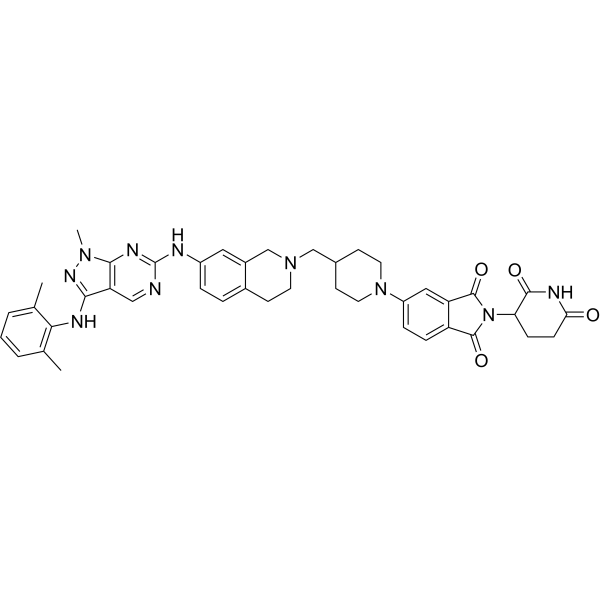
UBX-382 is an orally active BTK PROTAC degrader with a DC50 of 4.56 nM. UBX-382 inhibits B-cell receptor signaling by targeting BTK. UBX-382 shows superior degradation activity for wild-type and mutant BTK proteins. UBX-382 inhibits tumor growth in murine xenograft models harboring wild-type or C481S mutant BTK-expressing TMD-8 cells. UBX-382 can be used for the study of B-cell-related blood cancers.
For research use only. We do not sell to patients.
- Purity : 96.20%
- CAS No.: 2884554-45-2
- Formula: C42H44N10O4
- Molecular Weight:752.86
-
Storage:Powder -20°C, 3 years , 4°C, 2 years ; In solvent -80°C, 6 months , -20°C, 1 month

To place orders, for customer services and technical support, please contact: MedChemExpress USA
Tel: 609-228-6898 E-mail: [email protected] [email protected]


-
 Biological Activity
-
Chemical Information
- Protocols
- Purity & Documentation
- References
-
 Help & FAQs


-
HY-10997
IbrutinibBtk Inhibitor -
HY-19834
FenebrutinibBtk Inhibitor -
HY-17600
AcalabrutinibBTK Inhibitor -
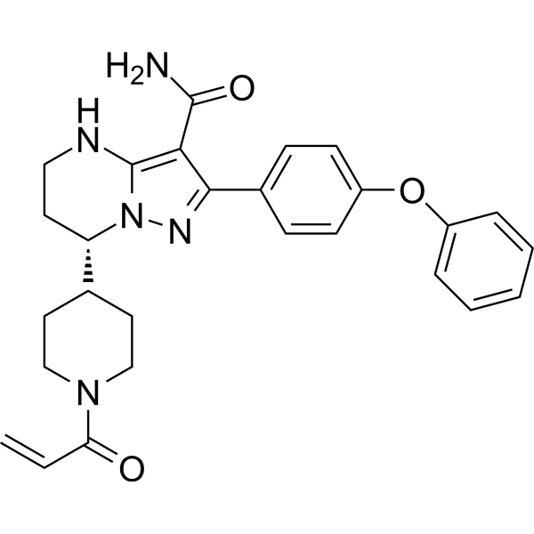
HY-101474A
ZanubrutinibBtk Inhibitor -
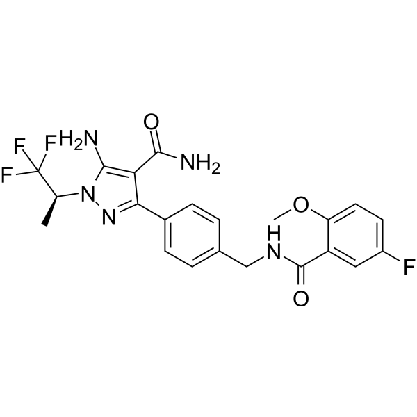
HY-131328
PirtobrutinibBTK Inhibitor -
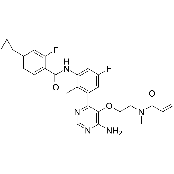
HY-128757
RemibrutinibBTK Inhibitor -
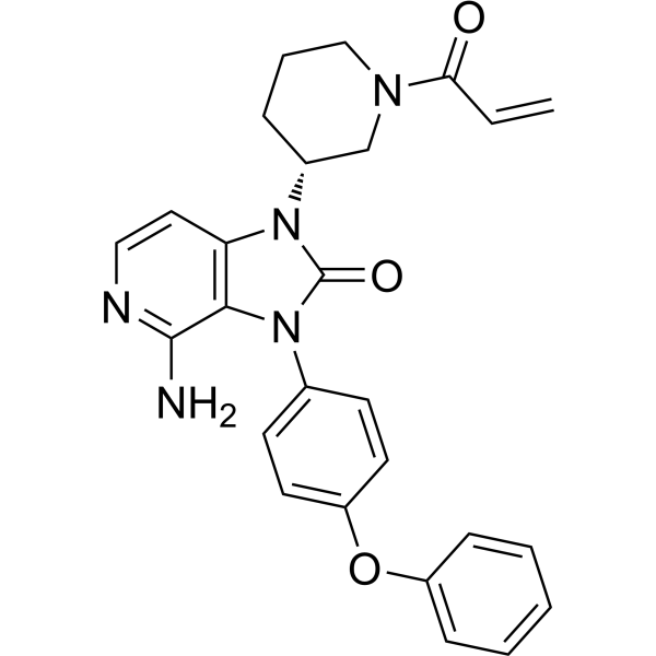
HY-109192
TolebrutinibBTK Inhibitor -
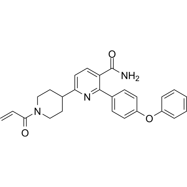
HY-129390
OrelabrutinibBTK Inhibitor -
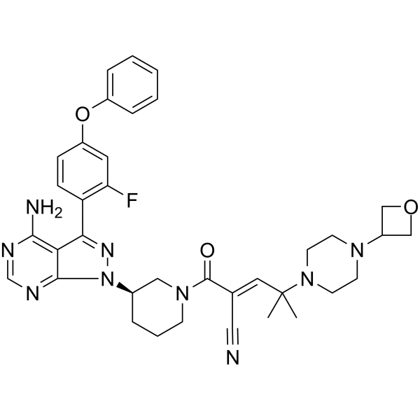
HY-112166
RilzabrutinibBTK Inhibitor -
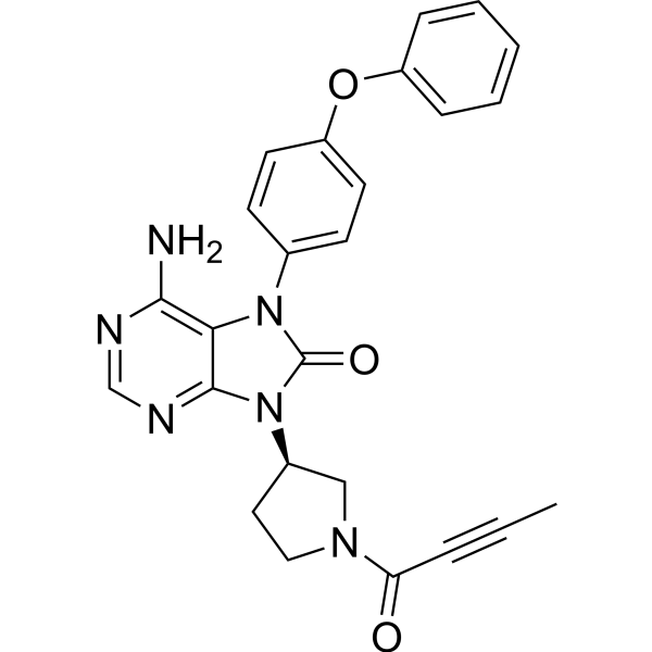
HY-15771
TirabrutinibBTK Inhibitor -
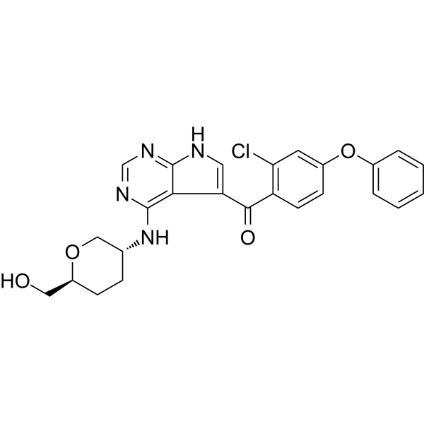
HY-112215
NemtabrutinibBTK Inhibitor -
HY-101215
Evobrutinib


Biological Activity
Description
IC50 & Target
[1]|
Cereblon |
In Vitro
UBX-382 (0.00001-10 nM, 0.5-48 h) induces potent BTK degradation with a DC 50 of 4.56 nM in TMD-8 cells[1].
UBX-382 (0.0001-10 μM, 3-5 days) inhibits the proliferation of TMD8 (IC50 = 14 nM), WSU-DLCL2 (IC50 = 18 nM), OCI-Ly3 (IC50 = 199 nM) and U2932 (IC50 = 21 nM) measured by the Cell Titer-Glo assay in duplicates[1].
UBX-382 (10 nM, 24 h) significantly inhibits CCL3 and CCL4 secretion induced by BCR signal activation in TMD-8 cells[1].
UBX-382 (100 nM, 6-24 h) inhibits the phosphorylation of BTK Y223, SYK, MEK, and ERK in U2932 cells[1].
UBX-382 (0.1-10 μM, 24 h) effectively degrades wild-type and various BTK mutants (E41K, C481S/R/T/Y/F, L528W) in HEK293 cells and strongly suppresses their phosphorylation[1].
MedChemExpress (MCE) has not independently confirmed the accuracy of these methods. They are for reference only.
-
Cell Line:U2932 cells
-
Concentration:100 nM
-
Incubation Time:6, 24 h
-
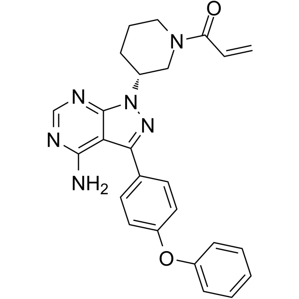
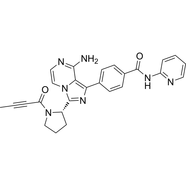
Result:Showed far more effective inhibition of BTK Y223 phosphorylation than Ibrutinib (HY-10997), Acalabrutinib (HY-17600), and Nemtabrutinib (ARQ-531) (HY-112215) in U2932 cells.
Inhibited the phosphorylation of SYK, MEK, and ERK.
In Vivo
MedChemExpress (MCE) has not independently confirmed the accuracy of these methods. They are for reference only.
-
Animal Model:TMD-8 cells (1 × 10⁷ in Matrigel) were subcutaneously injected into the flanks of 6-week-old CB17/severe combined immunodeficient mice[1]
-
Dosage:3, 10, 30 mg/kg
-
Administration:p.o. once daily for 21 days
-
Result:Achieved complete tumor regression in TMD-8 xenograft models.
Showed no significant changes in body weight and no other clinical toxicity signs during the experiment.
Reduced BTK levels in tumors of TMD-8 xenograft models.
-
Animal Model:TMD-8 BTK C481S cells were subcutaneously inoculated into the right flank of male CB17/severe combined immunodeficient mice[1]
-
Dosage:3, 10, 30 mg/kg
-
Administration:p.o. once daily for 21 days
-
Result:Induced remarkable dose-dependent tumor regression in TMD-8 BTK C481S xenograft models, while Ibrutinib failed to inhibit tumor growth.


Chemical Information
-
CAS No. 2884554-45-2
-
Appearance Solid
-
Molecular Weight 752.86
-
Formula C42H44N10O4
-
Color Light yellow to green yellow
-
SMILES
O=C1C2=CC=C(N3CCC(CC3)CN4CC5=CC(NC6=NC7=C(C(NC8=C(C)C=CC=C8C)=NN7C)C=N6)=CC=C5CC4)C=C2C(N1C9C(NC(CC9)=O)=O)=O
-
Shipping
Room temperature in continental US; may vary elsewhere.
-
Storage
Powder -20°C 3 years 4°C 2 years In solvent -80°C 6 months -20°C 1 month


Purity & Documentation
-
Data Sheet (271 KB)
-
SDS (251 KB)
- English - EN (251 KB)
- Français - FR (251 KB)
- Deutsch - DE (251 KB)
- Norwegian - NO (251 KB)
- Español - ES (251 KB)
- Swedish - SV (251 KB)
- Italian - IT (251 KB)
- Korean - KR (251 KB)
- Portuguese - PT (251 KB)
-
Handling Instructions (2659 KB)
References

Calculators
Concentration (start) × Volume (start) = Concentration (final) × Volume (final)


UBX-382 Related Classifications
HY-160142 Related Classifications
 Powered by Bioz