



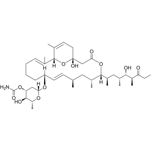
Venturicidin A

Based on 1 Customer Validation
Venturicidin A (Aabomycin A1) is a natural product derived from Streptomyces, possessing antifungal, anti-Trypanosoma brucei, anti-Leishmania donovani, and ATP synthase inhibitory activities. Venturicidin A acts as an antibiotic adjuvant, promoting the uptake of aminoglycoside antibiotics and enhancing bactericidal efficacy. Venturicidin A inhibits ATP synthesis by blocking proton translocation through the FO subunit. In bloodstream-form Trypanosoma brucei, Venturicidin A causes collapse of the mitochondrial membrane potential; in fungi, it disrupts cell membrane integrity, induces leakage of cytoplasmic contents, elevates ROS levels, and downregulates the expression of fungal pathogenicity-related genes. Venturicidin A exhibits antifungal activity against Botrytis cinerea. Venturicidin A is used in research on bacterial infections, trypanosome/leishmania infections, and gray mold disease.
For research use only. We do not sell to patients.
- Purity : 96.60%
- CAS No.: 33538-71-5
- Formula: C41H67NO11
- Molecular Weight:749.97
-
Storage:Powder -20°C, 3 years ; In solvent -80°C, 6 months , -20°C, 1 month

To place orders, for customer services and technical support, please contact: MedChemExpress USA
Tel: 609-228-6898 E-mail: [email protected] [email protected]


-
 Biological Activity
-
Chemical Information
-
Solvent & Solubility
- Protocols
- Purity & Documentation
- References
-
 Help & FAQs






Biological Activity
Description
IC50 & Target
|
Aminoglycoside |
Cellular Effect
|
Cell Line

|
Type | Value | Description | References |
|---|---|---|---|---|
| B16 | IC50 |
12 μM
Compound: 4
|
Cytotoxicity against mouse B16 cells assessed as inhibition of cell growth measured after 72 hrs by Celltiter-Glo assay
Cytotoxicity against mouse B16 cells assessed as inhibition of cell growth measured after 72 hrs by Celltiter-Glo assay
|
[PMID: 33356258] |
| Hep 3B2 | IC50 |
15 μM
Compound: 4
|
Cytotoxicity against human Hep3B cells assessed as inhibition of cell growth measured after 72 hrs by Celltiter-Glo assay
Cytotoxicity against human Hep3B cells assessed as inhibition of cell growth measured after 72 hrs by Celltiter-Glo assay
|
[PMID: 33356258] |
| K562 | IC50 |
5.8 μM
Compound: 4
|
Cytotoxicity against human K562 cells assessed as inhibition of cell growth measured after 72 hrs by Celltiter-Glo assay
Cytotoxicity against human K562 cells assessed as inhibition of cell growth measured after 72 hrs by Celltiter-Glo assay
|
[PMID: 33356258] |
| MDA-MB-468 | IC50 |
8.3 μM
Compound: 4
|
Cytotoxicity against human MDA-MB-468 cells assessed as inhibition of cell growth measured after 72 hrs by Celltiter-Glo assay
Cytotoxicity against human MDA-MB-468 cells assessed as inhibition of cell growth measured after 72 hrs by Celltiter-Glo assay
|
[PMID: 33356258] |
| MV4-11 | IC50 |
0.09 μM
Compound: 4
|
Cytotoxicity against human MV4-11 cells assessed as inhibition of cell growth measured after 72 hrs by Celltiter-Glo assay
Cytotoxicity against human MV4-11 cells assessed as inhibition of cell growth measured after 72 hrs by Celltiter-Glo assay
|
[PMID: 33356258] |
In Vitro
Venturicidin A (8-16 μg/mL; 24 h) enhances the activity of Gentamicin (HY-A0276A) against MDR isolates of MRSA, VRE, P. aeruginosa, A. baumannii, E. coli, and K. pneumoniae, with fractional inhibitory concentration indices below 0.5, but shows no enhancement against isolates carrying the ArmA ribosomal methyltransferase[1].
Venturicidin A in combination with Gentamicin (16-32 μg/mL; 0-24 h) exhibits rapid bactericidal activity against aminoglycoside-resistant MRSA C1014, reducing viable cells by >3 log CFU/mL within 4 h[1].
Venturicidin A does not inhibit the bifunctional aminoglycoside-modifying enzyme AAC (6′)-Ie-APH (2′′)-Ia, but instead acts as a Type 1b adjuvant through an indirect mechanism to enhance the effects of aminoglycosides[1].
Venturicidin A (48 h) exhibits in vitro toxicity against human embryonic kidney (HEK) cells with an IC50 of 31 μg/mL[1].
Venturicidin A is an inhibitor of ATP synthesis in MRSA C1014, depleting intracellular ATP levels by approximately 10-fold; and it induces membrane depolarization in MRSA C1014 in a dose-dependent manner[1].
Venturicidin A (VentA) (1.56-50 μg/mL; 5 days) effectively inhibits the mycelial growth of Botrytis cinerea on PDA medium, with an EC50 of 1.08 μg/mL[2].
Venturicidin A (1.08-12.5 μg/mL; 6-24 h) significantly inhibits Botrytis cinerea spore germination and germ tube elongation in a concentration-dependent manner; it also disrupts the morphology of spores and hyphae; it disrupts the integrity of the hyphal plasma membrane, leading to loss of cell viability[2].
Venturicidin A (1.08-12.5 μg/mL; 12-24 hpi) impairs the penetration ability of Botrytis cinerea into onion epidermal cells[2].
Venturicidin A (1.08-12.5 μg/mL; 2-10 h) severely disrupts the cell membrane integrity of Botrytis cinerea, leading to massive leakage of nucleic acids and proteins; it also increases MDA content in mycelia, indicating lipid peroxidation and membrane damage[2].
Venturicidin A (1.08-12.5 μg/mL; 24 h) induces ROS accumulation in Botrytis cinerea hyphae; downregulates the expression of pathogenicity-related genes; and upregulates the expression of NADPH oxidase subunit genes[2].
Venturicidin A (1.08-12.5 μg/mL; spray; once; 24 h before inoculation) inhibits spore germination and mycelial growth of Botrytis cinerea, disrupts fungal cell membrane integrity, induces ROS burst, and downregulates pathogenesis-related genes; it exhibits both protective and curative activity against tomato gray mold[2].
Venturicidin A exhibits high activity against Trypanosoma brucei brucei 2T1 BSF, Trypanosoma brucei rhodesiense BSF, and Leishmania donovani promastigotes, axenic amastigotes, and intracellular amastigotes, but shows markedly reduced activity against Trypanosoma evansi STIB806 BSF, with IC50 values of 21.49 nM, 5.05 nM, 24.10 nM, 2.28 nM, 267.6 nM, and 5260 nM, respectively[3].
Venturicidin A (21-172 nM; 24 h) treatment of Trypanosoma brucei brucei 2T1 bloodstream forms leads to collapse of the mitochondrial membrane potential and induces loss of kDNA[3].
MedChemExpress (MCE) has not independently confirmed the accuracy of these methods. They are for reference only. Further protocols information, click here.
-
Cell Line:Botrytis cinerea hyphae
-
Concentration:1.08, 6.25, 12.5 μg/mL
-
Incubation Time:24 h
-
Result:Downregulated the expression of pathogenicity-related genes; and upregulated the expression of NADPH oxidase subunit genes.
-
Cell Line:Trypanosoma brucei brucei 2T1 bloodstream forms
-
Concentration:21, 107, 172 nM
-
Incubation Time:24 h
-
Result:Induced a loss of kDNA in approximately 40% of cells at 21 nM and 107 nM.
Induced a loss of kDNA in up to 50% of cells at 172 nM.
Induced abnormal cell division with an abnormal number of kDNA (1K2N) at all tested concentrations.


Chemical Information
-
CAS No. 33538-71-5
-
Appearance Solid
-
Molecular Weight 749.97
-
Formula C41H67NO11
-
Color White to off-white
-
SMILES
CC1=CC[C@](O)(C2)O[C@]1([H])/C(C)=C\CCC[C@](O[C@@]3([H])C[C@H]([C@H](O)[C@@H](C)O3)OC(N)=O)([H])/C=C/[C@H](C)C[C@@H](C)[C@]([C@H](C)C[C@@H](C)[C@H](O)[C@H](C)C(CC)=O)([H])OC2=O
-
Synonyms
Aabomycin A1
-
Structure Classification
-
Initial Source
actinomycetes
-
Shipping
Room temperature in continental US; may vary elsewhere.
-
Storage
Powder -20°C 3 years In solvent -80°C 6 months -20°C 1 month


Solvent & Solubility
In Vitro:
DMSO : 8 mg/mL (10.67 mM; Need ultrasonic and warming; Hygroscopic DMSO has a significant impact on the solubility of product, please use newly opened DMSO)
Please refer to the solubility information to select the appropriate solvent. Once prepared, please aliquot and store the solution to prevent product inactivation from repeated freeze-thaw cycles.
Storage method and period of stock solution: -80°C, 6 months; -20°C, 1 month. When stored at -80°C, please use it within 6 months. When stored at -20°C, please use it within 1 month.
Please refer to the solubility information to select the appropriate solvent. Once prepared, please aliquot and store the solution to prevent product inactivation from repeated freeze-thaw cycles.
Storage method and period of stock solution: -80°C, 6 months; -20°C, 1 month. When stored at -80°C, please use it within 6 months. When stored at -20°C, please use it within 1 month.
Concentration (start) × Volume (start) = Concentration (final) × Volume (final)

Protocols
-
RNA extraction experimental
By lysing cells, releasing RNA, and removing impurities such as proteins and DNA, high-purity RNA products are finally obtained. The commonly used traditional method is the guanidine isothiocyanate/phenol/chloroform method (Trizol), which is suitable for a variety of animal materials including animal tissues, microorganisms, cultured cells, etc., and most plant materials.
-
Mitochondrial membrane-potential fluorescent assay
Mitochondrial membrane potential fluorescent assays estimate ΔΨm in living cells using lipophilic cationic dyes such as TMRM, TMRE, rhodamine 123, and JC-1, which accumulate in mitochondria according to membrane polarization; loss of signal after FCCP or CCCP treatment is interpreted as mitochondrial depolarization. TMRM/TMRE and rhodamine 123 are commonly used for semi-quantitative live-cell microscopy or flow cytometry, while JC-1 can report a shift from red aggregate fluorescence to green monomer fluorescence during depolarization; interpretation requires controls because dye concentration, quenching mode, cell type, dye efflux, and mitochondrial mass can affect fluorescence independently of ΔΨm.
-
ROS/oxidative-stress fluorescent staining
ROS/oxidative-stress fluorescent staining uses cell-permeant fluorogenic probes that become fluorescent after oxidation inside cells or tissues; commonly used examples include DCFH-DA/DCFDA for broad cellular oxidant detection, DHE for superoxide-related signal detection, MitoSOX for mitochondrial superoxide-related signal detection, and CellROX probes for oxidative-stress-associated fluorescence readouts. The assay detects probe oxidation rather than a single ROS species unless the probe and analysis method have been chemically validated for that species. DCFH-DA enters cells, is deacetylated by intracellular esterases to DCFH, and produces fluorescent DCF after oxidation, so the readout is used as an operational measure of total cellular oxidative stress rather than a species-specific ROS measurement. DHE and MitoSOX can report superoxide-related oxidation, but red fluorescence alone can include non-specific ethidium-like oxidation products; HPLC or optimized spectral approaches are
-
Fluorescent plasma-membrane potential dye assay
Fluorescent plasma-membrane potential dye assays measure changes in cell membrane potential using voltage-sensitive dyes whose fluorescence changes when cells depolarize or hyperpolarize. Anionic bis-oxonol dyes such as DiBAC4(3) enter depolarized cells more readily and show increased fluorescence after intracellular binding, while hyperpolarization reduces dye accumulation and fluorescence. FMP/FLIPR membrane-potential dyes are used for faster, homogeneous microplate assays of ion-channel or receptor-mediated membrane-potential changes.
-
Mitochondrial membrane-potential and mitochondrial mass staining
Mitochondrial membrane potential staining measures the electrochemical polarization across the mitochondrial inner membrane in live cells using lipophilic cationic fluorescent probes; early rhodamine-based work showed that selective mitochondrial dye accumulation is lost when the mitochondrial transmembrane potential is dissipated. JC-1 reports mitochondrial polarization by shifting from green monomer fluorescence to red J-aggregate fluorescence as dye concentration increases within energized mitochondria; therefore, the red/green fluorescence ratio is used as a relative readout of mitochondrial membrane potential. TMRE or TMRM staining provides a single-channel relative readout because these cationic rhodamine esters accumulate in polarized mitochondria, and lower fluorescence indicates reduced mitochondrial polarization when acquisition and dye-loading conditions are controlled. Mitochondrial mass staining is commonly performed with MitoTracker Green FM or related MitoTracker dyes as


Purity & Documentation
-
Data Sheet (291 KB)
-
SDS (393 KB)
- English - EN (393 KB)
- Français - FR (393 KB)
- Deutsch - DE (393 KB)
- Norwegian - NO (393 KB)
- Español - ES (393 KB)
- Swedish - SV (393 KB)
- Italian - IT (393 KB)
- Korean - KR (393 KB)
- Portuguese - PT (393 KB)
-
Handling Instructions (2659 KB)
References


Complete Stock Solution Preparation Table
Please refer to the solubility information to select the appropriate solvent. Once prepared, please aliquot and store the solution to prevent product inactivation from repeated freeze-thaw cycles.
Storage method and period of stock solution: -80°C, 6 months; -20°C, 1 month. When stored at -80°C, please use it within 6 months. When stored at -20°C, please use it within 1 month.
| Optional Solvent | Concentration Solvent Mass | 1 mg | 5 mg | 10 mg | 25 mg |
|---|---|---|---|---|---|
| DMSO | 1 mM | 1.3334 mL | 6.6669 mL | 13.3339 mL | 33.3347 mL |
| 5 mM | 0.2667 mL | 1.3334 mL | 2.6668 mL | 6.6669 mL | |
| 10 mM | 0.1333 mL | 0.6667 mL | 1.3334 mL | 3.3335 mL |


Venturicidin A Related Classifications
HY-N125722 Related Classifications
 Powered by Bioz
Keywords
- Venturicidin A
- 33538-71-5
- Aabomycin A1
- Aabomycin A 1
- Aabomycin A-1
- Endogenous Metabolite
- ATP Synthase
- Parasite
- Fungal
- Reactive Oxygen Species (ROS)
- Trypanosoma brucei
- Leishmania donovani
- Botrytis cinerea
- multidrug-resistant bacteria
- ATP synthase inhibitor
- mitochondrial membrane potential collapse
- gray mould
- antifungal activity
- aminoglycosides
- FO subunit
- Inhibitor
- inhibitor
- inhibit