



VPC-70063

Based on 1 Customer Validation
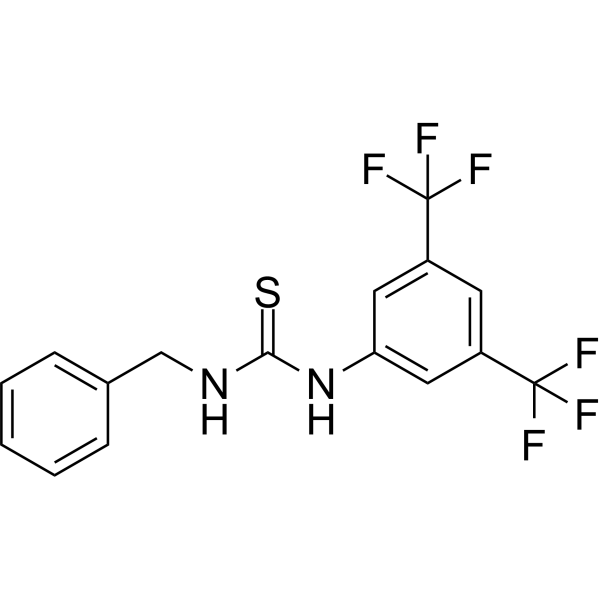
VPC-70063 is a potent Myc-Max inhibitor with an IC50 value of 8.9 μM for Myc-Max transcriptional activity inhibition. VPC-70063 reduces UBE2C promotor activity and AR-V7 levels, and induces PARP cleavage. VPC-70063 induces apoptosis and blocks Myc-Max interactions with DNA. VPC-70063 can be used for researching anticancer.
商品は「研究用試薬」です。人や動物の医療用・臨床診断用・食品用の製品ではありません。
研究用途以外に使用した場合、当社は一切の責任を負いかねます。
- 純度 : 99.92%
- CAS 番号: 13571-44-3
- 分子式: C16H12F6N2S
- 分子量:378.34
-
保管条件:Powder -20°C, 3 years , 4°C, 2 years ; In solvent -80°C, 6 months , -20°C, 1 month

To place orders, for customer services and technical support, please contact: MedChemExpress USA
Tel: 609-228-6898 E-mail: [email protected] [email protected]


-
 生物活性
生物活性
-
化学情報
-
溶剤 & 溶解度
- プロトコル
- 純度とドキュメンテーション
- 参考文献
-
 Help & FAQs


-
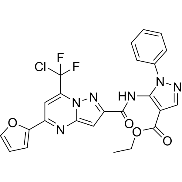
HY-100669
Mycro 3Myc-Max Inhibitor -
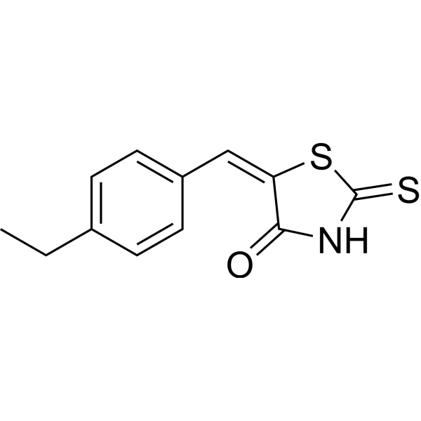
HY-12702
10058-F4c-Myc Inhibitor -
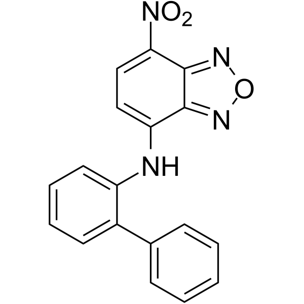
HY-100996
10074-G5c-Myc-Max Inhibitor -
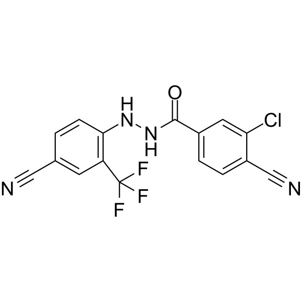
HY-144878
VPC-70619N-Myc Inhibitor -
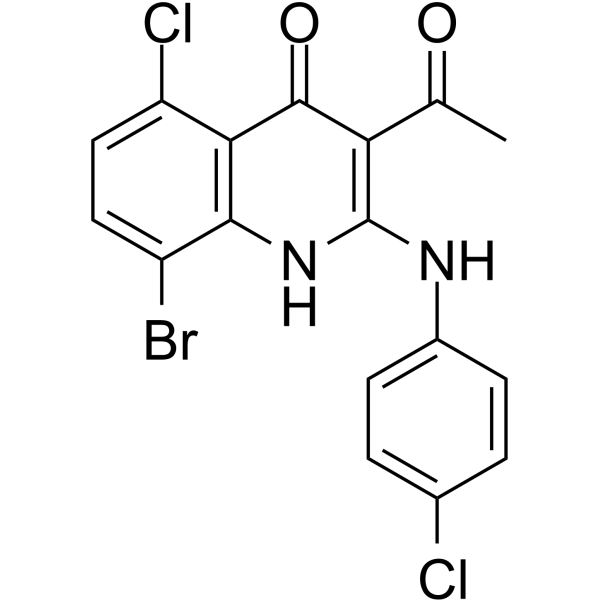
HY-12703
KSI-3716c-Myc Inhibitor -
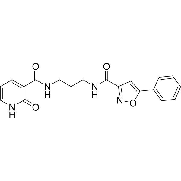
HY-103038
ML327MYC Blocker -
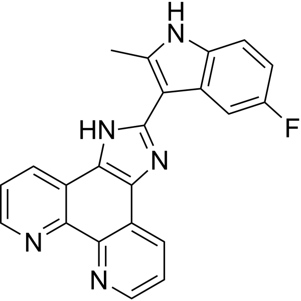
HY-16291
APTO-253 -

HY-103038R
ML327 (Standard)Reference Standard -

HY-100996R
10074-G5 (Standard)c-Myc-Max Inhibitor -
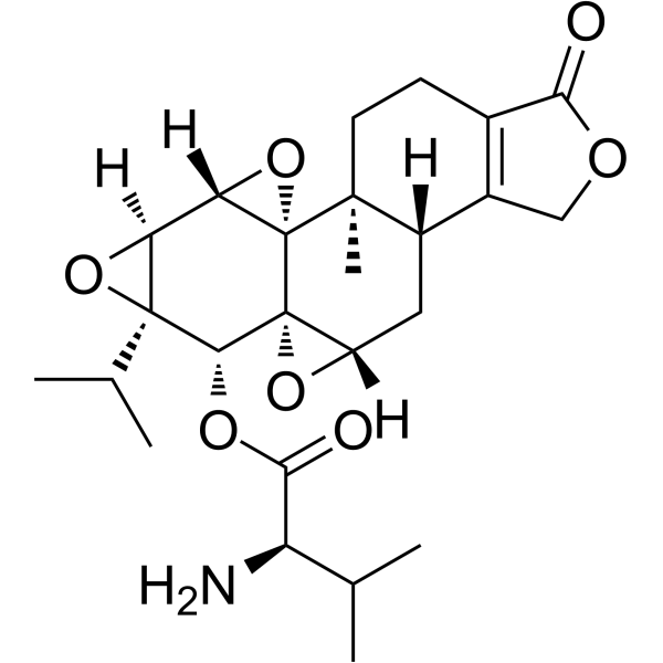
HY-145898
WBC100c-Myc Inhibitor -

HY-100669R
Mycro 3 (Standard)Myc-Max Inhibitor -
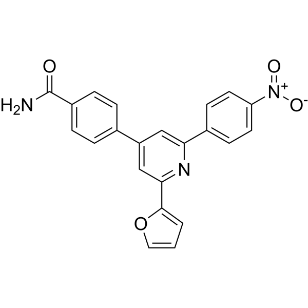
HY-19735
KJ Pyr 9MYC Inhibitor


生物活性
製品説明
IC50 & Target
IC50: 8.9 μM (Myc-Max)[1]
Cellular Effect
|
Cell Line

|
Type | Value | Description | References |
|---|---|---|---|---|
| LNCaP | IC50 |
2.5 μM
Compound: VPC-70063
|
Cytotoxicity against human LNCAP cells assessed as reduction in cell viability after 96 hrs by prestoblue assay
Cytotoxicity against human LNCAP cells assessed as reduction in cell viability after 96 hrs by prestoblue assay
|
[PMID: 30326371] |
| LNCaP | IC50 |
8.9 μM
Compound: VPC-70063
|
Inhibition of Myc-Max (unknown origin) expressed in human LNCAP cells assessed as reduction in transcriptional activity after 1 day by luciferase reporter gene assay
Inhibition of Myc-Max (unknown origin) expressed in human LNCAP cells assessed as reduction in transcriptional activity after 1 day by luciferase reporter gene assay
|
[PMID: 30326371] |
体外実験
VPC-70063 (25 μM; 96 h) shows Myc-Max transcriptional activity inhibition of 106% and Myc-Max/UBE2C downstream pathway inhibition of 94%[1].
VPC-70063 (6.25-25 μM, 48 h) causes apoptosis of LNCaP cells as indicated by cleavage of PARP[1].
VPC-70063 (0-500 μM; 0-600 s) disrupts the interaction of Myc-Max with DNA in a dose dependent manner[1].
MedChemExpress (MCE) has not independently confirmed the accuracy of these methods. They are for reference only. Further protocols information, click here.
-
Cell Line:LNCaP cells
-
Concentration:6.25 μM, 12.5 μM and 25 μM
-
Incubation Time:48 h
-
Result:Induced PARP cleavage.


化学情報
-
CAS 番号 13571-44-3
-
性状 Solid
-
分子量 378.34
-
分子式 C16H12F6N2S
-
Color White to off-white
-
SMILES
FC(F)(C1=CC(NC(NCC2=CC=CC=C2)=S)=CC(C(F)(F)F)=C1)F
-
輸送条件
Room temperature in continental US; may vary elsewhere.
-
保管条件
Powder -20°C 3 years 4°C 2 years In solvent -80°C 6 months -20°C 1 month


溶剤 & 溶解度
体外:
DMSO : 100 mg/mL (264.31 mM; Need ultrasonic; Hygroscopic DMSO has a significant impact on the solubility of product, please use newly opened DMSO)
Please refer to the solubility information to select the appropriate solvent. Once prepared, please aliquot and store the solution to prevent product inactivation from repeated freeze-thaw cycles.
Storage method and period of stock solution: -80°C, 6 months; -20°C, 1 month. When stored at -80°C, please use it within 6 months. When stored at -20°C, please use it within 1 month.
Please refer to the solubility information to select the appropriate solvent. Once prepared, please aliquot and store the solution to prevent product inactivation from repeated freeze-thaw cycles.
Storage method and period of stock solution: -80°C, 6 months; -20°C, 1 month. When stored at -80°C, please use it within 6 months. When stored at -20°C, please use it within 1 month.
濃度 (開始) × 体積 (開始) = 濃度 (終了) × 体積 (終了)

プロトコル
-
Dual Luciferin reporter gene assay
Luciferin reporter gene assay is a reporting system to detect the activity of Firefly Luciferase using luciferin as a substrate, which is often used in the research of miRNA target gene verification and promoter transcriptive activity regulation. Dual luciferase usually refers to Firefly luciferase and Renilla luciferase.
-
Apoptosis
Apoptosis, also called programmed cell death, is generally characterized by distinct morphological characteristics.
-
TUNEL staining for apoptotic DNA fragmentation
TUNEL staining detects DNA strand breaks by using terminal deoxynucleotidyl transferase to add labeled nucleotides to exposed 3′-OH DNA termini, generating either microscopic staining in fixed cells or tissue sections, or fluorescence/cytometric signal in cell suspensions. TUNEL positivity reflects DNA fragmentation but should not be interpreted alone as definitive apoptosis, because TUNEL can also label necrotic, autolytic, mechanically damaged, or DNA-repair-associated DNA breaks.
-
Cell Cytotoxicity Assay
Cytotoxicity assays are usually based on the assessment of cell membrane damage, which can also be indirectly detected by measuring cell viability. Detection methods include MTT assay, CKK-8 assay, LDH assay and ATP assay, etc.
-
Annexin V plus membrane-impermeant dye apoptosis staining
Annexin V-based apoptosis assays rely on the detection of phosphatidylserine (PS) externalization from the inner leaflet of the plasma membrane to the outer leaflet, an early biochemical hallmark of apoptosis. Fluorescently labeled Annexin V binds PS in a calcium-dependent manner, enabling identification of early apoptotic cells by flow cytometry or fluorescence microscopy. When combined with a membrane-impermeant DNA-binding dye (e. g. , propidium iodide), this approach allows discrimination between viable (Annexin V−/dye−), early apoptotic (Annexin V+/dye−), and late apoptotic or necrotic (Annexin V+/dye+) cell populations by assessing membrane integrity and PS exposure.
-
Apoptosis Solutions
Apoptosis is a regulated, generally non-lytic cell-death pathway that removes unwanted, damaged, infected, or abnormal cells through coordinated morphological changes, caspase activation, DNA fragmentation, and membrane remodeling. The intrinsic apoptosis pathway is controlled mainly by mitochondrial outer membrane permeabilization, BCL-2 family proteins, cytochrome c release, apoptosome formation, caspase-9 activation, and downstream executioner caspase-3/7 activation. The extrinsic apoptosis pathway is initiated by death receptors such as Fas, TNFR, and TRAIL receptors, which recruit adaptor proteins and activate caspase-8 before engaging executioner caspases or mitochondrial amplification through BID cleavage. Apoptosis is linked to many phenotypes, including cancer cell killing, tissue homeostasis, immune regulation, neurodegeneration, infection response, and treatment-induced cytotoxicity; unresolved questions include how apoptosis interacts with necroptosis, pyroptosis, ferroptos


純度とドキュメンテーション
-
データシート (271 KB)
-
SDS (252 KB)
- English - EN (252 KB)
- Français - FR (252 KB)
- Deutsch - DE (252 KB)
- Norwegian - NO (252 KB)
- Español - ES (252 KB)
- Swedish - SV (252 KB)
- Italian - IT (252 KB)
- Korean - KR (252 KB)
- Portuguese - PT (252 KB)
-
取扱説明書 (2659 KB)
参考文献


Complete Stock Solution Preparation Table
Please refer to the solubility information to select the appropriate solvent. Once prepared, please aliquot and store the solution to prevent product inactivation from repeated freeze-thaw cycles.
Storage method and period of stock solution: -80°C, 6 months; -20°C, 1 month. When stored at -80°C, please use it within 6 months. When stored at -20°C, please use it within 1 month.
| Optional Solvent | Concentration Solvent Mass | 1 mg | 5 mg | 10 mg | 25 mg |
|---|---|---|---|---|---|
| DMSO | 1 mM | 2.6431 mL | 13.2156 mL | 26.4313 mL | 66.0781 mL |
| 5 mM | 0.5286 mL | 2.6431 mL | 5.2863 mL | 13.2156 mL | |
| 10 mM | 0.2643 mL | 1.3216 mL | 2.6431 mL | 6.6078 mL | |
| 15 mM | 0.1762 mL | 0.8810 mL | 1.7621 mL | 4.4052 mL | |
| 20 mM | 0.1322 mL | 0.6608 mL | 1.3216 mL | 3.3039 mL | |
| 25 mM | 0.1057 mL | 0.5286 mL | 1.0573 mL | 2.6431 mL | |
| 30 mM | 0.0881 mL | 0.4405 mL | 0.8810 mL | 2.2026 mL | |
| 40 mM | 0.0661 mL | 0.3304 mL | 0.6608 mL | 1.6520 mL | |
| 50 mM | 0.0529 mL | 0.2643 mL | 0.5286 mL | 1.3216 mL | |
| 60 mM | 0.0441 mL | 0.2203 mL | 0.4405 mL | 1.1013 mL | |
| 80 mM | 0.0330 mL | 0.1652 mL | 0.3304 mL | 0.8260 mL | |
| 100 mM | 0.0264 mL | 0.1322 mL | 0.2643 mL | 0.6608 mL |


VPC-70063 関連分類
HY-147291 関連分類
 Powered by Bioz