



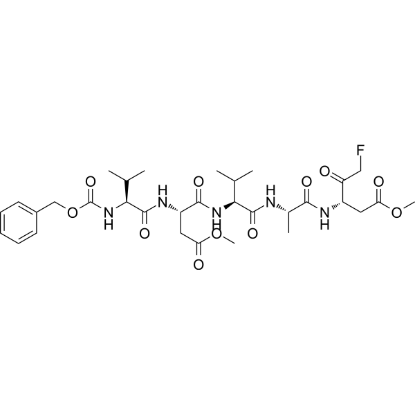
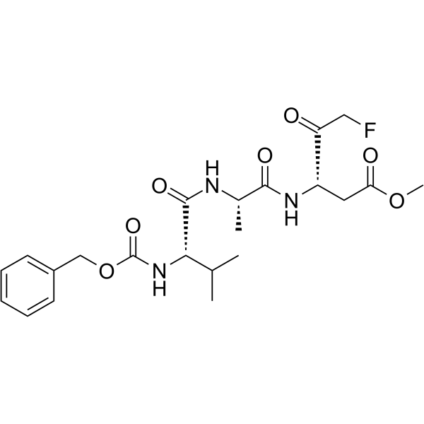
Z-VDVAD-FMK
Based on 5 publication(s) in Google Scholar
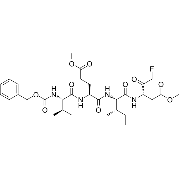
Z-VDVAD-FMK is a special inhibitor of caspase-2. Z-VDVAD-FMK produces a reduction in Lovastatin-induced apoptosis.
For research use only. We do not sell to patients.
- Assay : 98.90%
- CAS No.: 210344-92-6
- Formula: C32H46FN5O11
- Molecular Weight:695.73
-
Storage:
Sealed storage, away from moisture.
Powder -80°C, 2 years , -20°C, 1 year* In solvent : -80°C, 6 months; -20°C, 1 month (sealed storage, away from moisture)

To place orders, for customer services and technical support, please contact: MedChemExpress USA
Tel: 609-228-6898 E-mail: [email protected] [email protected]


-
 Biological Activity
-
Chemical Information
-
Solvent & Solubility
- Protocols
- Purity & Documentation
- References
-
 Help & FAQs


-
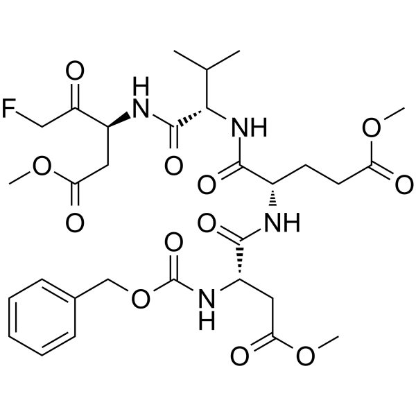
HY-12466
Z-DEVD-FMKCaspase-3 Inhibitor -
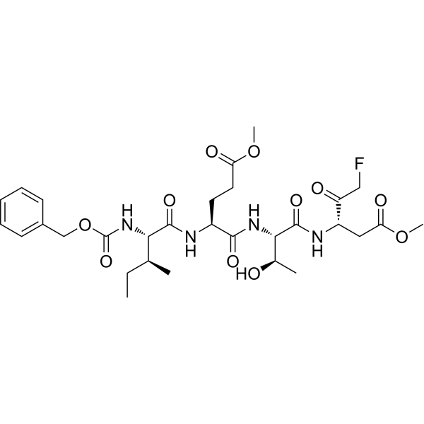
HY-101297
Z-IETD-FMKCaspase-8 Inhibitor -
HY-16658B
Z-VAD-FMKPan-Caspase Inhibitor -
HY-P1007
Z-VEID-FMKcaspase-6 Inhibitor -
HY-13205
BelnacasanCaspase-1 Inhibitor -
HY-16990
Ac-YVAD-cmkCaspase-1 Inhibitor -
HY-10396
Emricasanpan-Caspase Inhibitor -
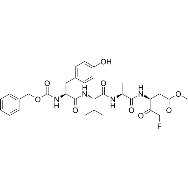
HY-P1009
Z-YVAD-FMKCaspase 1/4 Inhibitor -
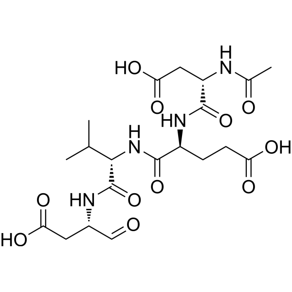
HY-P1001
Ac-DEVD-CHOCaspase-3 Inhibitor -
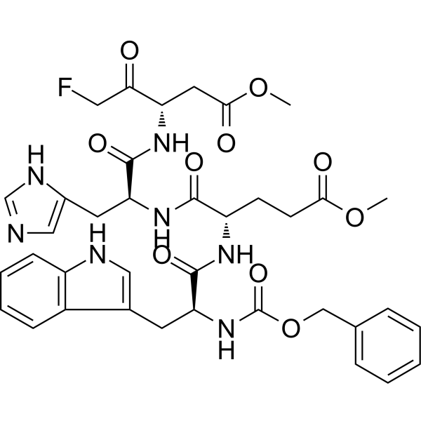
HY-P0111
Z-WEHD-FMKCaspase-1/5 Inhibitor -
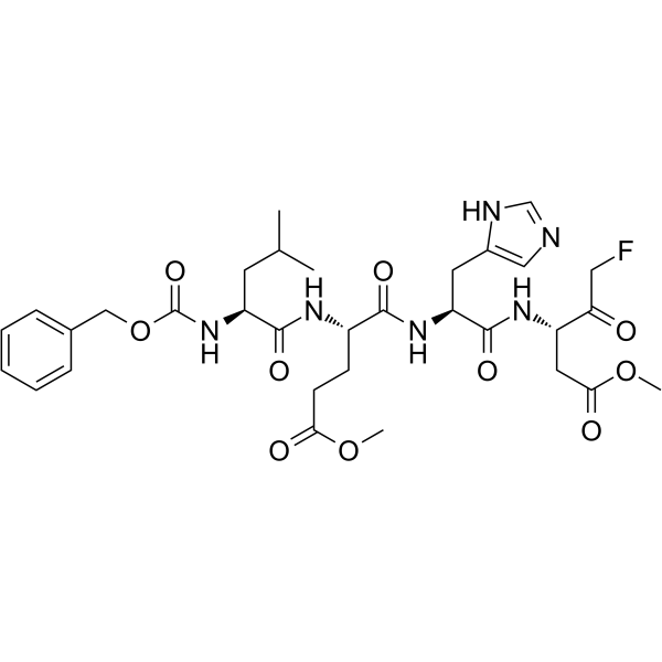
HY-P1010
Z-LEHD-FMKCaspase-9 Inhibitor -
HY-16658
Z-VAD(OMe)-FMKPan-caspase Inhibitor
Publications Citing Use of MedChemExpress (MCE) Z-VDVAD-FMK
More Customer Validation & Images

-
Cell Imaging/Staining

-
Bio/Physico-chemical Assay

-
Gel Electrophoresis

-
Gel Electrophoresis

All Caspase Isoforms
More

Biological Activity
Description
IC50 & Target
[1]|
Caspase-2 |
In Vitro
Cotreatment of cells with the caspase inhibitors Ac-DEVD-CHO, Z-VDVAD-FMK (100 μM), Z-IETD-fmk, and Z-LEHD-fmk alone or in combination, or overexpression of CrmA, prevents many morphological features of apoptosis but not loss of mitochondrial membrane potential (DCm), phospatidilserine exposure, and cell death[1].
Z-VDVAD-FMK (2 μM) greatly inhibits the Rho-kinase activity in HMEC-1 cells stimulated by Thrombin and displays no effect on control cells[2].
Z-VDVAD-FMK (zVDVAD-fmk) produces a reduction in Lovastatin-induced apoptosis. Z-VDVAD-FMK (100 μM) significantly reduces Lovastatin-induced loss of DNA by 19.1±8.3%[3].
MedChemExpress (MCE) has not independently confirmed the accuracy of these methods. They are for reference only. Further protocols information, click here.
-
Cell Line:The human T-cell leukemia Jurkat (Clone E6.1, ATCC TIB-152)
-
Concentration:100 μM
-
Incubation Time:22 hours
-
Result:Prevented Doxorubicin (1 μM)-induced nuclear apoptosis, but not cell death.


Chemical Information
-
CAS No. 210344-92-6
-
Appearance Solid
-
Molecular Weight 695.73
-
Formula C32H46FN5O11
-
Color White to off-white
-
Synonyms
Z-VD(OMe)VAD(OMe)-FMK
-
Sequence Shortening
{Cbz}-V{Asp(OMe)}-VA-{Asp(OMe)}-CH2F
-
Shipping
Room temperature in continental US; may vary elsewhere.
-
Storage
Sealed storage, away from moisture
Powder -80°C 2 years -20°C 1 year * In solvent : -80°C, 6 months; -20°C, 1 month (sealed storage, away from moisture)


Publications (5)
-
Journal Impact Factor
-
Most Recent
-
Signal Transduct Target Ther
Selective depletion of tumor-associated SAMHD1 enhances chemotherapeutic efficacy and antitumor immune responses. [Abstract]2025 Dec 15;10(1):406. PMID: 41392286 -
Cell
Neutrophil-derived vesicles control complement activation to facilitate inflammation resolution. [Abstract]2025 Mar 20;188(6):1623-1641.e26. PMID: 39938514
Z-VDVAD-FMK purchased from MedChemExpress. Usage Cited in: Cell. 2025 Mar 20;188(6):1623-1641.e26. [Abstract]
Percentage of live (annexin V−/PI−) neutrophils (from 4 × 10⁶ cells) from either untreated, treated with caspase-3 inhibitor Z-DEVD-FMK (10 μM), or caspase-3 inducer raptinal.
Z-VDVAD-FMK purchased from MedChemExpress. Usage Cited in: Cell. 2025 Mar 20;188(6):1623-1641.e26. [Abstract]
LAND-V production from neutrophils either untreated, treated with caspase-3 inhibitor Z-DEVD-FMK (10 μM), or caspase-3 inducer raptinal.
-
Int J Biol Macromol
Z-VAD(OMe)-FMK suppresses Seneca Valley Virus replication by targeting the active sites of the 3C protease. [Abstract]2025 May;306(Pt 3):141587. PMID: 40032110
Z-VDVAD-FMK purchased from MedChemExpress. Usage Cited in: Int J Biol Macromol. 2025 May;306(Pt 3):141587. [Abstract]
Inhibitory effect of inhibitors on SVV 3C-mediated cleavage of pro-IL-β in vitro. Z-VAD, VX-76, Z-VDVAD-FMK, Ac-DEVD-CHO, and Z-FA (0.25 mM) were incubated with SVV 3C recombinant protein (2.5 μg) in a reaction buffer at room temperature for 5 min, followed by the addition of pro-IL-β recombinant protein (15 μg). The mixture was then incubated at 37 ℃ for 2 h and the cleavage of pro-IL-β was analyzed using SDS-PAGE.
Z-VDVAD-FMK purchased from MedChemExpress. Usage Cited in: Int J Biol Macromol. 2025 May;306(Pt 3):141587. [Abstract]
Various concentrations of Z-VAD, Z-VDVAD-FMK (1, 2, 3, 4, 6, 8, 10, 20 and 30 μM) were incubated with SVV 3C in reaction buffer at room for a duration of 5 min. Subsequently pcGAS recombinant protein was added followed by incubation at a temperature of 37 ℃ for a period of 2 h. Samples were then subjected to SDS-PAGE analysis to assess pcGAS cleavage. The gray value of uncleaved pcGAS was quantified using ImageJ software to determine the IC50 values for different inhibitors against the activity of the SVV 3C protease.
-
Toxicology
Proteasome inhibition induces apoptosis through simultaneous inactivation of MCL-1/BCL-XL by NOXA independent of CHOP and JNK pathways. [Abstract]2024 Nov:508:153906. PMID: 39117261 -
Free Radic Res
Dioscin induces ferroptosis to suppress the metastasis of gastric cancer through the SLC7A11/GPX4 axis. [Abstract]2025 May;59(5):426-441. PMID: 40460255


Solvent & Solubility
In Vitro:
DMSO : 100 mg/mL (143.73 mM; Need ultrasonic; Hygroscopic DMSO has a significant impact on the solubility of product, please use newly opened DMSO)
Please refer to the solubility information to select the appropriate solvent. Once prepared, please aliquot and store the solution to prevent product inactivation from repeated freeze-thaw cycles.
Storage method and period of stock solution: -80°C, 6 months; -20°C, 1 month (sealed storage, away from moisture). When stored at -80°C, please use it within 6 months. When stored at -20°C, please use it within 1 month.
Please refer to the solubility information to select the appropriate solvent. Once prepared, please aliquot and store the solution to prevent product inactivation from repeated freeze-thaw cycles.
Storage method and period of stock solution: -80°C, 6 months; -20°C, 1 month (sealed storage, away from moisture). When stored at -80°C, please use it within 6 months. When stored at -20°C, please use it within 1 month.
Concentration (start) × Volume (start) = Concentration (final) × Volume (final)

Protocols
-
Apoptosis
Apoptosis, also called programmed cell death, is generally characterized by distinct morphological characteristics.
-
TUNEL staining for apoptotic DNA fragmentation
TUNEL staining detects DNA strand breaks by using terminal deoxynucleotidyl transferase to add labeled nucleotides to exposed 3′-OH DNA termini, generating either microscopic staining in fixed cells or tissue sections, or fluorescence/cytometric signal in cell suspensions. TUNEL positivity reflects DNA fragmentation but should not be interpreted alone as definitive apoptosis, because TUNEL can also label necrotic, autolytic, mechanically damaged, or DNA-repair-associated DNA breaks.
-
Annexin V plus membrane-impermeant dye apoptosis staining
Annexin V-based apoptosis assays rely on the detection of phosphatidylserine (PS) externalization from the inner leaflet of the plasma membrane to the outer leaflet, an early biochemical hallmark of apoptosis. Fluorescently labeled Annexin V binds PS in a calcium-dependent manner, enabling identification of early apoptotic cells by flow cytometry or fluorescence microscopy. When combined with a membrane-impermeant DNA-binding dye (e. g. , propidium iodide), this approach allows discrimination between viable (Annexin V−/dye−), early apoptotic (Annexin V+/dye−), and late apoptotic or necrotic (Annexin V+/dye+) cell populations by assessing membrane integrity and PS exposure.
-
Apoptosis Solutions
Apoptosis is a regulated, generally non-lytic cell-death pathway that removes unwanted, damaged, infected, or abnormal cells through coordinated morphological changes, caspase activation, DNA fragmentation, and membrane remodeling. The intrinsic apoptosis pathway is controlled mainly by mitochondrial outer membrane permeabilization, BCL-2 family proteins, cytochrome c release, apoptosome formation, caspase-9 activation, and downstream executioner caspase-3/7 activation. The extrinsic apoptosis pathway is initiated by death receptors such as Fas, TNFR, and TRAIL receptors, which recruit adaptor proteins and activate caspase-8 before engaging executioner caspases or mitochondrial amplification through BID cleavage. Apoptosis is linked to many phenotypes, including cancer cell killing, tissue homeostasis, immune regulation, neurodegeneration, infection response, and treatment-induced cytotoxicity; unresolved questions include how apoptosis interacts with necroptosis, pyroptosis, ferroptos


Purity & Documentation
-
Data Sheet (274 KB)
-
SDS (252 KB)
- English - EN (252 KB)
- Français - FR (252 KB)
- Deutsch - DE (252 KB)
- Norwegian - NO (252 KB)
- Español - ES (252 KB)
- Swedish - SV (252 KB)
- Italian - IT (252 KB)
- Korean - KR (252 KB)
- Portuguese - PT (252 KB)
-
Handling Instructions (2659 KB)
References
[1]. S Gamen, et al. Doxorubicin treatment activates a Z-VAD-sensitive caspase, which causes deltapsim loss, caspase-9 activity, and apoptosis in Jurkat cells. Exp Cell Res. 2000 Jul 10;258(1):223-35. [Content Brief]
[2]. Cédric Sapet, et al. Thrombin-induced endothelial microparticle generation: identification of a novel pathway involving ROCK-II activation by caspase-2. Blood. 2006 Sep 15;108(6):1868-76. [Content Brief]
[3]. Simon W Rabkin, et al. Lovastatin-induced cardiac toxicity involves both oncotic and apoptotic cell death with the apoptotic component blunted by both caspase-2 and caspase-3 inhibitors. Toxicol Appl Pharmacol. 2003 Dec 15;193(3):346-55. [Content Brief]


Complete Stock Solution Preparation Table
Please refer to the solubility information to select the appropriate solvent. Once prepared, please aliquot and store the solution to prevent product inactivation from repeated freeze-thaw cycles.
Storage method and period of stock solution: -80°C, 6 months; -20°C, 1 month (sealed storage, away from moisture). When stored at -80°C, please use it within 6 months. When stored at -20°C, please use it within 1 month.
| Optional Solvent | Concentration Solvent Mass | 1 mg | 5 mg | 10 mg | 25 mg |
|---|---|---|---|---|---|
| DMSO | 1 mM | 1.4373 mL | 7.1867 mL | 14.3734 mL | 35.9335 mL |
| 5 mM | 0.2875 mL | 1.4373 mL | 2.8747 mL | 7.1867 mL | |
| 10 mM | 0.1437 mL | 0.7187 mL | 1.4373 mL | 3.5933 mL | |
| 15 mM | 0.0958 mL | 0.4791 mL | 0.9582 mL | 2.3956 mL | |
| 20 mM | 0.0719 mL | 0.3593 mL | 0.7187 mL | 1.7967 mL | |
| 25 mM | 0.0575 mL | 0.2875 mL | 0.5749 mL | 1.4373 mL | |
| 30 mM | 0.0479 mL | 0.2396 mL | 0.4791 mL | 1.1978 mL | |
| 40 mM | 0.0359 mL | 0.1797 mL | 0.3593 mL | 0.8983 mL | |
| 50 mM | 0.0287 mL | 0.1437 mL | 0.2875 mL | 0.7187 mL | |
| 60 mM | 0.0240 mL | 0.1198 mL | 0.2396 mL | 0.5989 mL | |
| 80 mM | 0.0180 mL | 0.0898 mL | 0.1797 mL | 0.4492 mL | |
| 100 mM | 0.0144 mL | 0.0719 mL | 0.1437 mL | 0.3593 mL |


Z-VDVAD-FMK Related Classifications
HY-P1008 Related Classifications
 Powered by Bioz