



6-Iodonordihydrocapsaicin
6-Iodonordihydrocapsaicin is a TRPV1 antagonist. 6-Iodonordihydrocapsaicin functionally blocks TRPV1-mediated responses, including capsaicin-induced ion currents in dorsal root ganglion neurons and distension-induced firing of jejunal spinal afferent fibers in mice. 6-Iodonordihydrocapsaicin can be used in the research of visceral pain and anxiety disorders.
For research use only. We do not sell to patients.
- CAS No.: 859171-97-4
- Formula: C17H26INO3
- Molecular Weight:419.30
-
Storage:
Please store the product under the recommended conditions in the Certificate of Analysis.


To place orders, for customer services and technical support, please contact: MedChemExpress USA
Tel: 609-228-6898 E-mail: [email protected] [email protected]


-
 Biological Activity
-
Chemical Information
- Protocols
- Purity & Documentation
- References
-
 Help & FAQs


-
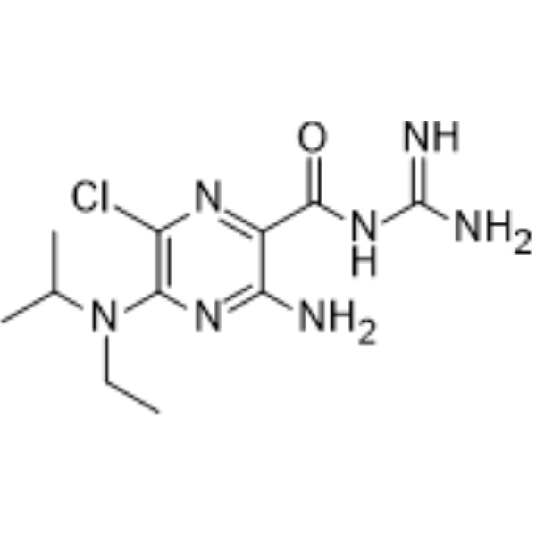
HY-101840
EIPATRPP3 Channel Inhibitor -
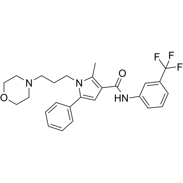
HY-100208
HC-067047TRPV4 Antagonist -
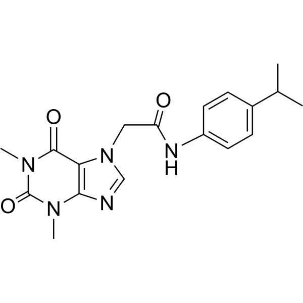
HY-15064
HC-030031TRP Channel Inhibitor -
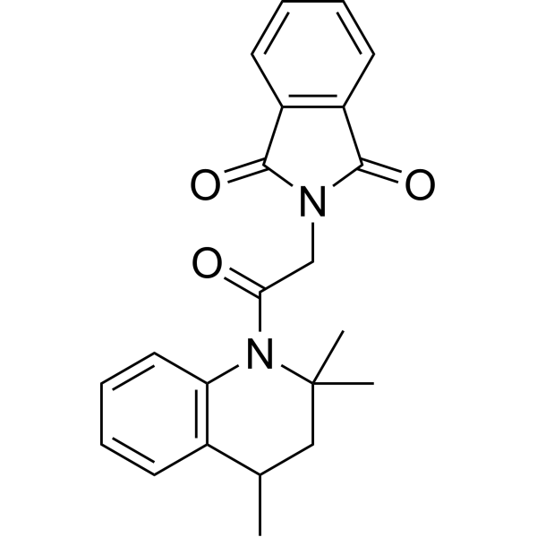
HY-108462
ML-SA1TRPML Agonist -
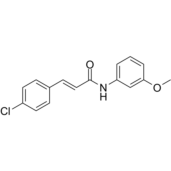
HY-12245
SB-366791VR1/TRPV1 Antagonist -
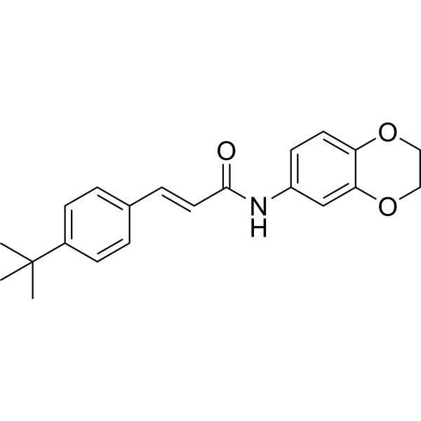
HY-101736
AMG9810TRPV1 Antagonist -
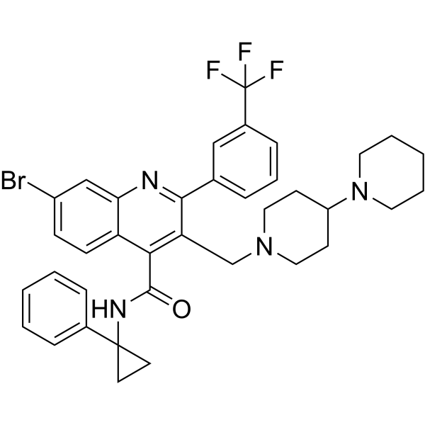
HY-100720
GSK2193874TRPV4 Antagonist -
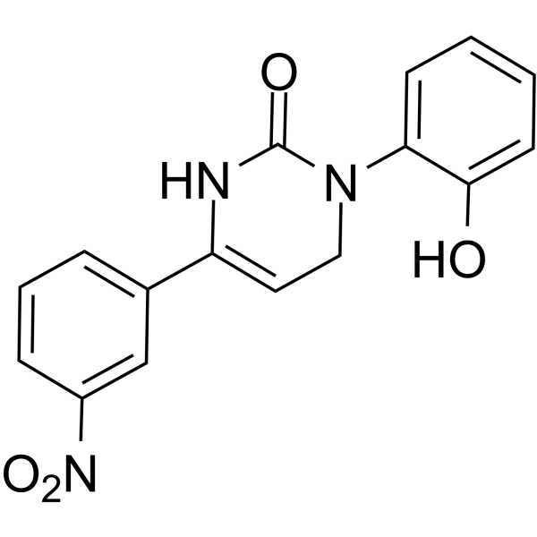
HY-11062
IcilinTRPM8 Agonist -
HY-108449
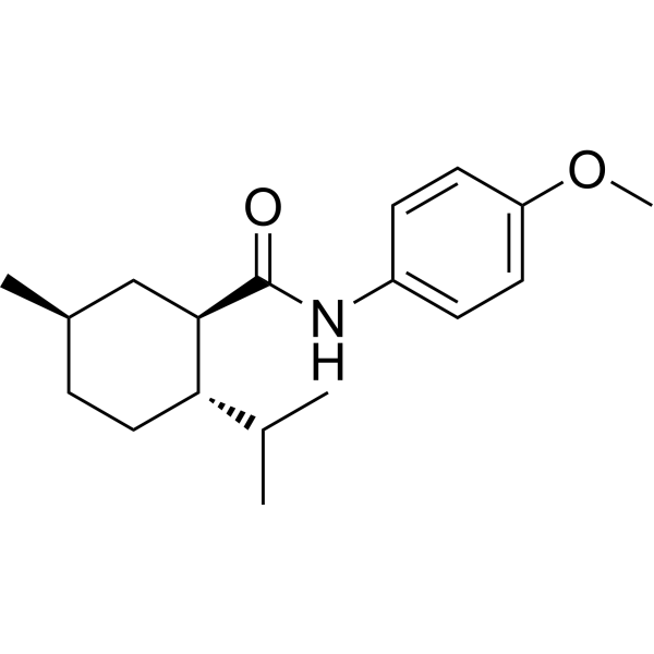
AcoltremonTRPM8 agonist -
HY-108457
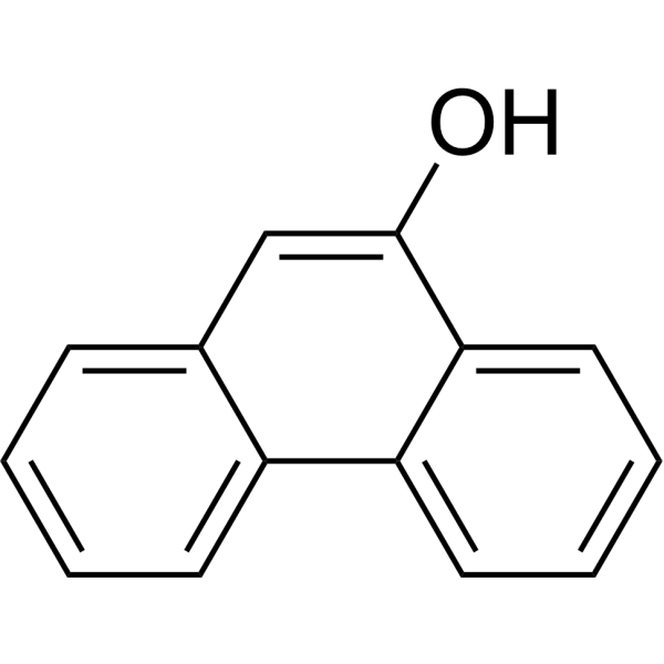
9-PhenanthrolTRPM4 Inhibitor -
HY-108463
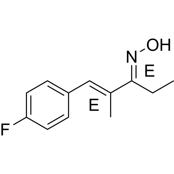
A-967079TRPA1 Receptor Antagonist -
HY-19960
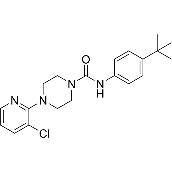
BCTCTRPV1/TRPM8 Antagonist


Biological Activity
Description
IC50 & Target
[1]|
TRPV1 |
Cellular Effect
|
Cell Line

|
Type | Value | Description | References |
|---|---|---|---|---|
| HEK293 | IC50 |
10 nM
Compound: 4b
|
Inhibition of 100 nM capsaicin effect on intracellular [Ca2+] concentration in HEK293 cells expressing human TRPV1
Inhibition of 100 nM capsaicin effect on intracellular [Ca2+] concentration in HEK293 cells expressing human TRPV1
|
[PMID: 16000002] |
In Vitro
6-Iodonordihydrocapsaicin (10 μM) inhibits capsaicin-evoked ionic currents in murine DRG primary cultures[1].
MedChemExpress (MCE) has not independently confirmed the accuracy of these methods. They are for reference only. Further protocols information, click here.
In Vivo
MedChemExpress (MCE) has not independently confirmed the accuracy of these methods. They are for reference only.
-
Animal Model:Wistar rats (male, 230-250 g)[2]
-
Dosage:1 nmol
-
Administration:intracerebroventricular (i.c.v.); single administration
-
Result:Abolished the anxiogenic-like effect of intracerebroventricular hemopressin, as evidenced by reversal of hemopressin-induced decreases in percentage of open arm entries and percentage of time spent in open arms (restored to levels comparable to vehicle-treated controls).
Caused no significant changes in enclosed arm entry counts.


Chemical Information
-
CAS No. 859171-97-4
-
Molecular Weight 419.30
-
Formula C17H26INO3
-
SMILES
CCCCCCCCC(NCC1=CC(OC)=C(O)C=C1I)=O
-
Shipping
Room temperature in continental US; may vary elsewhere.
-
Storage
Please store the product under the recommended conditions in the Certificate of Analysis.

Protocols
-
Primary Dorsal Root Ganglion Sensory Neuron Culture
Primary dorsal root ganglion sensory neuron culture isolates DRG neuronal somata from rodent or human ganglia, dissociates tissue enzymatically and mechanically, and maintains post-mitotic sensory neurons in vitro for readouts such as neurite outgrowth, immunocytochemical marker expression, calcium imaging, electrophysiology, RNA/protein analysis, or neuropeptide release assays. The method reflects peripheral sensory neuron biology because DRG neurons are primary sensory neurons whose cell bodies reside in dorsal root ganglia and whose cultured dissociated cells can retain neuronal morphology, sensory-neuron marker expression, and stimulus-responsive properties depending on the downstream assay.


Purity & Documentation
References
[1]. Perez-Burgos A, et al. The TRPV1 channel in rodents is a major target for antinociceptive effect of the probiotic Lactobacillus reuteri DSM 17938. J Physiol. 2015;593(17):3943-3957. [Content Brief]
[2]. Fogaça MV, et al. Anxiogenic-like effects induced by hemopressin in rats. Pharmacol Biochem Behav. 2015;129:7-13. [Content Brief]

Calculators
Concentration (start) × Volume (start) = Concentration (final) × Volume (final)


6-Iodonordihydrocapsaicin Related Classifications
HY-108459 Related Classifications
 Powered by Bioz