



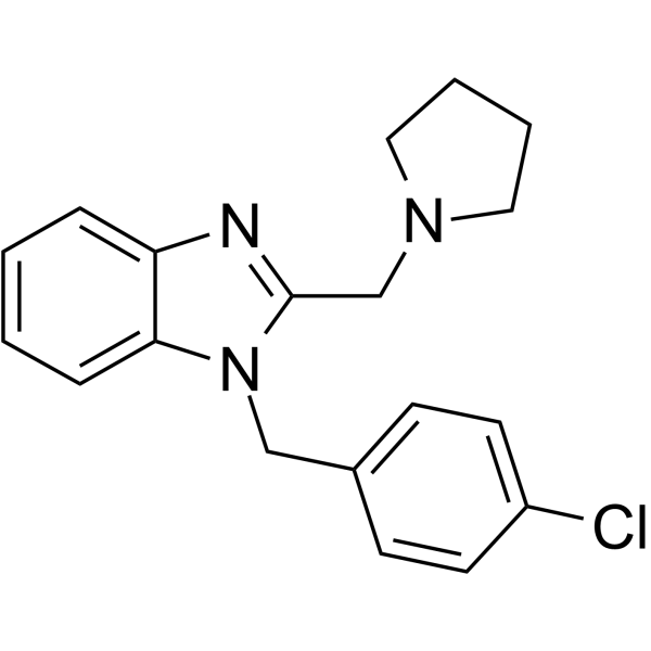
Clemizole
Based on 1 publication(s) in Google Scholar
Clemizole is an orally active, blood-brain barrier permeable TRPC5 inhibitor, with an IC50 value of 1.05-1.34 μM against mouse TRPC5. Clemizole blocks TRPC1:TRPC5, TRPC3, TRPC4, TRPC6, TRPC7, hERG, hKCNQ1/hKCNE1 and hKv1.5 channels, and activates TRPA1; it modulates 5HT-2B and HTR2A receptors; it inhibits HCV RNA replication, CrtN enzymatic activity, oxidative stress, neuroinflammation, cell apoptosis and bacterial virulence; it maintains blood-brain barrier (BBB) integrity; it enhances DNA repair capacity; it improves cell viability; and it alters cardiac electrophysiological properties. Clemizole can be used in the research of Dravet syndrome, hepatitis C virus infection, Staphylococcus aureus skin infection, Cisplatin (HY-17394)-induced nephrotoxicity, STXBP1-related diseases, traumatic brain injury and xeroderma pigmentosum type C.
For research use only. We do not sell to patients.
- Purity : 98.42%
- CAS No.: 442-52-4
- Formula: C19H20ClN3
- Molecular Weight:325.84
-
Storage:Powder -20°C, 3 years , 4°C, 2 years ; In solvent -80°C, 6 months , -20°C, 1 month

To place orders, for customer services and technical support, please contact: MedChemExpress USA
Tel: 609-228-6898 E-mail: [email protected] [email protected]


-
 Biological Activity
-
Chemical Information
-
Solvent & Solubility
- Protocols
- Purity & Documentation
- References
-
 Help & FAQs


-
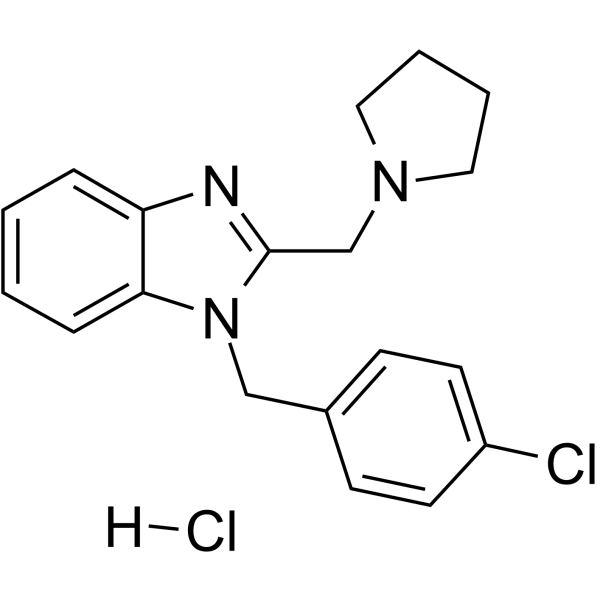
HY-30234A
Clemizole hydrochlorideTRPC5 Inhibitor -
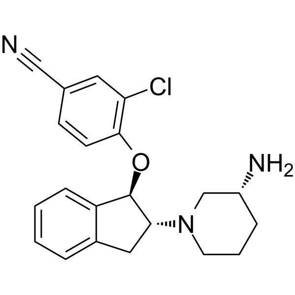
HY-15699
SAR7334TRPC6 Inhibitor -
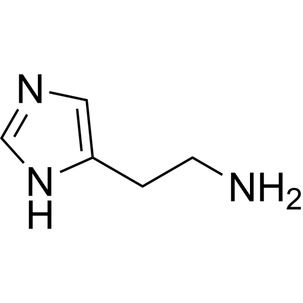
HY-B1204
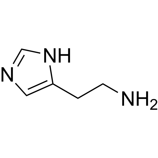
HistamineNeurotransmitter -
HY-N0054
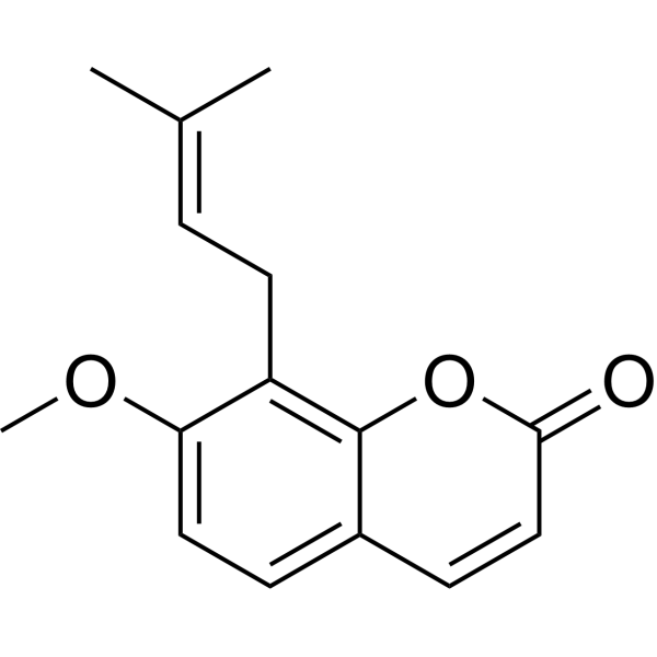
OstholeApoptosis Inducer -
HY-N0172
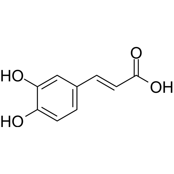
Caffeic acidEndogenous Metabolite -
HY-12532
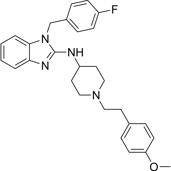
AstemizoleH1 Receptor Antagonist -
HY-B1204R
Histamine (Standard)Neurotransmitter -
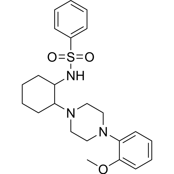
HY-139426
ML-SI3TRPML Modulator -

HY-N0172R
Caffeic acid (Standard)Endogenous Metabolite -
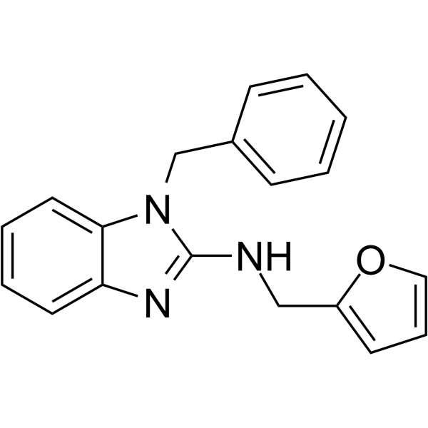
HY-122051
AC1903TRPC5 Inhibitor -
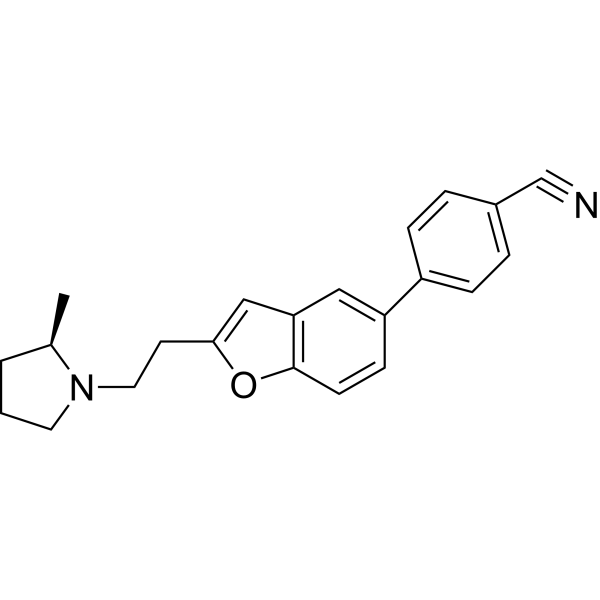
HY-12195
ABT-239 -

HY-N0054R
Osthole (Standard)Apoptosis Inducer
Publications Citing Use of MedChemExpress (MCE) Clemizole
More

Biological Activity
Description
IC50 & Target
|
TRPC5 1.05-1.34 μM (IC50) |
Cellular Effect
|
Cell Line

|
Type | Value | Description | References |
|---|---|---|---|---|
| HEK293 | IC50 |
>20 μM
Compound: 18
|
Inhibition of CFP-tagged rat TRPV1 expressed in HEK293 cells assessed as reduction in calcium level by fluo-4 dye based fluorescence assay
Inhibition of CFP-tagged rat TRPV1 expressed in HEK293 cells assessed as reduction in calcium level by fluo-4 dye based fluorescence assay
|
[PMID: 30943030] |
| HEK293 | IC50 |
>20 μM
Compound: 18
|
Inhibition of YFP-tagged mouse TRPV4 expressed in HEK293 cells assessed as reduction in calcium level by fluo-4 dye based fluorescence assay
Inhibition of YFP-tagged mouse TRPV4 expressed in HEK293 cells assessed as reduction in calcium level by fluo-4 dye based fluorescence assay
|
[PMID: 30943030] |
| HEK293 | IC50 |
>20 μM
Compound: 18
|
Inhibition of YFP-tagged rat TRPV2 expressed in HEK293 cells assessed as reduction in calcium level by fluo-4 dye based fluorescence assay
Inhibition of YFP-tagged rat TRPV2 expressed in HEK293 cells assessed as reduction in calcium level by fluo-4 dye based fluorescence assay
|
[PMID: 30943030] |
| HEK293 | IC50 |
>20 μM
Compound: 18
|
Inhibition of YFP-tagged rat TRPV3 expressed in HEK293 cells assessed as reduction in calcium level by fluo-4 dye based fluorescence assay
Inhibition of YFP-tagged rat TRPV3 expressed in HEK293 cells assessed as reduction in calcium level by fluo-4 dye based fluorescence assay
|
[PMID: 30943030] |
| HEK293 | IC50 |
11.3 μM
Compound: 18
|
Inhibition of YFP-tagged human TRPC6 expressed in thapsigargin treated HEK293 cells assessed as reduction in amix-induced calcium level by fluo-4 dye based fluorescence assay
Inhibition of YFP-tagged human TRPC6 expressed in thapsigargin treated HEK293 cells assessed as reduction in amix-induced calcium level by fluo-4 dye based fluorescence assay
|
[PMID: 30943030] |
| HEK293 | IC50 |
26.5 μM
Compound: 18
|
Inhibition of YFP-tagged mouse TRPC7 expressed in thapsigargin treated HEK293 cells assessed as reduction in amix-induced calcium level by fluo-4 dye based fluorescence assay
Inhibition of YFP-tagged mouse TRPC7 expressed in thapsigargin treated HEK293 cells assessed as reduction in amix-induced calcium level by fluo-4 dye based fluorescence assay
|
[PMID: 30943030] |
| HEK293 | IC50 |
6.4 μM
Compound: 18
|
Inhibition of YFP-tagged mouse TRPC4 beta expressed in thapsigargin treated HEK293 cells co-expressing M3 receptor assessed as reduction in carbachol-induced calcium level by fluo-4 dye based fluorescence assay
Inhibition of YFP-tagged mouse TRPC4 beta expressed in thapsigargin treated HEK293 cells co-expressing M3 receptor assessed as reduction in carbachol-induced calcium level by fluo-4 dye based fluorescence assay
|
[PMID: 30943030] |
| HEK293 | IC50 |
9.1 μM
Compound: 18
|
Inhibition of YFP-tagged human TRPC3 expressed in thapsigargin treated HEK293 cells assessed as reduction in amix-induced calcium level by fluo-4 dye based fluorescence assay
Inhibition of YFP-tagged human TRPC3 expressed in thapsigargin treated HEK293 cells assessed as reduction in amix-induced calcium level by fluo-4 dye based fluorescence assay
|
[PMID: 30943030] |
| Huh-7 | CC50 |
39.4 μM
Compound: Clemizole
|
Cytotoxicity against human Huh7.5 cells assessed as cell viability after 72 hrs by MTT assay
Cytotoxicity against human Huh7.5 cells assessed as cell viability after 72 hrs by MTT assay
|
[PMID: 24561671] |
| Huh-7 | EC50 |
8 μM
Compound: Clemizole
|
Antiviral activity against Hepatitis C virus JFH-1 J6 genotype 2a infected in human Huh7.5 cells assessed as inhibition of viral RNA replication after 72 hrs by luciferase reporter gene assay
Antiviral activity against Hepatitis C virus JFH-1 J6 genotype 2a infected in human Huh7.5 cells assessed as inhibition of viral RNA replication after 72 hrs by luciferase reporter gene assay
|
[PMID: 24561671] |
In Vitro
Clemizole exhibits strong, synergistic anti-HCV activity in Huh 7.5 cells when combined with Boceprevir (HY-10237), with a synergy volume of 230 μM2%, and does not induce cellular toxicity at tested concentrations[9].
Clemizole (as Clemizole Hydrochloride) (1-10 μM; 24 h) significantly increases the viability of XP-C patient-derived fibroblasts exposed to UVB irradiation, with a dose-dependent protective effect that enhances photo-resistance compared to DMSO-treated cells[11].
Clemizole (as Clemizole Hydrochloride) (5 μM; 24 h) significantly promotes repair of UVB-induced 6-4PP DNA lesions in XP-C patient-derived fibroblasts[11].
Clemizole (as Clemizole Hydrochloride) (0.1-10 μM; 24 h) mediates enhanced UVB photo-resistance in XP-C patient-derived fibroblasts through both pre- and post-UVB irradiation treatment, with the combined regimen yielding the highest bioactivity[11].
Clemizole (as Clemizole Hydrochloride) (10 μM; 2-4 h) does not affect DNA replication or cell proliferation in XP-C patient-derived fibroblasts exposed to UVB irradiation[11].
Clemizole (as Clemizole Hydrochloride) (10 μM; 24 h) does not reduce apoptosis or necrosis to mediate its protective effect in XP-C patient-derived fibroblasts exposed to UVB irradiation[11].
MedChemExpress (MCE) has not independently confirmed the accuracy of these methods. They are for reference only. Further protocols information, click here.
-
Cell Line:Xeroderma Pigmentosum C (XP-C) patient-derived immortalized fibroblasts (GM15983)
-
Concentration:10 μM (24 h pre-irradiation treatment); 10 μM (24 h post-irradiation treatment); 1-10 μM (both pre- and post-irradiation treatments)
-
Incubation Time:24 h (pre-irradiation treatment); 24 h (post-irradiation treatment)
-
Result:Showed ≥25% photo-resistance and a robust Z score above 2.6 in primary screen.
Induced a significant 20 to 40% increase in XP-C cell viability depending on UVB dose and reagent concentration, with increased bioactivity at higher concentrations.
Significantly enhanced photo-resistance in XP-C cells across increasing UVB doses, with viability remaining higher than DMSO-treated controls at all tested UVB doses.
-
Cell Line:Xeroderma Pigmentosum C (XP-C) patient-derived immortalized fibroblasts (GM15983)
-
Concentration:0.1-10 μM (pre-irradiation only; post-irradiation only; combined pre- and post-irradiation treatments)
-
Incubation Time:24 h post-UV
-
Result:Reached ~20% increased cell viability at 10 μM with pre-irradiation treatment alone.
Reached an average of ~50% increased viability at 10 μM with post-irradiation treatment alone.
Showed the highest bioactivity with combined pre- and post-irradiation treatment, with a significant increase compared to pre-treatment alone at both 5 μM and 10 μM, and no significant difference compared to post-treatment alone.
-
Cell Line:Xeroderma Pigmentosum C (XP-C) patient-derived immortalized fibroblasts (GM15983)
-
Concentration:10 μM (both pre- and post-irradiation treatments)
-
Incubation Time:2 h post-UV; 4 h post-UV
-
Result:Showed no significant increase in EdU mean fluorescence intensity (MFI) compared to DMSO-treated cells at 2 h or 4 h post-UV.
Showed no significant difference in the percentage of EdU-positive cells from DMSO-treated cells at either time point, indicating no enhancement of DNA replication or cell proliferation.
-
Cell Line:Xeroderma Pigmentosum C (XP-C) patient-derived immortalized fibroblasts (GM15983)
-
Concentration:10 μM (both pre- and post-irradiation treatments)
-
Incubation Time:24 h post-UV incubation
-
Result:Did not show a significant decrease in apoptotic (CellEvent-positive) or necrotic (PI-positive) cell populations compared to DMSO-treated cells.
Did not significantly increase the live cell population.
In Vivo
Clemizole (100-400 μM; p.o.; 30-min exposure; 90-min exposure) suppresses seizure-associated locomotion in scn1Lab mutant zebrafish larvae at doses of 100 μM (90-min exposure), 300 μM, and 400 μM (30-min exposure), with toxicity observed at higher concentrations during prolonged exposure[10].
Clemizole (250 μM; p.o.; single exposure) significantly suppresses seizure-associated locomotion in scn1Laa mutant zebrafish larvae, a second Dravet syndrome model[10].
MedChemExpress (MCE) has not independently confirmed the accuracy of these methods. They are for reference only.
-
Animal Model:Sprague-Dawley Rat (6-8 weeks old)[8]
-
Dosage:10 mg/kg; 30 mg/kg
-
Administration:i.p.; single dose
-
Result:Promoted neurological function recovery after TBI in a dose-dependent manner.
Improved sensorimotor dysfunction after TBI.
Reversed TBI-related anxiety-like behaviors.
Improved spatial learning and memory deficits after TBI without affecting motor function.
Alleviated TBI-induced oxidative stress and restored antioxidant defense capabilities.
Alleviated TBI-induced cortical cell damage.
Inhibited microglia and astrocyte activation, reduced pro-inflammatory cytokine levels, and restored tight junction protein expression, thus protecting the integrity of the blood-brain barrier.
Activated the PI3K-Akt pro-survival pathway, downregulated the Bax/Bcl-2 ratio, and inhibited the caspase cascade, thus exerting an anti-apoptotic effect.
-
Animal Model:TL strain (5 days post fertilization, undifferentiated sex)[10]
-
Dosage:100 μM (90-min exposure); 300 μM (30-min exposure); 400 μM (30-min exposure)
-
Administration:p.o.; 30-min exposure; 90-min exposure
-
Result:Suppressed seizure-related locomotion at 300 μM and 400 μM (30-min exposure) and at 100 μM (90-min exposure), reducing mean swim velocity by ≥40%.
Caused toxicity at 300 μM and 400 μM during prolonged 90-min exposure.
-
Animal Model:TL strain (5 days post fertilization, undifferentiated sex)[10]
-
Dosage:250 μM
-
Administration:p.o.; single exposure
-
Result:Significantly reduced mean swim velocity in scn1Laa mutant larvae, suppressing seizure behaviour similarly to known antiepileptic agents stiripentol and diazepam.
Clinical Trial
| NCT Number | Sponsor | Condition | Start Date |
Phase
|
|---|---|---|---|---|
| NCT01329991 | Plexxikon| | 2011-05 | PHASE1 |


Chemical Information
-
CAS No. 442-52-4
-
Appearance Powder
-
Molecular Weight 325.84
-
Formula C19H20ClN3
-
Color White to off-white
-
SMILES
ClC1=CC=C(C=C1)CN2C(CN3CCCC3)=NC4=CC=CC=C24
-
Shipping
Room temperature in continental US; may vary elsewhere.
-
Storage
Powder -20°C 3 years 4°C 2 years In solvent -80°C 6 months -20°C 1 month


Publications (1)
-
Journal Impact Factor
-
Most Recent
-
bioRxiv
2024 Aug 1:2024.08.01.606235. PMID: 39211253


Solvent & Solubility
In Vitro:
DMSO : 50 mg/mL (153.45 mM; Need ultrasonic; Hygroscopic DMSO has a significant impact on the solubility of product, please use newly opened DMSO)
Please refer to the solubility information to select the appropriate solvent. Once prepared, please aliquot and store the solution to prevent product inactivation from repeated freeze-thaw cycles.
Storage method and period of stock solution: -80°C, 6 months; -20°C, 1 month. When stored at -80°C, please use it within 6 months. When stored at -20°C, please use it within 1 month.
Please refer to the solubility information to select the appropriate solvent. Once prepared, please aliquot and store the solution to prevent product inactivation from repeated freeze-thaw cycles.
Storage method and period of stock solution: -80°C, 6 months; -20°C, 1 month. When stored at -80°C, please use it within 6 months. When stored at -20°C, please use it within 1 month.
Concentration (start) × Volume (start) = Concentration (final) × Volume (final)
In Vivo:
Select the appropriate dissolution method based on your experimental animal and administration route.
- For the following dissolution methods, please ensure to first prepare a clear stock solution using an In Vitro approach and then sequentially add co-solvents:
- To ensure reliable experimental results, the clarified stock solution can be appropriately stored based on storage conditions. As for the working solution for In Vivo experiments, it is recommended to prepare freshly and use it on the same day.
- The percentages shown for the solvents indicate their volumetric ratio in the final prepared solution. If precipitation or phase separation occurs during preparation, heat and/or sonication can be used to aid dissolution.
Add each solvent one by one: 10% DMSO 40% PEG300 5% Tween-80 45% Saline
Solubility: ≥ 2.5 mg/mL (7.67 mM); Clear solution
This protocol yields a clear solution of ≥ 2.5 mg/mL (saturation unknown).
Taking 1 mL working solution as an example, add 100 μL DMSO stock solution (25.0 mg/mL) to 400 μL PEG300, and mix evenly; then add 50 μL Tween-80 and mix evenly; then add 450 μL Saline to adjust the volume to 1 mL.
Preparation of Saline: Dissolve 0.9 g sodium chloride in ddH₂O and dilute to 100 mL to obtain a clear Saline solution.
In Vivo Dissolution Calculator
Please enter the basic information of animal experiments:
-
-
-
-
Recommended: Prepare an additional quantity of animals to account for potential losses during experiments.
Please enter your animal formula composition:
-
%DMSO +
Recommended: Keep the proportion of DMSO in working solution below 2% if your animal is weak.
-
%+
-
+%Tween-80 + +
-
%Saline +
The co-solvents required include: DMSO, . All of co-solvents are available by MedChemExpress (MCE). , Tween 80. All of co-solvents are available by MedChemExpress (MCE).
Working solution concentration: 0.22 mg/mL
Method for preparing stock solution: mg drug dissolved in μL DMSO. Stock solution concentration: mg/mL.
1. Take μL DMSO stock solution;
2. Add μL .
μL , mix evenly;
3. Then add μL Tween 80, mix evenly;
4. Then add μL
Please ensure that the stock solution in the first step is dissolved to a clear state, and add co-solvents in sequence. You can use ultrasonic heating (ultrasonic cleaner, recommended frequency 20-40 kHz), vortexing, etc. to assist dissolution.

Protocols
-
Research Protocol for Infectious Diseases
Infectious-disease experiments test how pathogens interact with host barriers, innate immune receptors, inflammatory signaling, pathogen replication, and tissue injury; pattern-recognition receptors such as TLRs, RIG-I-like receptors, NOD-like receptors, and inflammasomes detect microbial molecules and activate NF-κB, interferon, and cytokine responses. The central hypothesis is that infection severity reflects the balance between pathogen burden and host response: protective inflammation restricts pathogen growth, whereas excessive or mislocalized inflammation contributes to tissue damage and disease phenotype. Unresolved questions include which host pathways are protective versus pathogenic, why some infection models fail to translate to human disease, and which combined readouts best predict clinically relevant infection outcomes.
-
Nephrotoxicity Study
This protocol assesses nephrotoxicity by combining functional kidney injury readouts, urinary/tissue injury biomarkers, and renal histopathology. Serum creatinine and BUN reflect impaired kidney function, while KIM-1, NGAL, clusterin, osteopontin, IL-18, cystatin C, nephrin, Oat5, urinary protein, glucose, and alkaline phosphatase have been used to detect tubular injury in cisplatin-, gentamicin-, and acetaminophen-induced nephrotoxicity models.
-
Mammalian live/dead viability and cytotoxicity staining
Live/dead viability and cytotoxicity staining assays are based on the simultaneous detection of intracellular esterase activity in metabolically active (viable) cells and membrane integrity loss in non-viable cells. In commonly used dual-staining approaches, membrane-permeant fluorogenic substrates are converted by intracellular esterases into fluorescent products in live cells, while impermeant DNA-binding dyes selectively enter cells with compromised plasma membranes and label nucleic acids in dead or dying cells, enabling discrimination between viable and non-viable populations by fluorescence microscopy or flow cytometry.
-
Annexin V plus membrane-impermeant dye apoptosis staining
Annexin V-based apoptosis assays rely on the detection of phosphatidylserine (PS) externalization from the inner leaflet of the plasma membrane to the outer leaflet, an early biochemical hallmark of apoptosis. Fluorescently labeled Annexin V binds PS in a calcium-dependent manner, enabling identification of early apoptotic cells by flow cytometry or fluorescence microscopy. When combined with a membrane-impermeant DNA-binding dye (e. g. , propidium iodide), this approach allows discrimination between viable (Annexin V−/dye−), early apoptotic (Annexin V+/dye−), and late apoptotic or necrotic (Annexin V+/dye+) cell populations by assessing membrane integrity and PS exposure.
-
ROS/oxidative-stress fluorescent staining
ROS/oxidative-stress fluorescent staining uses cell-permeant fluorogenic probes that become fluorescent after oxidation inside cells or tissues; commonly used examples include DCFH-DA/DCFDA for broad cellular oxidant detection, DHE for superoxide-related signal detection, MitoSOX for mitochondrial superoxide-related signal detection, and CellROX probes for oxidative-stress-associated fluorescence readouts. The assay detects probe oxidation rather than a single ROS species unless the probe and analysis method have been chemically validated for that species. DCFH-DA enters cells, is deacetylated by intracellular esterases to DCFH, and produces fluorescent DCF after oxidation, so the readout is used as an operational measure of total cellular oxidative stress rather than a species-specific ROS measurement. DHE and MitoSOX can report superoxide-related oxidation, but red fluorescence alone can include non-specific ethidium-like oxidation products; HPLC or optimized spectral approaches are
-
CCK-8/WST-8 Cell Proliferation Assay
The CCK-8/WST-8 assay is based on the reduction of the water-soluble tetrazolium salt WST-8 to a water-soluble formazan product by cellular dehydrogenases in metabolically active cells, where the generated formazan amount is proportional to the number of living cells and is quantified by measuring absorbance in the visible range, providing a colorimetric readout for cell viability and proliferation assessment. This class of tetrazolium-based assays improves upon earlier MTT-based systems by producing a water-soluble formazan, eliminating the need for organic solubilization steps and enabling direct spectrophotometric measurement in culture medium.
-
Research Protocol for Neurological Diseases
PINK1/Parkin-mediated mitophagy pathway is a mitochondrial quality-control signaling axis in which mitochondrial depolarization stabilizes PINK1 on damaged mitochondria, activates Parkin recruitment and E3 ubiquitin ligase activity, promotes ubiquitination of outer mitochondrial membrane proteins, recruits selective autophagy adaptors, and drives lysosomal degradation of damaged mitochondria. In neurological disease research, this pathway is experimentally important because neurons, especially dopaminergic neurons, are highly dependent on mitochondrial integrity, and defective mitochondrial turnover can lead to mitochondrial dysfunction, oxidative stress, impaired neuronal survival, α-synuclein accumulation, and neuroinflammatory damage-associated signals. The genetic disease link is strongest in Parkinson’s disease because mutations in PRKN/parkin cause autosomal recessive juvenile parkinsonism, mutations in PINK1 cause hereditary early-onset Parkinson’s disease, and Drosophila studie
-
Cell Viability Determination by MTT Colorimetric Assay
The following protocol uses the MTT colorimetric assay as a classic literature-established method for assessing cell viability/metabolic activity in cultured mammalian cells. MTT[3-(4,5-dimethylthiazol-2-yl)-2,5-diphenyltetrazolium bromide] is reduced by metabolically active cells to a colored formazan product; the amount of formazan is quantified spectrophotometrically and provides an indirect measure of metabolically active viable cells. Importantly, MTT reduction reflects cellular oxidoreductase/metabolic activity rather than an absolute direct count of living cells, so changes in cellular metabolism can alter the signal independently of cell number.
-
Apoptosis
Apoptosis, also called programmed cell death, is generally characterized by distinct morphological characteristics.
-
TUNEL staining for apoptotic DNA fragmentation
TUNEL staining detects DNA strand breaks by using terminal deoxynucleotidyl transferase to add labeled nucleotides to exposed 3′-OH DNA termini, generating either microscopic staining in fixed cells or tissue sections, or fluorescence/cytometric signal in cell suspensions. TUNEL positivity reflects DNA fragmentation but should not be interpreted alone as definitive apoptosis, because TUNEL can also label necrotic, autolytic, mechanically damaged, or DNA-repair-associated DNA breaks.
-
Cell Cytotoxicity Assay
Cytotoxicity assays are usually based on the assessment of cell membrane damage, which can also be indirectly detected by measuring cell viability. Detection methods include MTT assay, CKK-8 assay, LDH assay and ATP assay, etc.
-
Apoptosis Solutions
Apoptosis is a regulated, generally non-lytic cell-death pathway that removes unwanted, damaged, infected, or abnormal cells through coordinated morphological changes, caspase activation, DNA fragmentation, and membrane remodeling. The intrinsic apoptosis pathway is controlled mainly by mitochondrial outer membrane permeabilization, BCL-2 family proteins, cytochrome c release, apoptosome formation, caspase-9 activation, and downstream executioner caspase-3/7 activation. The extrinsic apoptosis pathway is initiated by death receptors such as Fas, TNFR, and TRAIL receptors, which recruit adaptor proteins and activate caspase-8 before engaging executioner caspases or mitochondrial amplification through BID cleavage. Apoptosis is linked to many phenotypes, including cancer cell killing, tissue homeostasis, immune regulation, neurodegeneration, infection response, and treatment-induced cytotoxicity; unresolved questions include how apoptosis interacts with necroptosis, pyroptosis, ferroptos
-
MTT Cell Proliferation Assay
The MTT assay is a colorimetric endpoint assay for estimating viable cell number, cell growth, cytotoxicity, or cell activation in cultured mammalian cells. Living cells reduce the yellow tetrazolium salt MTT into purple/blue formazan, while dead cells do not generate the same signal; the resulting color can be quantified with a multiwell spectrophotometer. MTT reduction is commonly interpreted as a readout of metabolic activity that often correlates with viable cell number, but it should not be treated as a direct cell-counting method unless the assay is optimized for the cell type and experimental condition. Studies show that MTT reduction can involve mitochondrial and non-mitochondrial reducing systems, and formazan may accumulate in intracellular lipid droplets rather than simply marking mitochondria.


Purity & Documentation
-
Data Sheet (291 KB)
-
SDS (251 KB)
- English - EN (251 KB)
- Français - FR (251 KB)
- Deutsch - DE (251 KB)
- Norwegian - NO (251 KB)
- Español - ES (251 KB)
- Swedish - SV (251 KB)
- Italian - IT (251 KB)
- Korean - KR (251 KB)
- Portuguese - PT (251 KB)
-
Handling Instructions (2659 KB)
References
[1]. Richter JM, et al. Clemizole hydrochloride is a novel and potent inhibitor of transient receptor potential channel TRPC5. Molecular pharmacology. 2014 Nov;86(5):514-21. [Content Brief]
[2]. Baraban SC, et al. Drug screening in Scn1a zebrafish mutant identifies clemizole as a potential Dravet syndrome treatment. Nature communications. 2013;4:2410. [Content Brief]
[3]. Jie LJ, et al. Clemizole hydrochloride blocks cardiac potassium currents stably expressed in HEK 293 cells. British journal of pharmacology. 2017 Feb;174(3):254-266. [Content Brief]
[4]. Einav S, et al. Discovery of a hepatitis C target and its pharmacological inhibitors by microfluidic affinity analysis. Nature biotechnology. 2008 Sep;26(9):1019-27. [Content Brief]
[5]. Yu H, et al. Clemizole inhibits CrtN-driven staphyloxanthin biosynthesis in Staphylococcus aureus to enhance host immune clearance. Communications biology. 2026 Feb 25;9(1):484. [Content Brief]
[6]. Kumaş-Kulualp M, et al. Clemizole hydrochloride, a potent TRPC5 calcium channel inhibitor, prevents cisplatin-induced nephrotoxicity in Spraque-Dawley rats. Journal of biochemical and molecular toxicology. 2023 Jul;37(7):e23372. [Content Brief]
[7]. Moog M, et al. Clemizole and trazodone are effective antiseizure treatments in a zebrafish model of STXBP1 disorder. Epilepsia open. 2022 Sep;7(3):504-511. [Content Brief]
[8]. Chauhan C, et al. Clemizole Mitigates Traumatic Brain Injury by Inhibiting Oxidative Stress, Neuroinflammation, and Apoptosis. ACS chemical neuroscience. 2026 May 06;17(9):1787-1801. [Content Brief]
[9]. Nishimura T, et al. Using chimeric mice with humanized livers to predict human drug metabolism and a drug-drug interaction. The Journal of pharmacology and experimental therapeutics. 2013 Feb;344(2):388-96. [Content Brief]
[10]. Griffin A, et al. Clemizole and modulators of serotonin signalling suppress seizures in Dravet syndrome. Brain : a journal of neurology. 2017 Mar 01;140(3):669-683. [Content Brief]
[11]. Kobaisi F, et al. Isoconazole and Clemizole Hydrochloride Partially Reverse the Xeroderma Pigmentosum C Phenotype. International journal of molecular sciences. 2021 Jul 29;22(15):8156. [Content Brief]


Complete Stock Solution Preparation Table
Please refer to the solubility information to select the appropriate solvent. Once prepared, please aliquot and store the solution to prevent product inactivation from repeated freeze-thaw cycles.
Storage method and period of stock solution: -80°C, 6 months; -20°C, 1 month. When stored at -80°C, please use it within 6 months. When stored at -20°C, please use it within 1 month.
| Optional Solvent | Concentration Solvent Mass | 1 mg | 5 mg | 10 mg | 25 mg |
|---|---|---|---|---|---|
| DMSO | 1 mM | 3.0690 mL | 15.3450 mL | 30.6899 mL | 76.7248 mL |
| 5 mM | 0.6138 mL | 3.0690 mL | 6.1380 mL | 15.3450 mL | |
| 10 mM | 0.3069 mL | 1.5345 mL | 3.0690 mL | 7.6725 mL | |
| 15 mM | 0.2046 mL | 1.0230 mL | 2.0460 mL | 5.1150 mL | |
| 20 mM | 0.1534 mL | 0.7672 mL | 1.5345 mL | 3.8362 mL | |
| 25 mM | 0.1228 mL | 0.6138 mL | 1.2276 mL | 3.0690 mL | |
| 30 mM | 0.1023 mL | 0.5115 mL | 1.0230 mL | 2.5575 mL | |
| 40 mM | 0.0767 mL | 0.3836 mL | 0.7672 mL | 1.9181 mL | |
| 50 mM | 0.0614 mL | 0.3069 mL | 0.6138 mL | 1.5345 mL | |
| 60 mM | 0.0511 mL | 0.2557 mL | 0.5115 mL | 1.2787 mL | |
| 80 mM | 0.0384 mL | 0.1918 mL | 0.3836 mL | 0.9591 mL | |
| 100 mM | 0.0307 mL | 0.1534 mL | 0.3069 mL | 0.7672 mL |


Clemizole Related Classifications
HY-30234 Related Classifications
 Powered by Bioz