



Fusidic acid sodium salt
Based on 2 publication(s) in Google Scholar
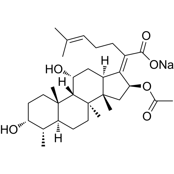
Fusidic acid sodium salt is an orally available antibacterial agent that inhibits bacterial protein synthesis by preventing the release of translation elongation factor G (EF-G) from ribosomes. Fusidic acid sodium salt inhibits the inhibitory and activating effects of interleukins IL-1 and IL-6 on glucose-induced insulin production and exhibits antidiabetic effects in a rat model. Fusidic acid sodium salt improves the symptoms of colitis in rats and inhibits the growth of Toxoplasma gondii and Listeria monocytogenes EGD in vitro, but not in mice.
For research use only. We do not sell to patients.
- Purity : 99.64%
- CAS No.: 751-94-0
- Formula: C31H47NaO6
- Molecular Weight:538.69
-
Storage:
4°C, sealed storage, away from moisture
* In solvent : -80°C, 6 months; -20°C, 1 month (sealed storage, away from moisture)

To place orders, for customer services and technical support, please contact: MedChemExpress USA
Tel: 609-228-6898 E-mail: [email protected] [email protected]


-
 Biological Activity
-
Chemical Information
-
Solvent & Solubility
- Protocols
- Purity & Documentation
- References
-
 Help & FAQs


-

HY-15763
ErastinFerroptosis Inducer -
HY-17362
Vancomycin hydrochlorideAntibiotic -
HY-W016420
Fosfomycin sodiumAntibiotic -

HY-B1350AR
Fusidic acid sodium salt (Standard)Bacteriostatic Antibiotic -
HY-N0565B
Doxycycline hyclateMMP Inhibitor -
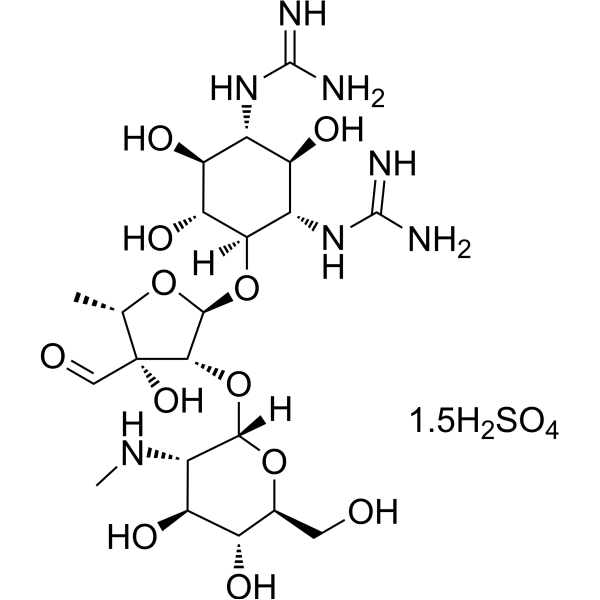
HY-B0472
Streptomycin sulfateBacterial Inhibitor -
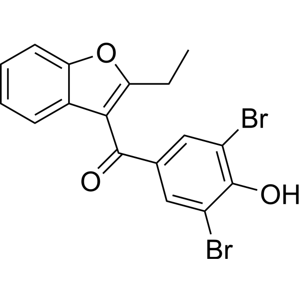
HY-B1135
BenzbromaroneApoptosis Inducer -
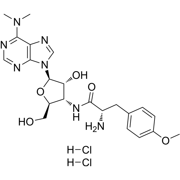
HY-B1743A
Puromycin dihydrochlorideProtein Synthesis Inhibitor -
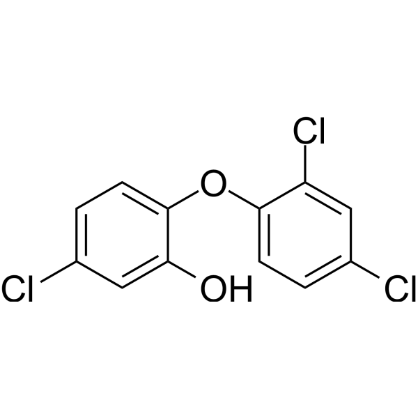
HY-B1119
TriclosanAntibacterial Agent -
HY-B0958
MupirocinAntibiotic
Publications Citing Use of MedChemExpress (MCE) Fusidic acid sodium salt
MoreAll Antibiotic Isoforms
More

Biological Activity
Description
IC50 & Target
|
IL-6 |
IL-1β |
IL-1 |
In Vitro
Fusidic acid sodium salt has good in vitro activity (usual MIC 1mg/L) against Staphylococcus species and has modest activity (usual MIC≤ 8 mg/L) against Streptococcus and Enterococcus species[2].
At an extracellular concentration of 100 mg/L in 90% human serum, fusidic acid is bacteriostatic[2].
Fusidic acid sodium salt (0-30 μg/mL, 1-72 h) inhibits the inhibitory and activating effects of interleukin IL-1β and IL-6 on glucose-induced insulin production in rat pancreas[3].
Fusidic acid sodium salt (0-100 μg/ml, 1 h) inhibits the growth of Toxoplasma gondii in human foreskin fibroblasts (HFFs) cells with an IC50 of 4 μg/ml and a MIC of 2 μg/mL for Listeria monocytogenes EGD[4].
MedChemExpress (MCE) has not independently confirmed the accuracy of these methods. They are for reference only. Further protocols information, click here.
In Vivo
Fusidic acid sodium salt (2.5 mg/ml, p.o., 200 days) has anti-diabetic effect on diabetic rats. [5].
Fusidic acid sodium salt (80 mg/kg, p.o., single dose) improves clinical, histological and serum immunological symptoms in rats with colitis[6].
MedChemExpress (MCE) has not independently confirmed the accuracy of these methods. They are for reference only.
-
Animal Model:Diabetes prone BB rats[5]
-
Dosage:Dissolved every second day in tap water at a concentration of 2.5 mg/ml and given to the rats as drinking water ad libitum; 200 days
-
Administration:Oral gavage (p.o.)
-
Result:Reduced the incidence of diabetes.
-
Animal Model:Mouse Model of Toxoplasma Gondii Infection; Mouse Model of L. monocytogenes EGD Infection[4]
-
Dosage:20 mg/kg, three times a day, 3 days
-
Administration:Subcutaneous injection (s.c.)
-
Result:Reduced the survival rate of mice and caused skin eczema at the inoculation site, but had no effect on Toxoplasma and EGD in mice.
-
Animal Model:Dinitrobenzenesulfonic acid (DNB)-induced colitis in rats[6]
-
Dosage:40, 80 mg/kg; single dose
-
Administration:p.o. for 40 and 80 mg/kg; Intracolonic injection for 80 mg/kg
-
Result:Reduced body weight losses, histological scores, as well as increased colon weights and mucosal damage. Decreased TNF-α and IFN-γ levels.


Chemical Information
-
CAS No. 751-94-0
-
Appearance Solid
-
Molecular Weight 538.69
-
Formula C31H47NaO6
-
Color White to off-white
-
SMILES
C[C@@H]([C@]1([H])CC[C@]([C@@](C[C@@H]/2OC(C)=O)3C)4C)[C@H](O)CC[C@]1(C)[C@]4([H])[C@H](O)C[C@@]3([H])C2=C(C(O[Na])=O)/CC/C=C(C)\C
-
Synonyms
Sodium fusidate; SQ-16360
-
Structure Classification
-
Initial Source
Fusidium coccineum
-
Shipping
Room temperature in continental US; may vary elsewhere.
-
Storage
4°C, sealed storage, away from moisture
* In solvent : -80°C, 6 months; -20°C, 1 month (sealed storage, away from moisture)


Publications (2)
-
Journal Impact Factor
-
Most Recent


Solvent & Solubility
In Vitro:
H2O : ≥ 100 mg/mL (185.64 mM)
DMSO : ≥ 100 mg/mL (185.64 mM; Hygroscopic DMSO has a significant impact on the solubility of product, please use newly opened DMSO)
* "≥" means soluble, but saturation unknown.
Please refer to the solubility information to select the appropriate solvent. Once prepared, please aliquot and store the solution to prevent product inactivation from repeated freeze-thaw cycles.
Storage method and period of stock solution: -80°C, 6 months; -20°C, 1 month (sealed storage, away from moisture). When stored at -80°C, please use it within 6 months. When stored at -20°C, please use it within 1 month.
* Note: If you choose water as the stock solution, please dilute it to the working solution, then filter and sterilize it with a 0.22 μm filter before use.
Please refer to the solubility information to select the appropriate solvent. Once prepared, please aliquot and store the solution to prevent product inactivation from repeated freeze-thaw cycles.
Storage method and period of stock solution: -80°C, 6 months; -20°C, 1 month (sealed storage, away from moisture). When stored at -80°C, please use it within 6 months. When stored at -20°C, please use it within 1 month.
* Note: If you choose water as the stock solution, please dilute it to the working solution, then filter and sterilize it with a 0.22 μm filter before use.
Concentration (start) × Volume (start) = Concentration (final) × Volume (final)
In Vivo:
Select the appropriate dissolution method based on your experimental animal and administration route.
- For the following dissolution methods, please ensure to first prepare a clear stock solution using an In Vitro approach and then sequentially add co-solvents:
- To ensure reliable experimental results, the clarified stock solution can be appropriately stored based on storage conditions. As for the working solution for In Vivo experiments, it is recommended to prepare freshly and use it on the same day.
- The percentages shown for the solvents indicate their volumetric ratio in the final prepared solution. If precipitation or phase separation occurs during preparation, heat and/or sonication can be used to aid dissolution.
Add each solvent one by one: 10% DMSO 40% PEG300 5% Tween-80 45% Saline
Solubility: ≥ 2.5 mg/mL (4.64 mM); Clear solution
This protocol yields a clear solution of ≥ 2.5 mg/mL (saturation unknown).
Taking 1 mL working solution as an example, add 100 μL DMSO stock solution (25.0 mg/mL) to 400 μL PEG300, and mix evenly; then add 50 μL Tween-80 and mix evenly; then add 450 μL Saline to adjust the volume to 1 mL.
Preparation of Saline: Dissolve 0.9 g sodium chloride in ddH₂O and dilute to 100 mL to obtain a clear Saline solution.
Add each solvent one by one: 10% DMSO 90% (20% SBE-β-CD in Saline)
Solubility: ≥ 2.5 mg/mL (4.64 mM); Clear solution
This protocol yields a clear solution of ≥ 2.5 mg/mL (saturation unknown).
Taking 1 mL working solution as an example, add 100 μL DMSO stock solution (25.0 mg/mL) to 900 μL 20% SBE-β-CD in Saline, and mix evenly.
Preparation of 20% SBE-β-CD in Saline (4°C, storage for one week): 2 g SBE-β-CD powder is dissolved in 10 mL Saline, completely dissolve until clear.
For the following dissolution methods, please prepare the working solution directly:
It is recommended to prepare fresh solutions and use them promptly within a short period of time.
The percentages shown for the solvents indicate their volumetric ratio in the final prepared solution. If precipitation or phase separation occurs during preparation, heat and/or sonication can be used to aid dissolution.
Add each solvent one by one: PBS
Solubility: 100 mg/mL (185.64 mM); Clear solution; Need ultrasonic
In Vivo Dissolution Calculator
Please enter the basic information of animal experiments:
-
-
-
-
Recommended: Prepare an additional quantity of animals to account for potential losses during experiments.
Working solution concentration: 0.22 mg/mL
This product has good water solubility, please refer to the measured solubility data in water/PBS/Saline for details.

Protocols
-
Research Protocol for Infectious Diseases
Infectious-disease experiments test how pathogens interact with host barriers, innate immune receptors, inflammatory signaling, pathogen replication, and tissue injury; pattern-recognition receptors such as TLRs, RIG-I-like receptors, NOD-like receptors, and inflammasomes detect microbial molecules and activate NF-κB, interferon, and cytokine responses. The central hypothesis is that infection severity reflects the balance between pathogen burden and host response: protective inflammation restricts pathogen growth, whereas excessive or mislocalized inflammation contributes to tissue damage and disease phenotype. Unresolved questions include which host pathways are protective versus pathogenic, why some infection models fail to translate to human disease, and which combined readouts best predict clinically relevant infection outcomes.
-
Research Protocol for Endocrine Diseases
Endocrine diseases often arise from disrupted hormone production, hormone signaling, or target-tissue responsiveness; for diabetes-focused endocrine disease models, insulin signaling regulates glucose uptake, hepatic glucose output, lipid metabolism, and β-cell compensation. Type 2 diabetes develops through interacting defects in insulin resistance, β-cell dysfunction, adipose inflammation, hepatic glucose overproduction, altered incretin signaling, and ectopic lipid metabolism. A major unresolved question is whether endocrine dysfunction is driven primarily by target-tissue insulin resistance, intrinsic β-cell failure, immune/inflammatory stress, or combined multi-organ failure that differs by disease stage.
-
DSS-Induced Colitis
Dextran sulfate sodium (DSS)-induced colitis is generated by administering DSS in mouse drinking water, producing epithelial injury, barrier disruption, weight loss, diarrhea, fecal blood, colon shortening, histologic mucosal damage, and inflammatory mediator changes; the model is mainly used to study acute or chronic intestinal inflammation resembling selected features of ulcerative colitis. DSS injury is interpreted through clinical and tissue readouts rather than a single molecular endpoint: daily body weight, stool consistency, and bleeding are combined into a disease activity index, while colon length, histology, cytokines, myeloperoxidase activity, intestinal permeability, and tight-junction markers provide complementary measures of inflammation and barrier damage.
-
How to Select a Suitable Non-Mouse Animal Model
Selecting a suitable non-mouse animal model is a structured decision based on the research question, required anatomy or physiology, disease mechanism, endpoint feasibility, translational relevance, and ethical justification. Non-mouse models are preferred when mice cannot reproduce key human-relevant features, such as organ size, surgical anatomy, cardiovascular physiology, neuroanatomy, immune features, pharmacology, toxicology, or long-term clinical procedures. Candidate species may include rats, rabbits, guinea pigs, ferrets, zebrafish, pigs, sheep, goats, dogs, cats, horses, and non-human primates, but each species must be justified by its specific scientific advantage rather than convenience or tradition. Unresolved questions include how to quantify translational superiority across species, how to balance increased biological relevance against higher ethical burden, and when human-derived systems or new approach methodologies should replace animal use.
-
TNBS-Induced Colitis
TNBS-induced colitis is produced by intrarectal delivery of 2,4,6-trinitrobenzene sulfonic acid in ethanol, where ethanol disrupts the mucosal barrier and TNBS haptenates colonic proteins, generating immune-mediated colonic inflammation with weight loss, diarrhea, ulceration, transmural injury, inflammatory-cell infiltration, and cytokine responses. The model is used as an experimental intestinal inflammation model with Crohn’s disease–like features, especially when Th1-type responses, IL-12–dependent inflammation, chronic relapsing inflammation, or fibrosis-related endpoints are studied.
-
How to Choose the Right Model Animal
Choosing the right model animal is a validity-driven decision in which the species, strain, sex, age, genetic background, disease-induction method, outcome measures, and welfare burden must match the scientific question rather than laboratory tradition or convenience. A model should be selected by judging face validity, construct validity, and predictive validity: whether it resembles the human phenotype, whether it reproduces relevant mechanisms, and whether results are likely to predict human biology or treatment response. Animal studies often fail to translate because of species differences, weak disease resemblance, poor experimental design, inadequate reporting, publication bias, and underuse of randomization, blinding, and sample-size justification. Unresolved questions include how to rank competing models objectively, how much human-disease complexity must be reproduced for a given objective, and when non-animal systems such as organoids, ex vivo tissue, or computational models


Purity & Documentation
-
Data Sheet (285 KB)
-
SDS (394 KB)
- English - EN (394 KB)
- Français - FR (394 KB)
- Deutsch - DE (394 KB)
- Norwegian - NO (394 KB)
- Español - ES (394 KB)
- Swedish - SV (394 KB)
- Italian - IT (394 KB)
- Korean - KR (394 KB)
- Portuguese - PT (394 KB)
-
Handling Instructions (2659 KB)
References
[1]. J Turnidge, et al. Fusidic acid pharmacology, pharmacokinetics and pharmacodynamics. Int J Antimicrob Agents. 1999 Aug:12 Suppl 2:S23-34. [Content Brief]
[2]. P Collignon, et al. Fusidic acid in vitro activity. Int J Antimicrob Agents. 1999 Aug:12 Suppl 2:S45-58. [Content Brief]
[3]. K Bendtzen, et al. Effect of fusidic acid on interleukin-1 (IL-1)- and IL-6-induced pancreatic beta-cell functions in rats. J Endocrinol. 1992 Mar;132(3):345-52. [Content Brief]
[4]. Amanda J Payne, et al. Fusidic acid is an effective treatment against Toxoplasma gondii and Listeria monocytogenes in vitro, but not in mice. Parasitol Res. 2013 Nov;112(11):3859-63. [Content Brief]
[5]. K Buschard, et al. Anti-diabetogenic effect of fusidic acid in diabetes prone BB rats. Autoimmunity. 1992;14(2):101-4. [Content Brief]
[6]. Roberto Di Marco, et al. Curative effects of sodium fusidate on the development of dinitrobenzenesulfonic acid-induced colitis in rats. Clin Immunol. 2003 Dec;109(3):266-71. [Content Brief]


Complete Stock Solution Preparation Table
Please refer to the solubility information to select the appropriate solvent. Once prepared, please aliquot and store the solution to prevent product inactivation from repeated freeze-thaw cycles.
Storage method and period of stock solution: -80°C, 6 months; -20°C, 1 month (sealed storage, away from moisture). When stored at -80°C, please use it within 6 months. When stored at -20°C, please use it within 1 month.
| Optional Solvent | Concentration Solvent Mass | 1 mg | 5 mg | 10 mg | 25 mg |
|---|---|---|---|---|---|
| H2O / DMSO | 1 mM | 1.8564 mL | 9.2818 mL | 18.5636 mL | 46.4089 mL |
| 5 mM | 0.3713 mL | 1.8564 mL | 3.7127 mL | 9.2818 mL | |
| 10 mM | 0.1856 mL | 0.9282 mL | 1.8564 mL | 4.6409 mL | |
| 15 mM | 0.1238 mL | 0.6188 mL | 1.2376 mL | 3.0939 mL | |
| 20 mM | 0.0928 mL | 0.4641 mL | 0.9282 mL | 2.3204 mL | |
| 25 mM | 0.0743 mL | 0.3713 mL | 0.7425 mL | 1.8564 mL | |
| 30 mM | 0.0619 mL | 0.3094 mL | 0.6188 mL | 1.5470 mL | |
| 40 mM | 0.0464 mL | 0.2320 mL | 0.4641 mL | 1.1602 mL | |
| 50 mM | 0.0371 mL | 0.1856 mL | 0.3713 mL | 0.9282 mL | |
| 60 mM | 0.0309 mL | 0.1547 mL | 0.3094 mL | 0.7735 mL | |
| 80 mM | 0.0232 mL | 0.1160 mL | 0.2320 mL | 0.5801 mL | |
| 100 mM | 0.0186 mL | 0.0928 mL | 0.1856 mL | 0.4641 mL |
* Note: If you choose water as the stock solution, please dilute it to the working solution, then filter and sterilize it with a 0.22 μm filter before use.


Fusidic acid sodium salt Related Classifications
HY-B1350A Related Classifications
 Powered by Bioz