



PRT062607
Based on 5 publication(s) in Google Scholar
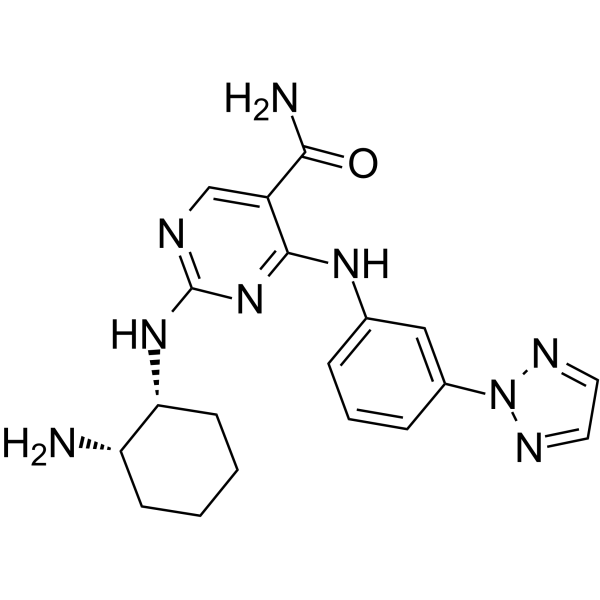
PRT062607 (P505-15; PRT-2607) is an orally active ATP-competitive Syk inhibitor with an IC50 value of 1 nM, and exhibits at least 80-fold selectivity over other kinases. PRT062607 blocks B cell antigen receptor-mediated activation, Fcε receptor 1-mediated basophil degranulation and microglial phagocytosis, and induces caspase-dependent apoptosis and microglial death. PRT062607 inhibits tumor growth and peripheral nerve injury-induced mechanical allodynia, and prevents neuronal loss. PRT062607 can be used in research related to rheumatoid arthritis, chronic lymphocytic leukemia, non-Hodgkin's lymphoma, neurodegenerative diseases and neuropathic pain.
For research use only. We do not sell to patients.
- Purity : 99.38%
- CAS No.: 1370261-96-3
- Formula: C19H23N9O
- Molecular Weight:393.45
-
Storage:Powder -20°C, 3 years , 4°C, 2 years ; In solvent -80°C, 6 months , -20°C, 1 month

To place orders, for customer services and technical support, please contact: MedChemExpress USA
Tel: 609-228-6898 E-mail: [email protected] [email protected]


-
 Biological Activity
-
Chemical Information
-
Solvent & Solubility
- Protocols
- Purity & Documentation
- References
-
 Help & FAQs


-
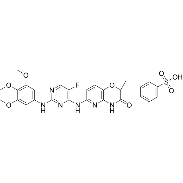
HY-12067
R406Syk/FLT3 Inhibitor -
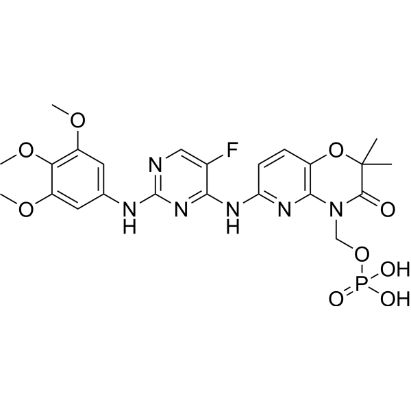
HY-13038A
FostamatinibSyk/FLT3 Inhibitor -
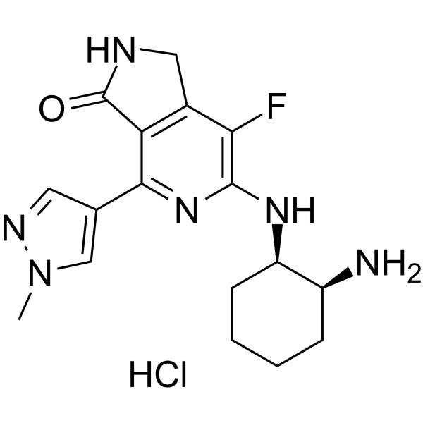
HY-100867A
Mivavotinib monohydrochlorideSYK/FLT3 Inhibitor -
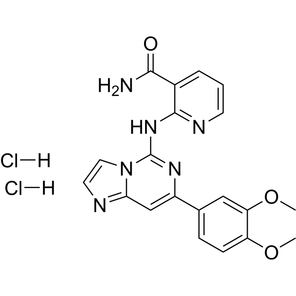
HY-14985
BAY 61-3606 dihydrochlorideApoptosis Inducer -
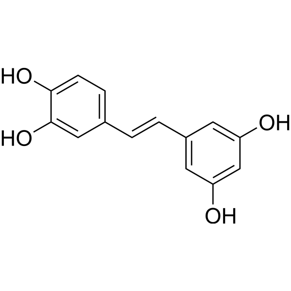
HY-13518
PiceatannolSyk Inhibitor -
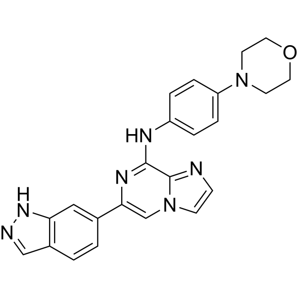
HY-15968
EntospletinibSyk Inhibitor -
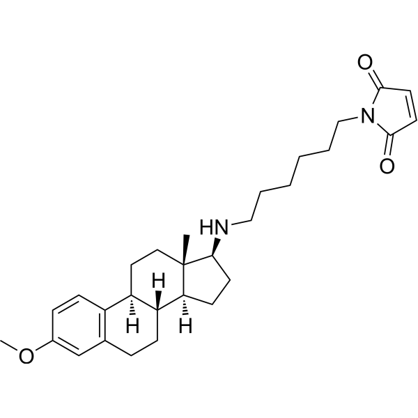
HY-13419
U-73122Phospholipase C Inhibitor -

HY-15141
StaurosporinePKC/PKA Inhibitor -
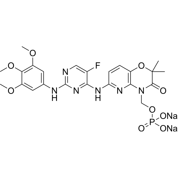
HY-13038
Fostamatinib DisodiumSyk/FLT3 Inhibitor -
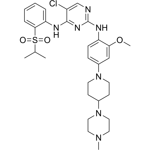
HY-10192
NVP-TAE 684ALK Inhibitor -
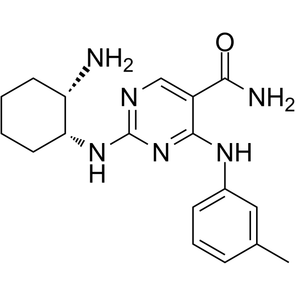
HY-12974
PRT-060318Syk Inhibitor -
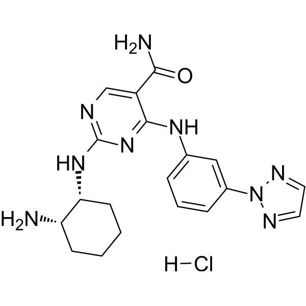
HY-15323
PRT062607 HydrochlorideSyk Inhibitor
Publications Citing Use of MedChemExpress (MCE) PRT062607
MoreAll Caspase Isoforms
More

Biological Activity
Description
Cellular Effect
|
Cell Line

|
Type | Value | Description | References |
|---|---|---|---|---|
| Bone marrow cell | IC50 |
5853 nM
Compound: 1, BIB-057
|
Cytotoxicity against C57BL/6 mouse bone marrow cells assessed as growth inhibition preincubated for 4 days followed by [3H]-thymidine addition measured after 5 hrs by betaplate counting analysis
Cytotoxicity against C57BL/6 mouse bone marrow cells assessed as growth inhibition preincubated for 4 days followed by [3H]-thymidine addition measured after 5 hrs by betaplate counting analysis
|
[PMID: 25633741] |
| Bone marrow cell | IC50 |
5853 nM
Compound: 1, BIIB-057
|
Inhibition of IL3 dependent proliferation in C57/B16 mouse bone marrow cells using [3H]thymidine by liquid scintillation counting
Inhibition of IL3 dependent proliferation in C57/B16 mouse bone marrow cells using [3H]thymidine by liquid scintillation counting
|
[PMID: 24726806] |
| CHO | IC50 |
8600 nM
Compound: 1, BIB-057
|
Inhibition of human ERG expressed in CHO cells by automated Qpatch clamp assay
Inhibition of human ERG expressed in CHO cells by automated Qpatch clamp assay
|
[PMID: 25633741] |
| Jurkat | IC50 |
1805 nM
Compound: 1, BIB-057
|
Inhibition of ZAP70 in anti-CD3 stimulated human Jurkat T cells assessed as SLP76 phosphorylation at Y128 preincubated for 30 mins followed by anti-CD3 stimulation measured after 2 mins by FACS analysis
Inhibition of ZAP70 in anti-CD3 stimulated human Jurkat T cells assessed as SLP76 phosphorylation at Y128 preincubated for 30 mins followed by anti-CD3 stimulation measured after 2 mins by FACS analysis
|
[PMID: 25633741] |
| Ramos | IC50 |
0.223 μM
Compound: 2
|
Inhibition of SYK in human Ramos cells
Inhibition of SYK in human Ramos cells
|
[PMID: 23350847] |
| Ramos | IC50 |
0.223 μM
Compound: PRT062607
|
Cytotoxicity against human Ramos cells
Cytotoxicity against human Ramos cells
|
[PMID: 23151054] |
| Ramos | IC50 |
178 nM
Compound: 1, BIIB-057
|
Inhibition of Syk in anti IgM-stimulated human Ramos cells assessed as BLNK phosphorylation by cellular assay
Inhibition of Syk in anti IgM-stimulated human Ramos cells assessed as BLNK phosphorylation by cellular assay
|
[PMID: 24726806] |
In Vitro
PRT062607 potently inhibits purified Syk in FRET kinase assays containing 15 and 100 μM ATP, with IC50 values of 2.1 and 6 nM[1].
PRT062607 (0.01-2.5 μM; 1.5 h) specifically inhibits BCR-mediated Syk-dependent BLNK Tyr84 phosphorylation in SUDHL4 cells, with an IC50 of 0.16-0.4 μM[1].
PRT062607 potently inhibits BCR-mediated Syk-dependent ERK Tyr204 phosphorylation in Ramos cells with an IC50 of 50 nM; at concentrations up to 2 μM, this compound does not affect non-Syk-dependent, PMA-mediated ERK phosphorylation[1].
PRT062607 (P505-15) (0-6 μM; 3 days) selectively inhibits the proliferation of Syk-dependent TEL-Syk Ba/F3 cells, with an IC50 of 120 nM[1].
PRT062607 (0-4 μM; 40 min) potently inhibits Syk-dependent BCR-mediated ERK Tyr204 phosphorylation in human whole blood B cells, with a mean IC50 of 0.27 μM[1].
PRT062607 (0-4 μM; 16.5 h) potently inhibits Syk-dependent BCR-mediated B cell activation (CD69 upregulation) in human whole blood, with a mean IC50 of 0.28 μM[1].
PRT062607 (0-4 μM; 1.25 h) potently inhibits Syk-dependent FcεR1-mediated basophil degranulation in human whole blood, with a mean IC50 of 0.15 μM[1].
PRT062607 (2 μM; 30 min) inhibits anti-IgM-induced autophosphorylation of SYK in Ramos human non-Hodgkin's lymphoma B cells[2].
PRT062607 (0.02-2 μM; 30 min) inhibits anti-IgM-induced phosphorylation of ERK and AKT in the human non-Hodgkin's lymphoma B cell line Ramos in a concentration-dependent manner, with an IC50 of approximately 50 nM, and achieves complete inhibition at 250 nM[2].
PRT062607 (1-3 μM; 72 hours) induces apoptosis in human non-Hodgkin's lymphoma B cell lines SU-DHL4, SU-DHL6 and Ramos with functional BCR signaling, but exerts no such effect in BCR/BLNK-deficient Toledo/Karpas-422 cell lines or primary human B cells; PRT062607 (1 μM; 24 hours) induces apoptosis in SU-DHL6 cells mixed with whole blood, while primary B cells remain unaffected[2].
PRT062607 (0.01-1 μM; 30 min) concentration-dependently inhibits BCR-induced AKT phosphorylation in primary human chronic lymphocytic leukemia (CLL) cells, with complete inhibition achieved at concentrations ranging from 0.3 to 1 μM[2].
PRT062607 (10 nM-10 μM; 72 h) reduces cell viability by 36% in primary human chronic lymphocytic leukemia (CLL) samples, including high-risk samples with 17p deletion, 11q deletion or unmutated IgVh, with an IC50 < 3 μM in responsive samples[2].
PRT062607 (10 nM-10 μM; 72 h) acts synergistically with fludarabine to reduce the viability of primary human chronic lymphocytic leukemia (CLL) cells, and a fludarabine hyporeactive effect is observed at lower fludarabine concentrations[2].
PRT062607 (10 μM; 3 d) completely blocks LPS (HY-D1056)-induced neuronal loss as well as spontaneous age-related neuronal loss in primary rat cerebellar neuron-glia cultures[3].
PRT062607 (10 μM; 3 d) significantly depletes microglia and induces microglial death in primary rat cerebellar neuron-glia cultures without LPS treatment, but does not deplete microglia in LPS-treated cultures[3].
PRT062607 (10 μM; 24 h) induces significant microglial necrosis and apoptotic cell death, and reduces total cell density in primary rat cortical microglia[3].
PRT062607 (10 μM; overnight) reduces the phagocytosis of 5 μm microspheres by primary rat cortical microglia under both resting and LPS-activated conditions[3].
MedChemExpress (MCE) has not independently confirmed the accuracy of these methods. They are for reference only. Further protocols information, click here.
-
Cell Line:SUDHL4 human non-Hodgkin's lymphoma B cell line
-
Concentration:0.01, 0.025, 0.064, 0.16, 0.4, 1 and 2.5 μM
-
Incubation Time:1 h (pretreatment); 30 min (stimulation)
-
Result:Inhibited BCR-mediated BLNK Tyr84 phosphorylation in a concentration-dependent manner, with an IC50 range of 0.16-0.4 μM.
Did not inhibit BCR-mediated Syk Tyr352 phosphorylation at concentrations up to 2.5 μM.
-
Cell Line:Ba/F3 mouse pro-B cell line stably expressing TEL-Syk
-
Concentration:0.074, 0.22, 0.66, 2 and 6 μM
-
Incubation Time:3 days
-
Result:Selectively inhibited proliferation of the TEL-Syk-expressing Ba/F3 cell line with an IC50 of 120 ± 5 nM.
Did not inhibit proliferation of Ba/F3 cell lines expressing TEL-JAK1, TEL-JAK2, TEL-JAK3, TEL-Tyk2, or TEL-Zap70 at 2 μM.
-
Cell Line:Ramos human non-Hodgkin lymphoma B-cell line
-
Concentration:2 μM
-
Incubation Time:30 minutes (preincubation)
-
Result:Significantly reduced SYK autophosphorylation at the Y525/526 sites compared to stimulated, untreated cells.
-
Cell Line:SU-DHL4, SU-DHL6, Ramos, Toledo, and Karpas-422 human non-Hodgkin lymphoma B-cell lines; primary human B-cells
-
Concentration:1 μM (24 h whole blood experiment); 1 μM, 3 μM (72 h apoptosis assay)
-
Incubation Time:24 h (whole blood experiment); 72 h (apoptosis assay)
-
Result:Induced apoptosis (measured by active caspase-3 cleavage) in BCR-signaling competent cell lines SU-DHL4, SU-DHL6, and Ramos, but not in BCR-deficient Toledo or BLNK-deficient Karpas-422 lines.
Induced apoptosis in SU-DHL6 cells mixed with heparinized human whole blood, while primary B-cells did not undergo apoptosis.
Parmacokinetics
In Vivo
PRT062607 (15 mg/kg; p.o.; twice daily; 5 days) significantly inhibits B cell receptor (BCR)-induced splenomegaly and marginal B cell zone expansion in mice[2].
PRT062607 (10-20 mg/kg; p.o.; twice daily; 5 weeks) significantly inhibits the growth of Ramos NHL tumors in NOD/SCID mice[2].
PRT062607 (4.5 mg per rat; intrathecal injection; single administration) significantly reverses mechanical allodynia induced by spared nerve injury in rats[4].
MedChemExpress (MCE) has not independently confirmed the accuracy of these methods. They are for reference only.
-
Animal Model:BALB/c (female)[1]
-
Dosage:5 mg/kg; 15 mg/kg; 30 mg/kg
-
Administration:p.o.; b.i.d.; 10 days
-
Result:Produced average paw inflammation score inhibitions of 12%, 44%, and 87% respectively, with corresponding average plasma concentrations of 0.38, 0.95, and 1.47 μM.
Significantly reduced blinded histopathology scores and hind paw ankle thickness in the 15 and 30 mg/kg dose groups compared with vehicle controls.
Rendered joint structure indistinguishable from normal naive mice at 30 mg/kg, with no intra-articular neutrophils, fibrin, or synovial tissue damage.
-
Animal Model:Lewis (female, 7-week-old)[1]
-
Dosage:5 mg/kg; 10 mg/kg; 15 mg/kg
-
Administration:p.o.; b.i.d.; 15 days
-
Result:Completely suppressed inflammation in 7 of 8 rats at 15 mg/kg, resulting in a mean inflammation score of 0.63 with an average plasma concentration of 1.34 μM.
Produced a significantly efficacious reduction in inflammation at 10 mg/kg with an average plasma concentration of 0.72 μM.
Significantly reduced blinded histopathology scores and hind paw ankle thickness in the 15 mg/kg dose group compared with vehicle controls.
Rendered joint morphology indistinguishable from normal naive rats at 15 mg/kg, with no inflammatory infiltrate, synovial hyperplasia, or cartilage erosion.
-
Animal Model:Balb/c (5 per group)[2]
-
Dosage:15 mg/kg
-
Administration:p.o.; twice daily; 5 days
-
Result:Reduced average spleen weight from ~0.17 g to ~0.12 g.
Corrected the expansion of the marginal B-cell zone observed in vehicle-treated anti-IgD mice.
-
Animal Model:NOD/SCID (15 per group)[2]
-
Dosage:10 mg/kg; 15 mg/kg; 20 mg/kg
-
Administration:p.o.; twice daily; until study termination
-
Result:Reduced average tumor weight.
Sustained steady-state tumor concentrations above 63 nM (10 mg/kg), 176 nM (15 mg/kg), and 325 nM (20 mg/kg) over the entire dosing interval.
Increased lymphocyte counts in 15 mg/kg group; no reductions in any leukocyte subsets across all dose groups.
-
Animal Model:Sprague Dawley (male and female, 7-8 weeks old, n=6 per sex per treatment group)[4]
-
Dosage:4.5 mg
-
Administration:intrathecal injection; single injection
-
Result:Significantly reversed SNI-induced mechanical pain hypersensitivity in both male and female rats.
Clinical Trial
| NCT Number | Sponsor | Condition | Start Date |
Phase
|
|---|---|---|---|---|
| NCT01329991 | Plexxikon| | 2011-05 | PHASE1 |


Chemical Information
-
CAS No. 1370261-96-3
-
Appearance Solid
-
Molecular Weight 393.45
-
Formula C19H23N9O
-
Color White to off-white
-
SMILES
NC(C1=CN=C(N[C@H]2[C@@H](N)CCCC2)N=C1NC3=CC=CC(N4N=CC=N4)=C3)=O
-
Synonyms
P505-15; PRT-2607; BIIB-057
-
Shipping
Room temperature in continental US; may vary elsewhere.
-
Storage
Powder -20°C 3 years 4°C 2 years In solvent -80°C 6 months -20°C 1 month


Publications (5)
-
Journal Impact Factor
-
Most Recent
-
Nat Commun
Human iPSC-based Modeling of Pulmonary Fibrosis Reveals p300/CBP Inhibition Suppresses Alveolar Transitional Cell State. [Abstract]2026 Feb 12;17(1):1214. PMID: 41680175 -
Cell Death Dis
Activation of AKT via a dual mechanism enhances the susceptibility of melanoma cells to glucose deprivation. [Abstract]2025 Aug 7;16(1):595. PMID: 40774947 -
Proc Natl Acad Sci U S A
Endocytosis triggers V-ATPase-SYK-mediated priming of cGAS activation and innate immune response. [Abstract]2022 Oct 25;119(43):e2207280119. PMID: 36252040 -
Cell Rep
2026 Mar 27;45(4):117183. PMID: 41903135 -
Int J Ophthalmol
NADPH oxidase 2 plays a protective role in experimental Aspergillus fumigatus keratitis in mice through killing fungi and limiting the degree of inflammation. [Abstract]2022 Jul 18;15(7):1044-1052. PMID: 35919314


Solvent & Solubility
In Vitro:
DMSO : 100 mg/mL (254.16 mM; Need ultrasonic; Hygroscopic DMSO has a significant impact on the solubility of product, please use newly opened DMSO)
Please refer to the solubility information to select the appropriate solvent. Once prepared, please aliquot and store the solution to prevent product inactivation from repeated freeze-thaw cycles.
Storage method and period of stock solution: -80°C, 6 months; -20°C, 1 month. When stored at -80°C, please use it within 6 months. When stored at -20°C, please use it within 1 month.
Please refer to the solubility information to select the appropriate solvent. Once prepared, please aliquot and store the solution to prevent product inactivation from repeated freeze-thaw cycles.
Storage method and period of stock solution: -80°C, 6 months; -20°C, 1 month. When stored at -80°C, please use it within 6 months. When stored at -20°C, please use it within 1 month.
Concentration (start) × Volume (start) = Concentration (final) × Volume (final)
In Vivo:
Select the appropriate dissolution method based on your experimental animal and administration route.
- For the following dissolution methods, please ensure to first prepare a clear stock solution using an In Vitro approach and then sequentially add co-solvents:
- To ensure reliable experimental results, the clarified stock solution can be appropriately stored based on storage conditions. As for the working solution for In Vivo experiments, it is recommended to prepare freshly and use it on the same day.
- The percentages shown for the solvents indicate their volumetric ratio in the final prepared solution. If precipitation or phase separation occurs during preparation, heat and/or sonication can be used to aid dissolution.
Add each solvent one by one: 10% DMSO 40% PEG300 5% Tween-80 45% Saline
Solubility: ≥ 5 mg/mL (12.71 mM); Clear solution
This protocol yields a clear solution of ≥ 5 mg/mL (saturation unknown).
Taking 1 mL working solution as an example, add 100 μL DMSO stock solution (50.0 mg/mL) to 400 μL PEG300, and mix evenly; then add 50 μL Tween-80 and mix evenly; then add 450 μL Saline to adjust the volume to 1 mL.
Preparation of Saline: Dissolve 0.9 g sodium chloride in ddH₂O and dilute to 100 mL to obtain a clear Saline solution.
Add each solvent one by one: 10% DMSO 90% (20% SBE-β-CD in Saline)
Solubility: ≥ 2.5 mg/mL (6.35 mM); Clear solution
This protocol yields a clear solution of ≥ 2.5 mg/mL (saturation unknown).
Taking 1 mL working solution as an example, add 100 μL DMSO stock solution (25.0 mg/mL) to 900 μL 20% SBE-β-CD in Saline, and mix evenly.
Preparation of 20% SBE-β-CD in Saline (4°C, storage for one week): 2 g SBE-β-CD powder is dissolved in 10 mL Saline, completely dissolve until clear.
In Vivo Dissolution Calculator
Please enter the basic information of animal experiments:
-
-
-
-
Recommended: Prepare an additional quantity of animals to account for potential losses during experiments.
Please enter your animal formula composition:
-
%DMSO +
Recommended: Keep the proportion of DMSO in working solution below 2% if your animal is weak.
-
%+
-
+%Tween-80 + +
-
%Saline +
The co-solvents required include: DMSO, . All of co-solvents are available by MedChemExpress (MCE). , Tween 80. All of co-solvents are available by MedChemExpress (MCE).
Working solution concentration: 0.22 mg/mL
Method for preparing stock solution: mg drug dissolved in μL DMSO. Stock solution concentration: mg/mL.
1. Take μL DMSO stock solution;
2. Add μL .
μL , mix evenly;
3. Then add μL Tween 80, mix evenly;
4. Then add μL
Please ensure that the stock solution in the first step is dissolved to a clear state, and add co-solvents in sequence. You can use ultrasonic heating (ultrasonic cleaner, recommended frequency 20-40 kHz), vortexing, etc. to assist dissolution.

Protocols
-
Phagocytosis Functional Assay
A phagocytosis functional assay measures the ability of phagocytic cells, such as neutrophils, macrophages, monocytes, or microglia/macrophages, to bind and internalize particulate targets including bacteria, yeast particles, beads, or myelin particles. Fluorescent flow-cytometry assays detect target uptake as fluorescence associated with gated phagocytes, while pH-sensitive dyes such as pHrodo increase signal in acidic phagosomal compartments and therefore preferentially report internalized particles rather than particles remaining outside the cell. Microscopy or high-content imaging can be used to confirm intracellular localization and, in some protocols, to follow uptake kinetics.
-
Collagen-Induced Arthritis
Collagen-induced arthritis (CIA) is an autoimmune murine model of rheumatoid arthritis in which immunization with type II collagen (CII) emulsified in an adjuvant induces a T cell- and autoantibody-driven inflammatory arthritis characterized by synovial hyperplasia, immune cell infiltration, and joint destruction. The model typically relies on genetically susceptible mouse strains (e. g. , DBA/1) and reproduces key features of human rheumatoid arthritis, including anti-collagen immune responses and progressive joint inflammation. Disease onset generally occurs within ~3-4 weeks after immunization, depending on antigen/adjuvant combinations and protocol variation. The immunopathology is driven by adaptive immune activation against CII, leading to systemic and local joint inflammation mediated by pro-inflammatory cytokines and effector immune cells, making CIA a standard preclinical platform for evaluating immunomodulatory and anti-arthritic interventions.
-
Apoptosis
Apoptosis, also called programmed cell death, is generally characterized by distinct morphological characteristics.
-
TUNEL staining for apoptotic DNA fragmentation
TUNEL staining detects DNA strand breaks by using terminal deoxynucleotidyl transferase to add labeled nucleotides to exposed 3′-OH DNA termini, generating either microscopic staining in fixed cells or tissue sections, or fluorescence/cytometric signal in cell suspensions. TUNEL positivity reflects DNA fragmentation but should not be interpreted alone as definitive apoptosis, because TUNEL can also label necrotic, autolytic, mechanically damaged, or DNA-repair-associated DNA breaks.
-
Cell Cytotoxicity Assay
Cytotoxicity assays are usually based on the assessment of cell membrane damage, which can also be indirectly detected by measuring cell viability. Detection methods include MTT assay, CKK-8 assay, LDH assay and ATP assay, etc.
-
Annexin V plus membrane-impermeant dye apoptosis staining
Annexin V-based apoptosis assays rely on the detection of phosphatidylserine (PS) externalization from the inner leaflet of the plasma membrane to the outer leaflet, an early biochemical hallmark of apoptosis. Fluorescently labeled Annexin V binds PS in a calcium-dependent manner, enabling identification of early apoptotic cells by flow cytometry or fluorescence microscopy. When combined with a membrane-impermeant DNA-binding dye (e. g. , propidium iodide), this approach allows discrimination between viable (Annexin V−/dye−), early apoptotic (Annexin V+/dye−), and late apoptotic or necrotic (Annexin V+/dye+) cell populations by assessing membrane integrity and PS exposure.
-
Patient-Derived Xenograft (PDX)
Patient-derived xenograft (PDX) models are generated by engrafting primary human tumor tissue directly into immunodeficient mice, allowing in vivo propagation of patient tumor biology without initial in vitro adaptation. These models are used to preserve key histopathological and molecular characteristics of the original tumor and enable assessment of tumor growth dynamics and therapeutic response in a living organism. The biological readout is tumor engraftment and subsequent growth in the murine host, which reflects the ability of human tumor cells to survive, vascularize, and expand in an immunocompromised microenvironment.
-
Apoptosis Solutions
Apoptosis is a regulated, generally non-lytic cell-death pathway that removes unwanted, damaged, infected, or abnormal cells through coordinated morphological changes, caspase activation, DNA fragmentation, and membrane remodeling. The intrinsic apoptosis pathway is controlled mainly by mitochondrial outer membrane permeabilization, BCL-2 family proteins, cytochrome c release, apoptosome formation, caspase-9 activation, and downstream executioner caspase-3/7 activation. The extrinsic apoptosis pathway is initiated by death receptors such as Fas, TNFR, and TRAIL receptors, which recruit adaptor proteins and activate caspase-8 before engaging executioner caspases or mitochondrial amplification through BID cleavage. Apoptosis is linked to many phenotypes, including cancer cell killing, tissue homeostasis, immune regulation, neurodegeneration, infection response, and treatment-induced cytotoxicity; unresolved questions include how apoptosis interacts with necroptosis, pyroptosis, ferroptos
-
Patient-Derived Orthotopic Xenograft (PDOX)
Patient-derived orthotopic xenograft (PDOX) modeling implants fresh patient tumor tissue or patient-derived tumor cells into the anatomically corresponding organ or tissue site of immunodeficient mice, usually by surgical orthotopic implantation, to preserve patient tumor histology, local microenvironmental context, invasion, metastatic behavior, and treatment-response features better than subcutaneous implantation. PDOX readouts include tumor engraftment, orthotopic tumor growth, local invasion, metastasis, recurrence after resection, histologic similarity to the donor tumor, biomarker retention, molecular concordance, survival, and response or resistance to therapy. PDOX models are used for preclinical drug testing and individualized therapy evaluation, but engraftment success varies by tumor type and specimen quality.
-
Subcutaneous Cell-Line-Derived Xenograft
Subcutaneous cell-line-derived xenograft (CDX) models are established by implanting cultured human cancer cell lines into immunodeficient mice, where the injected cells form localized tumors that can be monitored in vivo as a measure of tumorigenic potential, growth kinetics, and treatment response. These models are widely used in oncology research because they allow reproducible tumor formation and enable comparative assessment of tumor growth between different cell lines or genetic manipulations in a controlled in vivo microenvironment. Subcutaneous implantation of cancer cells in immunodeficient mice is a standard approach for evaluating tumor growth behavior and therapeutic response across multiple cancer types, including prostate, esophageal, pancreatic, and colon cancer models.
-
Orthotopic Cell-Line Xenograft
Orthotopic cell-line xenograft models involve implantation of human cancer cell lines into the anatomically corresponding organ of immunodeficient mice to reproduce tumor growth within a native microenvironment, enabling more clinically relevant tumor behavior compared with subcutaneous models. These models are widely used because orthotopic placement better recapitulates tumor progression, including invasion and metastatic spread, which are often underrepresented in heterotopic implantation systems. Compared with conventional xenografts, orthotopic implantation is described as more technically complex but provides improved simulation of tumor-microenvironment interactions and metastatic behavior, making it particularly valuable for translational oncology research. Surgical orthotopic implantation approaches have been emphasized as enabling faithful reproduction of clinical cancer features, including metastasis and disease progression patterns that align with the tumor’s organ of origi


Purity & Documentation
-
Data Sheet (295 KB)
-
SDS (393 KB)
- English - EN (393 KB)
- Français - FR (393 KB)
- Deutsch - DE (393 KB)
- Norwegian - NO (393 KB)
- Español - ES (393 KB)
- Swedish - SV (393 KB)
- Italian - IT (393 KB)
- Korean - KR (393 KB)
- Portuguese - PT (393 KB)
-
Handling Instructions (2659 KB)
References
[1]. Coffey G, et al. Specific inhibition of spleen tyrosine kinase suppresses leukocyte immune function and inflammation in animal models of rheumatoid arthritis. J Pharmacol Exp Ther. 2012;340(2):350-359. [Content Brief]
[2]. Spurgeon SE, et al. The selective SYK inhibitor P505-15 (PRT062607) inhibits B cell signaling and function in vitro and in vivo and augments the activity of fludarabine in chronic lymphocytic leukemia. J Pharmacol Exp Ther. 2013;344(2):378-387. [Content Brief]
[3]. Birkle TJY, et al. Syk inhibitors protect against microglia-mediated neuronal loss in culture. Front Aging Neurosci. 2023;15:1120952. Published 2023 Mar 15. [Content Brief]
[4]. Ghazisaeidi S, et al. Conserved transcriptional programming across sex and species after peripheral nerve injury predicts treatments for neuropathic pain. Br J Pharmacol. 2023;180(21):2822-2836. [Content Brief]


Complete Stock Solution Preparation Table
Please refer to the solubility information to select the appropriate solvent. Once prepared, please aliquot and store the solution to prevent product inactivation from repeated freeze-thaw cycles.
Storage method and period of stock solution: -80°C, 6 months; -20°C, 1 month. When stored at -80°C, please use it within 6 months. When stored at -20°C, please use it within 1 month.
| Optional Solvent | Concentration Solvent Mass | 1 mg | 5 mg | 10 mg | 25 mg |
|---|---|---|---|---|---|
| DMSO | 1 mM | 2.5416 mL | 12.7081 mL | 25.4162 mL | 63.5405 mL |
| 5 mM | 0.5083 mL | 2.5416 mL | 5.0832 mL | 12.7081 mL | |
| 10 mM | 0.2542 mL | 1.2708 mL | 2.5416 mL | 6.3540 mL | |
| 15 mM | 0.1694 mL | 0.8472 mL | 1.6944 mL | 4.2360 mL | |
| 20 mM | 0.1271 mL | 0.6354 mL | 1.2708 mL | 3.1770 mL | |
| 25 mM | 0.1017 mL | 0.5083 mL | 1.0166 mL | 2.5416 mL | |
| 30 mM | 0.0847 mL | 0.4236 mL | 0.8472 mL | 2.1180 mL | |
| 40 mM | 0.0635 mL | 0.3177 mL | 0.6354 mL | 1.5885 mL | |
| 50 mM | 0.0508 mL | 0.2542 mL | 0.5083 mL | 1.2708 mL | |
| 60 mM | 0.0424 mL | 0.2118 mL | 0.4236 mL | 1.0590 mL | |
| 80 mM | 0.0318 mL | 0.1589 mL | 0.3177 mL | 0.7943 mL | |
| 100 mM | 0.0254 mL | 0.1271 mL | 0.2542 mL | 0.6354 mL |


PRT062607 Related Classifications
HY-15322 Related Classifications
 Powered by Bioz
Keywords
- PRT062607
- 1370261-96-3
- P505-15
- PRT-2607
- BIIB-057
- PRT 062607
- PRT-062607
- PRT2607
- PRT 2607
- PRT-2607
- BIIB057
- BIIB 057
- BIIB-057
- Syk
- Apoptosis
- Caspase
- chronic lymphocytic leukemia
- SUDHL4 cells
- Fcε receptor 1
- B cell antigen receptor
- non-Hodgkin lymphoma
- rheumatoid arthritis
- neuropathic pain
- spleen tyrosine kinase
- neurodegenerative disease
- Ramos cells
- Inhibitor
- inhibitor
- inhibit