



Streptonigrin
Based on 2 publication(s) in Google Scholar
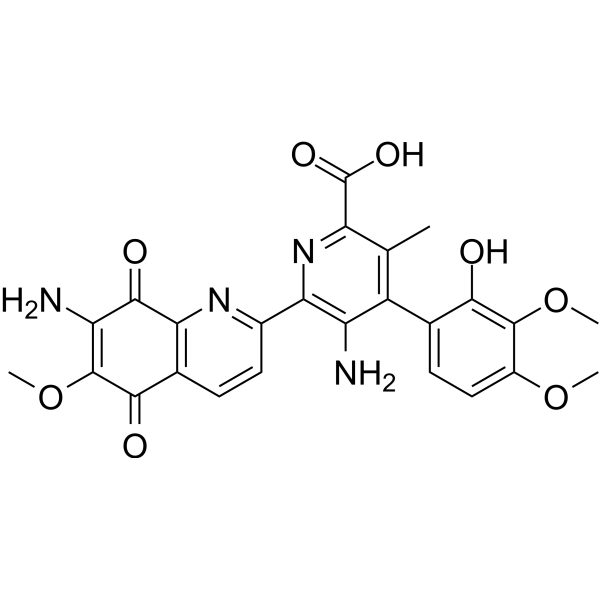
Streptonigrin (Bruneomycin) is an orally active antibiotic and pan-PAD inhibitor, inhibiting PAD1, PAD2, PAD3 and PAD4 with IC50 of 48.3 μM, 26.1 μM, 0.43 μM and 2.5 μM, respectively. Streptonigrin inhibits SENP1 (IC50 of 0.518 μM) and reduces HIF1α. Streptonigrin increases p53 and Apoptosis. Streptonigrin shows antiviral activity against Rauscher murine leukemia virus. Streptonigrin has immunosuppressive effects. Streptonigrin has antitumor activity against osteosarcoma.
For research use only. We do not sell to patients.
- Purity : 99.9%
- CAS No.: 3930-19-6
- Formula: C25H22N4O8
- Molecular Weight:506.46
-
Storage:Powder -20°C, 3 years , 4°C, 2 years ; In solvent -80°C, 6 months , -20°C, 1 month

To place orders, for customer services and technical support, please contact: MedChemExpress USA
Tel: 609-228-6898 E-mail: [email protected] [email protected]


-
 Biological Activity
-
Chemical Information
- Protocols
- Purity & Documentation
- References
-
 Help & FAQs


-
HY-111347
BB-Cl-AmidinePAD Inhibitor -
HY-100574A
Cl-amidine hydrochloridePAD Inhibitor -
HY-N2306
Aclacinomycin ATopoisomerase Inhibitor -
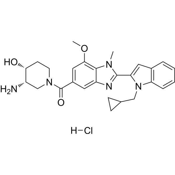
HY-100514
GSK484 hydrochloridePAD4 Inhibitor -
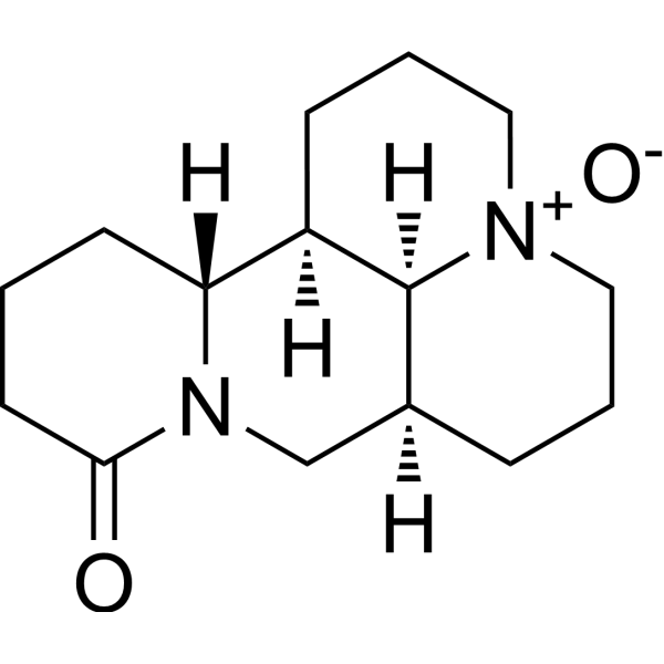
HY-N0158
OxymatrineAntitumor Agent -
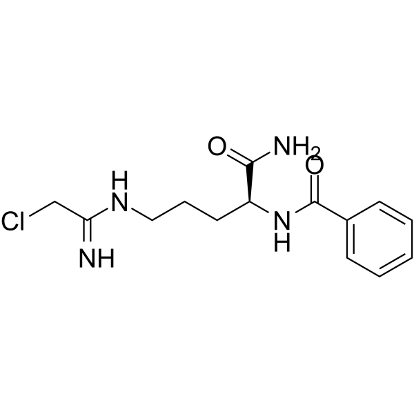
HY-100574
Cl-amidinePAD Inhibitor -

HY-100514R
GSK484 hydrochloride (Standard)PAD4 Inhibitor -
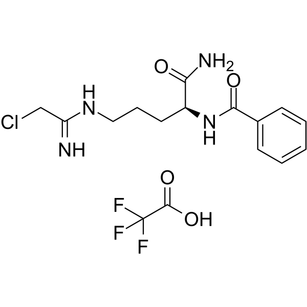
HY-100574B
Cl-amidine TFAPAD Inhibitor -

HY-N0158R
Oxymatrine (Standard)Antitumor Agent
Publications Citing Use of MedChemExpress (MCE) Streptonigrin
More Customer Validation & Images

-
WB
All Antibiotic Isoforms
More

Biological Activity
Description
IC50 & Target
[4]|
HIF-1α |
Cellular Effect
|
Cell Line

|
Type | Value | Description | References |
|---|---|---|---|---|
| A549 | IC50 |
0.33 μg/mL
Compound: Streptonigrin
|
In vitro cytotoxic activity against A 549 (non-small cell lung) using SRB (sulforhodamine B) assay
In vitro cytotoxic activity against A 549 (non-small cell lung) using SRB (sulforhodamine B) assay
|
[PMID: 10328288] |
| A549 | IC50 |
0.33 μg/mL
Compound: 1
|
Cytotoxicity against Homo sapiens (human) A549 cells after 72 hr by MTT assay
Cytotoxicity against Homo sapiens (human) A549 cells after 72 hr by MTT assay
|
10.1007/s00044-004-0032-8 |
| B16 | IC50 |
0.48 μg/mL
Compound: 1a
|
Cytotoxicity against mouse melanoma cell culture(B-16)
Cytotoxicity against mouse melanoma cell culture(B-16)
|
[PMID: 3656364] |
| CAKI-1 | IC50 |
0.4 nM
Compound: 1
|
Cytotoxicity against CD30 deficient and CD70 expressing human Caki1 cells after 96 hrs by resazurin conversion assay
Cytotoxicity against CD30 deficient and CD70 expressing human Caki1 cells after 96 hrs by resazurin conversion assay
|
[PMID: 19386499] |
| CCRF-CEM | IC50 |
0.00017 μg/mL
Compound: 1a
|
Cytotoxicity against human lymphoblastic leukemia cell culture (CCRF-CEM)
Cytotoxicity against human lymphoblastic leukemia cell culture (CCRF-CEM)
|
[PMID: 3656364] |
| CCRF-CEM | IC50 |
0.17 ng/mL
Compound: 1a
|
Cytotoxicity against human lymphoblastic leukemia cell culture (CCRF-CEM)
Cytotoxicity against human lymphoblastic leukemia cell culture (CCRF-CEM)
|
[PMID: 3656364] |
| Epidermoid carcinoma cell line | IC50 |
0.0025 μg/mL
Compound: 1a
|
Cytotoxicity against human epidermoid carcinoma of the nasopharynx (9KB)
Cytotoxicity against human epidermoid carcinoma of the nasopharynx (9KB)
|
[PMID: 3656364] |
| HCT-15 | IC50 |
0.02 μg/mL
Compound: Streptonigrin
|
In vitro cytotoxic activity against HCT-15 (colon) using SRB (sulforhodamine B) assay
In vitro cytotoxic activity against HCT-15 (colon) using SRB (sulforhodamine B) assay
|
[PMID: 10328288] |
| HCT-15 | IC50 |
0.02 μg/mL
Compound: 1
|
Cytotoxicity against Homo sapiens (human) HCT15 cells after 72 hr by MTT assay
Cytotoxicity against Homo sapiens (human) HCT15 cells after 72 hr by MTT assay
|
10.1007/s00044-004-0032-8 |
| KARPAS-299 | IC50 |
0.4 nM
Compound: 1
|
Cytotoxicity against CD30 expressing and CD70 deficient human KARPAS299 cells after 96 hrs by resazurin conversion assay
Cytotoxicity against CD30 expressing and CD70 deficient human KARPAS299 cells after 96 hrs by resazurin conversion assay
|
[PMID: 19386499] |
| L1210 | IC50 |
0.61 μg/mL
Compound: 1a
|
Cytotoxicity against mouse lymphoblastic leukemia cell culture (L-1210)
Cytotoxicity against mouse lymphoblastic leukemia cell culture (L-1210)
|
[PMID: 3656364] |
| MDA-MB-231 | IC50 |
30.5 μM
Compound: 8
|
Cytotoxicity against human MDA-MB-231 cells assessed as reduction in cell viability incubated for 48 hrs by MTT assay
Cytotoxicity against human MDA-MB-231 cells assessed as reduction in cell viability incubated for 48 hrs by MTT assay
|
[PMID: 38870832] |
| P388 | IC50 |
0.045 μg/mL
Compound: 1a
|
Cytotoxicity against P388 mouse leukemia cell culture (9PS(P388))
Cytotoxicity against P388 mouse leukemia cell culture (9PS(P388))
|
[PMID: 3656364] |
| SH-SY5Y | IC50 |
0.05 μM
Compound: 1
|
Cytotoxicity against human wild type SH-SY5Y cells
Cytotoxicity against human wild type SH-SY5Y cells
|
[PMID: 12027749] |
| SH-SY5Y | IC50 |
4.6 μM
Compound: 1
|
Cytotoxicity against human SH-SY5Y cells overexpressing dominant negative mutant of p53
Cytotoxicity against human SH-SY5Y cells overexpressing dominant negative mutant of p53
|
[PMID: 12027749] |
| SK-MEL-2 | IC50 |
0.02 μg/mL
Compound: Streptonigrin
|
In vitro cytotoxic activity against SK-MEL-2 (melanoma) using SRB (sulforhodamine B) assay
In vitro cytotoxic activity against SK-MEL-2 (melanoma) using SRB (sulforhodamine B) assay
|
[PMID: 10328288] |
| SK-MEL-2 | IC50 |
0.02 μg/mL
Compound: 1
|
Cytotoxicity against Homo sapiens (human) SK-MEL-2 cells after 72 hr by MTT assay
Cytotoxicity against Homo sapiens (human) SK-MEL-2 cells after 72 hr by MTT assay
|
10.1007/s00044-004-0032-8 |
| SK-OV-3 | IC50 |
0.28 μg/mL
Compound: Streptonigrin
|
In vitro cytotoxic activity against SK-OV-3 (ovarian) using SRB (sulforhodamine B) assay
In vitro cytotoxic activity against SK-OV-3 (ovarian) using SRB (sulforhodamine B) assay
|
[PMID: 10328288] |
| SK-OV-3 | IC50 |
0.28 μg/mL
Compound: 1
|
Cytotoxicity against Homo sapiens (human) SKOV3 cells after 72 hr by MTT assay
Cytotoxicity against Homo sapiens (human) SKOV3 cells after 72 hr by MTT assay
|
10.1007/s00044-004-0032-8 |
| T-cell | IC50 |
<20 μM
Compound: 1, NSC-45383
|
Inhibition of JAK-mediated IL10-induced Stat3 phosphorylation in BALB/c mouse CD4+ T cells by Phospho-Flow cytometry
Inhibition of JAK-mediated IL10-induced Stat3 phosphorylation in BALB/c mouse CD4+ T cells by Phospho-Flow cytometry
|
[PMID: 18157122] |
| T-cell | IC50 |
<20 μM
Compound: 1, NSC-45383
|
Inhibition of JAK-mediated IL10-induced Stat3 phosphorylation in BALB/c mouse CD8+ T cells by Phospho-Flow cytometry
Inhibition of JAK-mediated IL10-induced Stat3 phosphorylation in BALB/c mouse CD8+ T cells by Phospho-Flow cytometry
|
[PMID: 18157122] |
| T-cell | IC50 |
<20 μM
Compound: 1, NSC-45383
|
Inhibition of JAK-mediated IL15-induced Stat5 phosphorylation in BALB/c mouse CD4+ T cells by Phospho-Flow cytometry
Inhibition of JAK-mediated IL15-induced Stat5 phosphorylation in BALB/c mouse CD4+ T cells by Phospho-Flow cytometry
|
[PMID: 18157122] |
| T-cell | IC50 |
<20 μM
Compound: 1, NSC-45383
|
Inhibition of JAK-mediated IL15-induced Stat5 phosphorylation in BALB/c mouse CD8+ T cells by Phospho-Flow cytometry
Inhibition of JAK-mediated IL15-induced Stat5 phosphorylation in BALB/c mouse CD8+ T cells by Phospho-Flow cytometry
|
[PMID: 18157122] |
| T-cell | IC50 |
<20 μM
Compound: 1, NSC-45383
|
Inhibition of JAK-mediated IL4-induced Stat5 phosphorylation in BALB/c mouse CD4+ T cells by Phospho-Flow cytometry
Inhibition of JAK-mediated IL4-induced Stat5 phosphorylation in BALB/c mouse CD4+ T cells by Phospho-Flow cytometry
|
[PMID: 18157122] |
| T-cell | IC50 |
<20 μM
Compound: 1, NSC-45383
|
Inhibition of JAK-mediated IL4-induced Stat5 phosphorylation in BALB/c mouse CD8+ T cells by Phospho-Flow cytometry
Inhibition of JAK-mediated IL4-induced Stat5 phosphorylation in BALB/c mouse CD8+ T cells by Phospho-Flow cytometry
|
[PMID: 18157122] |
| T-cell | IC50 |
<20 μM
Compound: 1, NSC-45383
|
Inhibition of JAK-mediated IL4-induced Stat6 phosphorylation in BALB/c mouse CD4+ T cells by Phospho-Flow cytometry
Inhibition of JAK-mediated IL4-induced Stat6 phosphorylation in BALB/c mouse CD4+ T cells by Phospho-Flow cytometry
|
[PMID: 18157122] |
| T-cell | IC50 |
<20 μM
Compound: 1, NSC-45383
|
Inhibition of JAK-mediated IL4-induced Stat6 phosphorylation in BALB/c mouse CD8+ T cells by Phospho-Flow cytometry
Inhibition of JAK-mediated IL4-induced Stat6 phosphorylation in BALB/c mouse CD8+ T cells by Phospho-Flow cytometry
|
[PMID: 18157122] |
| T-cell | IC50 |
<20 μM
Compound: 1, NSC-45383
|
Inhibition of JAK-mediated IL6-induced Stat1 phosphorylation in BALB/c mouse CD4+ T cells by Phospho-Flow cytometry
Inhibition of JAK-mediated IL6-induced Stat1 phosphorylation in BALB/c mouse CD4+ T cells by Phospho-Flow cytometry
|
[PMID: 18157122] |
| T-cell | IC50 |
<20 μM
Compound: 1, NSC-45383
|
Inhibition of JAK-mediated IL6-induced Stat1 phosphorylation in BALB/c mouse CD8+ T cells by Phospho-Flow cytometry
Inhibition of JAK-mediated IL6-induced Stat1 phosphorylation in BALB/c mouse CD8+ T cells by Phospho-Flow cytometry
|
[PMID: 18157122] |
| T-cell | IC50 |
<20 μM
Compound: 1, NSC-45383
|
Inhibition of JAK-mediated IL6-induced Stat3 phosphorylation in BALB/c mouse CD4+ T cells by Phospho-Flow cytometry
Inhibition of JAK-mediated IL6-induced Stat3 phosphorylation in BALB/c mouse CD4+ T cells by Phospho-Flow cytometry
|
[PMID: 18157122] |
| T-cell | IC50 |
<20 μM
Compound: 1, NSC-45383
|
Inhibition of JAK-mediated IL6-induced Stat3 phosphorylation in BALB/c mouse CD8+ T cells by Phospho-Flow cytometry
Inhibition of JAK-mediated IL6-induced Stat3 phosphorylation in BALB/c mouse CD8+ T cells by Phospho-Flow cytometry
|
[PMID: 18157122] |
| T-cell | IC50 |
<20 μM
Compound: 1, NSC-45383
|
Inhibition of JAK-mediated IL7-induced Stat5 phosphorylation in BALB/c mouse CD4+ T cells by Phospho-Flow cytometry
Inhibition of JAK-mediated IL7-induced Stat5 phosphorylation in BALB/c mouse CD4+ T cells by Phospho-Flow cytometry
|
[PMID: 18157122] |
| T-cell | IC50 |
<20 μM
Compound: 1, NSC-45383
|
Inhibition of JAK-mediated IL7-induced Stat5 phosphorylation in BALB/c mouse CD8+ T cells by Phospho-Flow cytometry
Inhibition of JAK-mediated IL7-induced Stat5 phosphorylation in BALB/c mouse CD8+ T cells by Phospho-Flow cytometry
|
[PMID: 18157122] |
| T-cell | IC50 |
<20 μM
Compound: 1, NSC-45383
|
Inhibition of JAK-mediated interferon-gamma-induced Stat1 phosphorylation in BALB/c mouse CD4+ T cells by Phospho-Flow cytometry
Inhibition of JAK-mediated interferon-gamma-induced Stat1 phosphorylation in BALB/c mouse CD4+ T cells by Phospho-Flow cytometry
|
[PMID: 18157122] |
| XF498 | IC50 |
0.31 μg/mL
Compound: Streptonigrin
|
In vitro cytotoxic activity against XF 498 (CNS) using SRB (sulforhodamine B) assay
In vitro cytotoxic activity against XF 498 (CNS) using SRB (sulforhodamine B) assay
|
[PMID: 10328288] |
| XF498 | IC50 |
0.31 μg/mL
Compound: 1
|
Cytotoxicity against Homo sapiens (human) XF498 cells after 72 hr by MTT assay
Cytotoxicity against Homo sapiens (human) XF498 cells after 72 hr by MTT assay
|
10.1007/s00044-004-0032-8 |
In Vitro
Streptonigrin (10 μM) shows cytotoxicity towards both cancerous U2OS and normal NIH 3T3 cell lines[1].
Streptonigrin (0-5 μM; 24 h) inhibits the proliferation of SW480 cells, AGS cells and HEK293 cells transfected with β-catenin in a dose-dependent manner[2].
Streptonigrin (0.1-5 μM) does not influence the basal activity of soluble guanylate cyclase but causes concentration-dependent inhibition of enzyme activation by SNP (IC50 of 4.16 μM) and also by spermineNONO[3].
Streptonigrin (0-1 μM; 10 min) inhibits SENP1 activity with an IC50 of 0.518 μM when SUMO1 is used as a substrate[4].
Streptonigrin (2 μM; 16 h) reduces HIF1α protein level in HCT116 cells transfected with HIF1α expression plasmid[4].
Streptonigrin (1 nM-10 μM; 4 h) shows a concentration-dependent effect on nuclear morphology in NIH 3T3 cells, promoting heterochromatin formation[5].
Streptonigrin (2.5-500 μM; 60 min) inhibits the catalytic activity of purified calf thymus DNA topoisomerase II by relaxation of pBR322 DNA[6].
Streptonigrin (72 h) selectively suppresses B-cell proliferation induced by LPS, with an IC50 value of 0.29 ng/mL[8].
Streptonigrin (10-100 nM) increases p53 and Apoptosis in ACHN and CAKI-1 cells[9].
MedChemExpress (MCE) has not independently confirmed the accuracy of these methods. They are for reference only. Further protocols information, click here.
-
Cell Line:A20.2J, M12.4.5, YAC-I, P815, FDC.P2
-
Concentration:
-
Incubation Time:48 h
-
Result:Showed relatively low inhibitory effect on the proliferation of A20.2J cells, with an IC50 value of 10.9 ng/mL.
Showed relatively low inhibitory effect on the proliferation of M12.4.5 cells, with an IC50 value of 28.8 ng/mL.
Showed inhibitory effect on the proliferation of YAC-I cells, with an IC50 value of 0.3 ng/mL.
Showed a certain inhibitory effect on the proliferation of P815 cells, with an IC50value of 0.78 ng/mL.
Had a good inhibitory effect on the proliferation of FDC.P2 cells, with an IC50value of 0.07 ng/mL.
In Vivo
Streptonigrin (0.1-0.2 mg/kg; i.p.; on day 0, 2, 4, and 6; until day 7) significantly suppresses the proliferative response of T-cells rather than that of B-cells in C57BL/6 mice[8].
Streptonigrin (0.1 mg/kg; p.o.; once a day; 5 days a week) increases the expression of p53 in tumors, and decreases the expression of Ki67 in the xenograft model of human renal cell carcinoma in BALB/c nude mice[9].
MedChemExpress (MCE) has not independently confirmed the accuracy of these methods. They are for reference only.
-
Animal Model:Female BALB/c mice (5-week-old) infected with Rauscher murine leukemia virus[7]
-
Dosage:0.05 mg/kg, 0.2 mg/kg, 0.5 mg/kg
-
Administration:i.p. or p.o.
-
Result:Significantly reduced splenomegaly, with the maximum effect at 0.2 mg/kg i.p. or 0.5 mg/kg p.o..
Prolonged the survival time of infected mice.


Chemical Information
-
CAS No. 3930-19-6
-
Appearance Solid
-
Molecular Weight 506.46
-
Formula C25H22N4O8
-
Color Light brown to brown
-
SMILES
O=C(C1=NC(C2=NC3=C(C(C(OC)=C(N)C3=O)=O)C=C2)=C(N)C(C4=CC=C(OC)C(OC)=C4O)=C1C)O
-
Synonyms
Bruneomycin
-
Structure Classification
-
Initial Source
Streptomyces flocculus
-
Shipping
Room temperature in continental US; may vary elsewhere.
-
Storage
Powder -20°C 3 years 4°C 2 years In solvent -80°C 6 months -20°C 1 month


Publications (2)
-
Journal Impact Factor
-
Most Recent
-
Sci Signal
SUMOylated IL-33 in the nucleus stabilizes the transcription factor IRF1 in hepatocellular carcinoma cells to promote immune escape. [Abstract]2023 Mar 14;16(776):eabq3362. PMID: 36917642
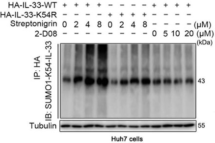
Streptonigrin purchased from MedChemExpress. Usage Cited in: Sci Signal. 2023 Mar 14;16(776):eabq3362. [Abstract]
Streptonigrin (SUMOylation agonist; 2, 4, 8 μM; 48 h) markedly increases the amount of IL-33 protein SUMOylation in Huh7 cells transfected with wild-type IL-33.
-

Protocols
-
Research Protocol for Infectious Diseases
Infectious-disease experiments test how pathogens interact with host barriers, innate immune receptors, inflammatory signaling, pathogen replication, and tissue injury; pattern-recognition receptors such as TLRs, RIG-I-like receptors, NOD-like receptors, and inflammasomes detect microbial molecules and activate NF-κB, interferon, and cytokine responses. The central hypothesis is that infection severity reflects the balance between pathogen burden and host response: protective inflammation restricts pathogen growth, whereas excessive or mislocalized inflammation contributes to tissue damage and disease phenotype. Unresolved questions include which host pathways are protective versus pathogenic, why some infection models fail to translate to human disease, and which combined readouts best predict clinically relevant infection outcomes.
-
Apoptosis
Apoptosis, also called programmed cell death, is generally characterized by distinct morphological characteristics.
-
TUNEL staining for apoptotic DNA fragmentation
TUNEL staining detects DNA strand breaks by using terminal deoxynucleotidyl transferase to add labeled nucleotides to exposed 3′-OH DNA termini, generating either microscopic staining in fixed cells or tissue sections, or fluorescence/cytometric signal in cell suspensions. TUNEL positivity reflects DNA fragmentation but should not be interpreted alone as definitive apoptosis, because TUNEL can also label necrotic, autolytic, mechanically damaged, or DNA-repair-associated DNA breaks.
-
Cell Cytotoxicity Assay
Cytotoxicity assays are usually based on the assessment of cell membrane damage, which can also be indirectly detected by measuring cell viability. Detection methods include MTT assay, CKK-8 assay, LDH assay and ATP assay, etc.
-
Annexin V plus membrane-impermeant dye apoptosis staining
Annexin V-based apoptosis assays rely on the detection of phosphatidylserine (PS) externalization from the inner leaflet of the plasma membrane to the outer leaflet, an early biochemical hallmark of apoptosis. Fluorescently labeled Annexin V binds PS in a calcium-dependent manner, enabling identification of early apoptotic cells by flow cytometry or fluorescence microscopy. When combined with a membrane-impermeant DNA-binding dye (e. g. , propidium iodide), this approach allows discrimination between viable (Annexin V−/dye−), early apoptotic (Annexin V+/dye−), and late apoptotic or necrotic (Annexin V+/dye+) cell populations by assessing membrane integrity and PS exposure.
-
Apoptosis Solutions
Apoptosis is a regulated, generally non-lytic cell-death pathway that removes unwanted, damaged, infected, or abnormal cells through coordinated morphological changes, caspase activation, DNA fragmentation, and membrane remodeling. The intrinsic apoptosis pathway is controlled mainly by mitochondrial outer membrane permeabilization, BCL-2 family proteins, cytochrome c release, apoptosome formation, caspase-9 activation, and downstream executioner caspase-3/7 activation. The extrinsic apoptosis pathway is initiated by death receptors such as Fas, TNFR, and TRAIL receptors, which recruit adaptor proteins and activate caspase-8 before engaging executioner caspases or mitochondrial amplification through BID cleavage. Apoptosis is linked to many phenotypes, including cancer cell killing, tissue homeostasis, immune regulation, neurodegeneration, infection response, and treatment-induced cytotoxicity; unresolved questions include how apoptosis interacts with necroptosis, pyroptosis, ferroptos
-
Subcutaneous Cell-Line-Derived Xenograft
Subcutaneous cell-line-derived xenograft (CDX) models are established by implanting cultured human cancer cell lines into immunodeficient mice, where the injected cells form localized tumors that can be monitored in vivo as a measure of tumorigenic potential, growth kinetics, and treatment response. These models are widely used in oncology research because they allow reproducible tumor formation and enable comparative assessment of tumor growth between different cell lines or genetic manipulations in a controlled in vivo microenvironment. Subcutaneous implantation of cancer cells in immunodeficient mice is a standard approach for evaluating tumor growth behavior and therapeutic response across multiple cancer types, including prostate, esophageal, pancreatic, and colon cancer models.


Purity & Documentation
-
Data Sheet (281 KB)
-
SDS (480 KB)
- English - EN (480 KB)
- Français - FR (480 KB)
- Deutsch - DE (480 KB)
- Norwegian - NO (480 KB)
- Español - ES (480 KB)
- Swedish - SV (480 KB)
- Italian - IT (480 KB)
- Korean - KR (480 KB)
- Portuguese - PT (480 KB)
-
Handling Instructions (2659 KB)
References
[1]. Dreyton CJ, et al. Insights into the mechanism of Streptonigrin-induced protein arginine deiminase inactivation. Bioorg Med Chem. 2014 Feb 15;22(4):1362-9. [Content Brief]
[2]. Park S, et al. Streptonigrin inhibits β-Catenin/Tcf signaling and shows cytotoxicity in β-catenin-activated cells. Biochim Biophys Acta. 2011 Dec;1810(12):1340-5. [Content Brief]
[3]. Piatakova NV, et al. Soluble guanylate cyclase in the molecular mechanism underlying the therapeutic action of drugs]. Biomed Khim. 2012 Jan-Feb;58(1):32-42. Russian. [Content Brief]
[4]. Ambaye N, et al. Streptonigrin Inhibits SENP1 and Reduces the Protein Level of Hypoxia-Inducible Factor 1α (HIF1α) in Cells. Biochemistry. 2018 Mar 20;57(11):1807-1813. [Content Brief]
[5]. Loyola AC, et al. Streptonigrin at low concentration promotes heterochromatin formation. Sci Rep. 2020 Feb 26;10(1):3478. [Content Brief]
[6]. Yamashita Y, et al. Induction of mammalian DNA topoisomerase II dependent DNA cleavage by antitumor antibiotic streptonigrin. Cancer Res. 1990 Sep 15;50(18):5841-4. [Content Brief]
[7]. McBride TJ, et al. The activity of streptonigrin against the Rauscher murine leukemia virus in vivo. Cancer Res. 1966 Apr;26(4):727-32. [Content Brief]
[8]. Suzuki H, et al. Immunosuppressive activity of streptonigrin in vitro and in vivo. Biosci Biotechnol Biochem. 1996 May;60(5):789-93. [Content Brief]
[9]. Kang JH, et al. Inhibition of Transglutaminase 2 but Not of MDM2 Has a Significant Therapeutic Effect on Renal Cell Carcinoma. Cells. 2020 Jun 16;9(6):1475. [Content Brief]
[10]. Long GV, et al. Interaction of the antitumour antibiotic streptonigrin with DNA and oligonucleotides. Anticancer Drug Des. 1997 Sep;12(6):453-72. [Content Brief]

Calculators
Concentration (start) × Volume (start) = Concentration (final) × Volume (final)


Streptonigrin Related Classifications
HY-124586 Related Classifications
 Powered by Bioz