



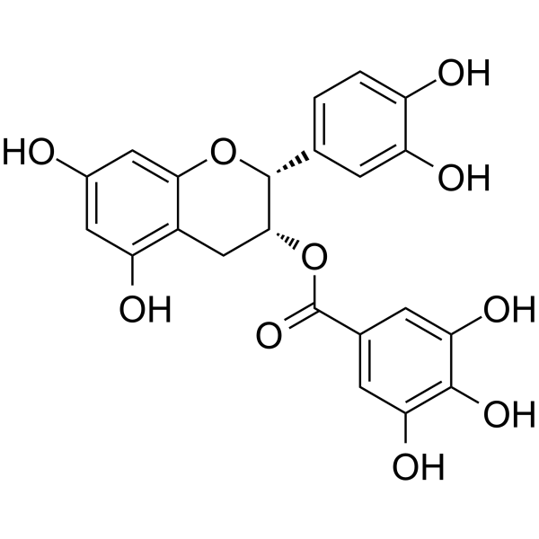
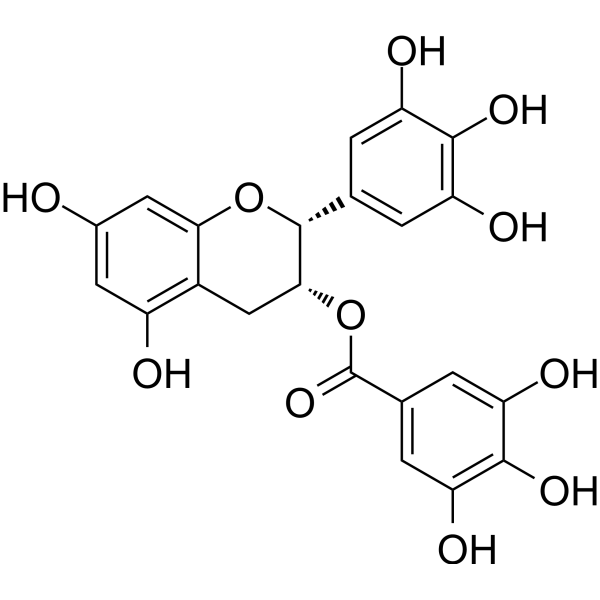
(-)-Epicatechin gallate
Based on 8 publication(s) in Google Scholar
(-)-Epicatechin gallate (Epicatechin gallate) inhibits cyclooxygenase-1 (COX-1) with an IC50 of 7.5 μM. (-)-Epicatechin gallate is a GLUT1 and GLUT5 inhibitor. (-)-Epicatechin gallate decreases cell growth of GLUT5-expressing hxt0 yeast cells.
For research use only. We do not sell to patients.
- Purity : 99.96%
- CAS No.: 1257-08-5
- Formula: C22H18O10
- Molecular Weight:442.37
-
Storage:
4°C, protect from light
* In solvent : -80°C, 6 months; -20°C, 1 month (protect from light)

To place orders, for customer services and technical support, please contact: MedChemExpress USA
Tel: 609-228-6898 E-mail: [email protected] [email protected]


-
 Biological Activity
-
Chemical Information
-
Solvent & Solubility
- Protocols
- Purity & Documentation
- References
-
 Help & FAQs


-
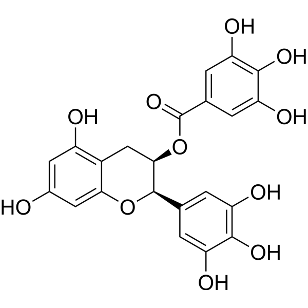
HY-13653
(-)-Epigallocatechin GallateAnticancer Agent -
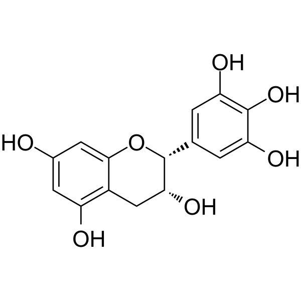
HY-N0225
(-)-EpigallocatechinAutophagy Inducer -
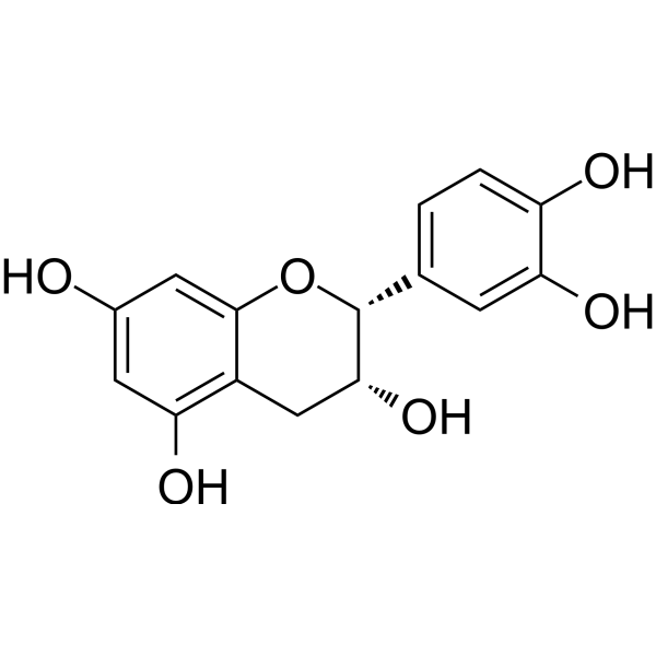
HY-N0001
(-)-EpicatechinCOX Inhibitor -
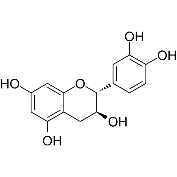
HY-N0898
CatechinCOX-1 Inhibitor -
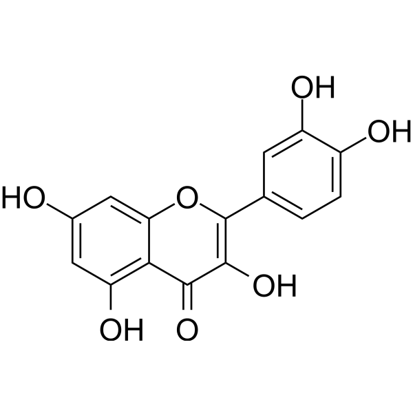
HY-18085
QuercetinFlavonoid Antioxidant -
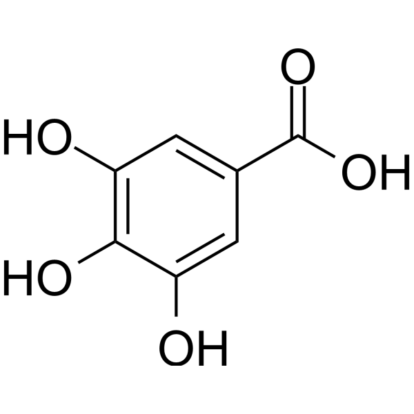
HY-N0523
Gallic acidBioactive Polyhydroxyphenolic Compound -
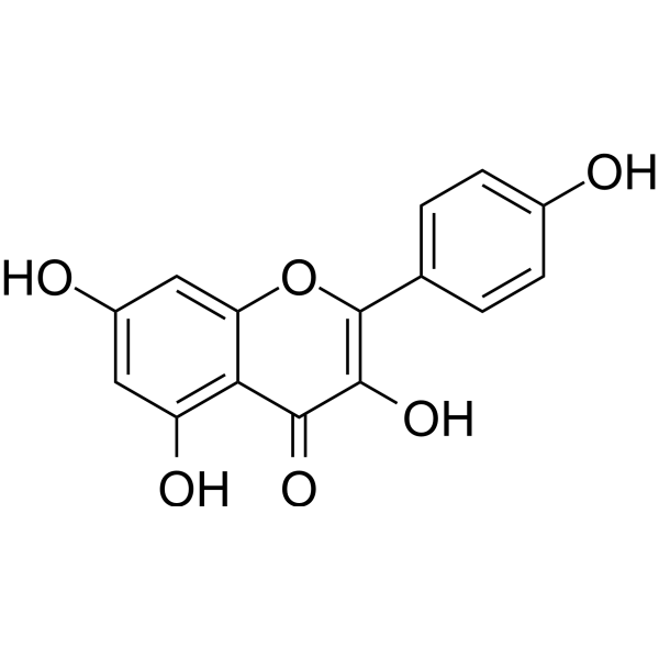
HY-14590
KaempferolEstrogen Receptor α Inhibitor -
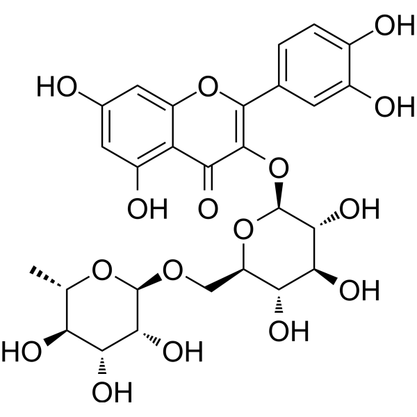
HY-N0148
RutinCBR1 Inhibitor -
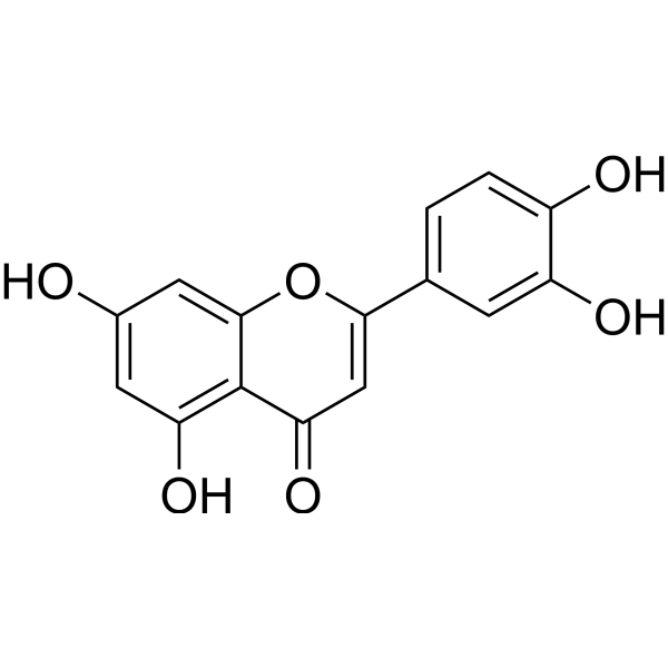
HY-N0162
LuteolinNrf2 Inhibitor -
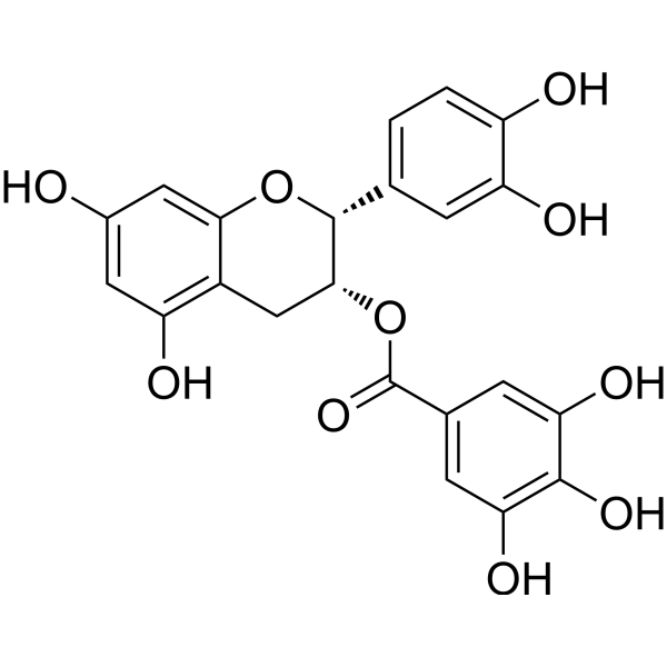
HY-N0002R
(-)-Epicatechin gallate (Standard)Analytical Standard -
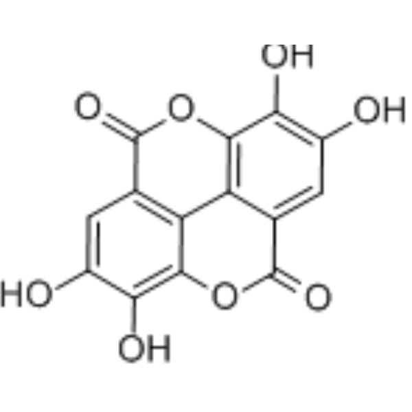
HY-B0183
Ellagic acidSHP2 Inhibitor -
HY-13653R
(-)-Epigallocatechin Gallate (Standard)
Publications Citing Use of MedChemExpress (MCE) (-)-Epicatechin gallate
More- Nat Commun. 2023 Mar 22;14(1):1594. [Abstract]
- Acta Pharm Sin B. 2021 Jan;11(1):143-155. [Abstract]
- Acta Biochim Biophys Sin (Shanghai). 2021 Dec 8;53(12):1662-1669. [Abstract]
- Pathogens. 2026 Mar 20;15(3):334. [Abstract]
- Int J Parasitol Drugs Drug Resist. 2026 Aug:31:100657.
- Arch Biochem Biophys. 2022 Oct 30:729:109393. [Abstract]
- Chem Biodivers. 2025 Oct;22(10):e00662. [Abstract]
- Vet Microbiol. 2026 May:316:110992. [Abstract]
 Customer Validation & Images

-
IF
-
Cell Proliferation/Viability Assay

-
Bio/Physico-chemical Assay

-
WB

-
Cell Migration/Invasion Assay


Biological Activity
Description
|
COX-1 7.5 μM (IC50) |
GLUT1 |
GLUT5 |
Cellular Effect
|
Cell Line

|
Type | Value | Description | References |
|---|---|---|---|---|
| 3T3-L1 | IC50 |
25 μM
Compound: ECG, (-)-epicatechin gallate
|
Inhibition of G6PD-mediated NADPH production in mouse 3T3-L1 cells
Inhibition of G6PD-mediated NADPH production in mouse 3T3-L1 cells
|
[PMID: 18313308] |
| A549 | ED50 |
>10 μg/mL
Compound: 50
|
Cytotoxicity against human A549 cells by tetrazolium salt-based colorimetric assay
Cytotoxicity against human A549 cells by tetrazolium salt-based colorimetric assay
|
[PMID: 1431932] |
| H9 | IC50 |
>10 μg/mL
Compound: 4
|
Inhibition of uninfected H9 lymphocytic cell growth
Inhibition of uninfected H9 lymphocytic cell growth
|
10.1016/0960-894X(96)00095-9 |
| HCT-116 | IC50 |
76 μM
Compound: Epicatechin gallate
|
Cytotoxicity against human HCT116 cells by MTT assay
Cytotoxicity against human HCT116 cells by MTT assay
|
[PMID: 23719279] |
| HCT-8 | ED50 |
>10 μg/mL
Compound: 50
|
Cytotoxicity against human HCT8 cells by tetrazolium salt-based colorimetric assay
Cytotoxicity against human HCT8 cells by tetrazolium salt-based colorimetric assay
|
[PMID: 1431932] |
| KB | ED50 |
>10 μg/mL
Compound: 50
|
Cytotoxicity against human KB cells by tetrazolium salt-based colorimetric assay
Cytotoxicity against human KB cells by tetrazolium salt-based colorimetric assay
|
[PMID: 1431932] |
| LNCaP | IC50 |
36 μM
Compound: ECG
|
Antiproliferative activity against human LNCaP assessed as reduction in cell viability incubated for 72 hrs by MTT assay
Antiproliferative activity against human LNCaP assessed as reduction in cell viability incubated for 72 hrs by MTT assay
|
[PMID: 37776575] |
| MDCK | CC50 |
525.9 μM
Compound: 57; ECG
|
Cytotoxicity against MDCK cells after 48 hrs by MTT assay
Cytotoxicity against MDCK cells after 48 hrs by MTT assay
|
[PMID: 28118010] |
| PC-3 | IC50 |
168.2 μM
Compound: 1
|
In vitro inhibitory concentration against PC-3 cell line in MTT assay was determined after 3 days incubation
In vitro inhibitory concentration against PC-3 cell line in MTT assay was determined after 3 days incubation
|
[PMID: 15380225] |
| SK-OV-3 | IC50 |
185.4 μM
Compound: 1
|
In vitro inhibitory concentration against SKOV3 cell line in MTT assay was determined after 3 days incubation
In vitro inhibitory concentration against SKOV3 cell line in MTT assay was determined after 3 days incubation
|
[PMID: 15380225] |
| TE-671 | ED50 |
>10 μg/mL
Compound: 50
|
Cytotoxicity against human TE671 cells by tetrazolium salt-based colorimetric assay
Cytotoxicity against human TE671 cells by tetrazolium salt-based colorimetric assay
|
[PMID: 1431932] |
| U-373MG ATCC | IC50 |
157 μM
Compound: 1
|
In vitro inhibitory concentration against U373MG cell line in MTT assay was determined after 3 days incubation
In vitro inhibitory concentration against U373MG cell line in MTT assay was determined after 3 days incubation
|
[PMID: 15380225] |
In Vitro
(-)-Epicatechin gallate (Epicatechin gallate) exhibits >95% inhibitory activity at 70 μg/mL against cyclooxygenase-1 (COX-1) with an IC50 of 7.5 μM[1].
MedChemExpress (MCE) has not independently confirmed the accuracy of these methods. They are for reference only. Further protocols information, click here.
In Vivo
MedChemExpress (MCE) has not independently confirmed the accuracy of these methods. They are for reference only.
Clinical Trial
| NCT Number | Sponsor | Condition | Start Date |
Phase
|
|---|---|---|---|---|
| NCT01329991 | Plexxikon| | 2011-05 | PHASE1 |


Chemical Information
-
CAS No. 1257-08-5
-
Appearance Solid
-
Molecular Weight 442.37
-
Formula C22H18O10
-
Color White to off-white
-
SMILES
O=C(O[C@H]1[C@@H](C2=CC=C(O)C(O)=C2)OC3=CC(O)=CC(O)=C3C1)C4=CC(O)=C(O)C(O)=C4
-
Synonyms
Epicatechin gallate; ECG; (-)-Epicatechin 3-O-gallate
-
Structure Classification
-
Initial Source
-
Shipping
Room temperature in continental US; may vary elsewhere.
-
Storage
4°C, protect from light
* In solvent : -80°C, 6 months; -20°C, 1 month (protect from light)


Publications (8)
-
Journal Impact Factor
-
Most Recent
-
Nat Commun
2023 Mar 22;14(1):1594. PMID: 36949052 -
Acta Pharm Sin B
Chrysin serves as a novel inhibitor of DGK α/FAK interaction to suppress the malignancy of esophageal squamous cell carcinoma (ESCC). [Abstract]2021 Jan;11(1):143-155. PMID: 33532186 -
Acta Biochim Biophys Sin (Shanghai)
Epicatechin gallate prevents the de novo synthesis of fatty acid and the migration of prostate cancer cells. [Abstract]2021 Dec 8;53(12):1662-1669. PMID: 34718375 -
Pathogens
2026 Mar 20;15(3):334. PMID: 41901787
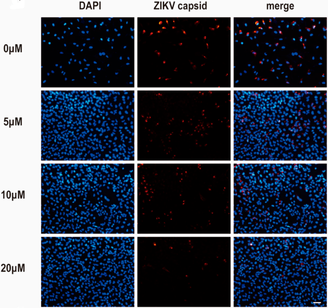
(-)-Epicatechin gallate purchased from MedChemExpress. Usage Cited in: Pathogens. 2026 Mar 20;15(3):334. [Abstract]
Immunofluorescence analysis indicating that ECG ((-)-Epicatechin gallate, 0-20 μM) remarkably suppresses the expression of ZIKV capsid protein (red) in A549 cells.
-
-
Arch Biochem Biophys
(-)-Epicatechin gallate prevents inflammatory response in hypoxia-activated microglia and cerebral edema by inhibiting NF-κB signaling. [Abstract]2022 Oct 30:729:109393. PMID: 36084697
(-)-Epicatechin gallate purchased from MedChemExpress. Usage Cited in: Arch Biochem Biophys. 2022 Oct 30:729:109393. [Abstract]
ECG and EGCG rather than EGC ((-)-Epicatechin gallate, 10-40 μM, 24 h) ameliorated the toxicity of LPS/ATP-induced microglia medium to neuronal cells in a concentration-dependent manner.
(-)-Epicatechin gallate purchased from MedChemExpress. Usage Cited in: Arch Biochem Biophys. 2022 Oct 30:729:109393. [Abstract]
ECG ((-)-Epicatechin gallate) and EGCG rather than EGC inhibited the LPS/ATP-induced nuclear translocation of p65; this outcome was consistent with the protein changes in the inflammatory factors.
(-)-Epicatechin gallate purchased from MedChemExpress. Usage Cited in: Arch Biochem Biophys. 2022 Oct 30:729:109393. [Abstract]
ECG ((-)-Epicatechin gallate, 40 μM, 24 h) significantly reduced the p-p65, NLRP3, and IL-1β in hypoxic microglia, similar to the function of Bay 11-7082.
(-)-Epicatechin gallate purchased from MedChemExpress. Usage Cited in: Arch Biochem Biophys. 2022 Oct 30:729:109393. [Abstract]
Co-cultured bEnd.3 cells and microglia were inoculated in 24-transwell inserts and co-treated with 40 μM ECG ((-)-Epicatechin gallate) and 1% O2 for 24 h. Endothelial tight junction integrity was tested by TEER.
-
Chem Biodivers
Anti-Inflammatory and Immunomodulatory Phytochemicals for Management of Oral Lichen Planus: A Multi-Omics System Biology and Experimental Assessment. [Abstract]2025 Oct;22(10):e00662. PMID: 40522753 -
Vet Microbiol
The Chinese medicine monomer Schisandrin C inhibits PRRSV infection by regulating the OGT-PI3K/AKT/mTOR signaling pathway. [Abstract]2026 May:316:110992. PMID: 41865607


Solvent & Solubility
In Vitro:
DMSO : 100 mg/mL (226.06 mM; Need ultrasonic; Hygroscopic DMSO has a significant impact on the solubility of product, please use newly opened DMSO)
Please refer to the solubility information to select the appropriate solvent. Once prepared, please aliquot and store the solution to prevent product inactivation from repeated freeze-thaw cycles.
Storage method and period of stock solution: -80°C, 6 months; -20°C, 1 month (protect from light). When stored at -80°C, please use it within 6 months. When stored at -20°C, please use it within 1 month.
Please refer to the solubility information to select the appropriate solvent. Once prepared, please aliquot and store the solution to prevent product inactivation from repeated freeze-thaw cycles.
Storage method and period of stock solution: -80°C, 6 months; -20°C, 1 month (protect from light). When stored at -80°C, please use it within 6 months. When stored at -20°C, please use it within 1 month.
Concentration (start) × Volume (start) = Concentration (final) × Volume (final)
In Vivo:
Select the appropriate dissolution method based on your experimental animal and administration route.
- For the following dissolution methods, please ensure to first prepare a clear stock solution using an In Vitro approach and then sequentially add co-solvents:
- To ensure reliable experimental results, the clarified stock solution can be appropriately stored based on storage conditions. As for the working solution for In Vivo experiments, it is recommended to prepare freshly and use it on the same day.
- The percentages shown for the solvents indicate their volumetric ratio in the final prepared solution. If precipitation or phase separation occurs during preparation, heat and/or sonication can be used to aid dissolution.
Add each solvent one by one: 10% DMSO 40% PEG300 5% Tween-80 45% Saline
Solubility: ≥ 2.17 mg/mL (4.91 mM); Clear solution
This protocol yields a clear solution of ≥ 2.17 mg/mL (saturation unknown).
Taking 1 mL working solution as an example, add 100 μL DMSO stock solution (21.7 mg/mL) to 400 μL PEG300, and mix evenly; then add 50 μL Tween-80 and mix evenly; then add 450 μL Saline to adjust the volume to 1 mL.
Preparation of Saline: Dissolve 0.9 g sodium chloride in ddH₂O and dilute to 100 mL to obtain a clear Saline solution.
Add each solvent one by one: 10% DMSO 90% (20% SBE-β-CD in Saline)
Solubility: ≥ 2.17 mg/mL (4.91 mM); Clear solution
This protocol yields a clear solution of ≥ 2.17 mg/mL (saturation unknown).
Taking 1 mL working solution as an example, add 100 μL DMSO stock solution (21.7 mg/mL) to 900 μL 20% SBE-β-CD in Saline, and mix evenly.
Preparation of 20% SBE-β-CD in Saline (4°C, storage for one week): 2 g SBE-β-CD powder is dissolved in 10 mL Saline, completely dissolve until clear.
In Vivo Dissolution Calculator
Please enter the basic information of animal experiments:
-
-
-
-
Recommended: Prepare an additional quantity of animals to account for potential losses during experiments.
Please enter your animal formula composition:
-
%DMSO +
Recommended: Keep the proportion of DMSO in working solution below 2% if your animal is weak.
-
%+
-
+%Tween-80 + +
-
%Saline +
The co-solvents required include: DMSO, . All of co-solvents are available by MedChemExpress (MCE). , Tween 80. All of co-solvents are available by MedChemExpress (MCE).
Working solution concentration: 0.22 mg/mL
Method for preparing stock solution: mg drug dissolved in μL DMSO. Stock solution concentration: mg/mL. * In solvent : -80°C, 6 months; -20°C, 1 month (protect from light)
1. Take μL DMSO stock solution;
2. Add μL .
μL , mix evenly;
3. Then add μL Tween 80, mix evenly;
4. Then add μL
Please ensure that the stock solution in the first step is dissolved to a clear state, and add co-solvents in sequence. You can use ultrasonic heating (ultrasonic cleaner, recommended frequency 20-40 kHz), vortexing, etc. to assist dissolution.

Protocols
-
Cell Cytotoxicity Assay
Cytotoxicity assays are usually based on the assessment of cell membrane damage, which can also be indirectly detected by measuring cell viability. Detection methods include MTT assay, CKK-8 assay, LDH assay and ATP assay, etc.
-
Protocol for Cell Counting and Cell Density Analysis
Cell counting and cell-density analysis estimate the number of cells in a known volume or field area. Manual hemocytometer counting uses a chamber of defined geometry to convert counted cells into cells/mL, while automated counters and image-analysis workflows detect cell objects from optical, brightfield, fluorescence, impedance, or digital-image features. Trypan blue viability counting is based on dye exclusion: viable cells with intact membranes exclude dye, while non-viable cells with compromised membranes stain blue. The readout is total cell density, viable-cell density, dead-cell density, and percent viability. Cell density can also be estimated from microscopy images by counting objects per image area, from flow cytometry using calibrated volume or reference particles, or from in situ microscopy in bioreactors after calibration against reference methods such as hemocytometer or flow cytometry.
-
Somatic Cell Culture
A method of simulating the in vivo environment in vitro to maintain the cell growth, differentation and main functions.
-
CCK-8/WST-8 Cell Proliferation Assay
The CCK-8/WST-8 assay is based on the reduction of the water-soluble tetrazolium salt WST-8 to a water-soluble formazan product by cellular dehydrogenases in metabolically active cells, where the generated formazan amount is proportional to the number of living cells and is quantified by measuring absorbance in the visible range, providing a colorimetric readout for cell viability and proliferation assessment. This class of tetrazolium-based assays improves upon earlier MTT-based systems by producing a water-soluble formazan, eliminating the need for organic solubilization steps and enabling direct spectrophotometric measurement in culture medium.
-
Cell Counting-Based Growth Curve Assay
Cell counting-based growth curve assays quantify cell proliferation by directly measuring changes in viable cell number over time using manual or automated counting methods such as hemocytometer-based counting or instrument-assisted cell enumeration, enabling construction of growth curves that reflect population expansion dynamics in response to culture conditions. A widely used approach is trypan blue exclusion with hemocytometer counting, where membrane-compromised (non-viable) cells take up the dye, allowing discrimination between viable and non-viable cells while simultaneously enabling total cell number quantification. Repeated sampling across time points allows estimation of proliferation rate, growth phases, and comparative growth kinetics between experimental conditions.
-
MTT Cell Proliferation Assay
The MTT assay is a colorimetric endpoint assay for estimating viable cell number, cell growth, cytotoxicity, or cell activation in cultured mammalian cells. Living cells reduce the yellow tetrazolium salt MTT into purple/blue formazan, while dead cells do not generate the same signal; the resulting color can be quantified with a multiwell spectrophotometer. MTT reduction is commonly interpreted as a readout of metabolic activity that often correlates with viable cell number, but it should not be treated as a direct cell-counting method unless the assay is optimized for the cell type and experimental condition. Studies show that MTT reduction can involve mitochondrial and non-mitochondrial reducing systems, and formazan may accumulate in intracellular lipid droplets rather than simply marking mitochondria.
-
Cell Viability Determination by MTT Colorimetric Assay
The following protocol uses the MTT colorimetric assay as a classic literature-established method for assessing cell viability/metabolic activity in cultured mammalian cells. MTT[3-(4,5-dimethylthiazol-2-yl)-2,5-diphenyltetrazolium bromide] is reduced by metabolically active cells to a colored formazan product; the amount of formazan is quantified spectrophotometrically and provides an indirect measure of metabolically active viable cells. Importantly, MTT reduction reflects cellular oxidoreductase/metabolic activity rather than an absolute direct count of living cells, so changes in cellular metabolism can alter the signal independently of cell number.


Purity & Documentation
-
Data Sheet (281 KB)
-
SDS (396 KB)
- English - EN (396 KB)
- Français - FR (396 KB)
- Deutsch - DE (396 KB)
- Norwegian - NO (396 KB)
- Español - ES (396 KB)
- Swedish - SV (396 KB)
- Italian - IT (396 KB)
- Korean - KR (396 KB)
- Portuguese - PT (396 KB)
-
Handling Instructions (2659 KB)
References
[1]. Waffo-Téguo P, et al. Potential cancer-chemopreventive activities of wine stilbenoids and flavans extracted from grape (Vitis vinifera) cell cultures. Nutr Cancer. 2001;40(2):173-9. [Content Brief]
[2]. Takizawa Y, et al. Pharmacokinetics of (-)-epicatechin-3-O-gallate, an active component of Onpi-to, in rats. Biol Pharm Bull. 2003 May;26(5):608-12. [Content Brief]
[3]. Tripp J, et al. Establishing a yeast-based screening system for discovery of human GLUT5 inhibitors and activators. Sci Rep. 2017 Jul 24;7(1):6197. [Content Brief]


Complete Stock Solution Preparation Table
Please refer to the solubility information to select the appropriate solvent. Once prepared, please aliquot and store the solution to prevent product inactivation from repeated freeze-thaw cycles.
Storage method and period of stock solution: -80°C, 6 months; -20°C, 1 month (protect from light). When stored at -80°C, please use it within 6 months. When stored at -20°C, please use it within 1 month.
| Optional Solvent | Concentration Solvent Mass | 1 mg | 5 mg | 10 mg | 25 mg |
|---|---|---|---|---|---|
| DMSO | 1 mM | 2.2606 mL | 11.3028 mL | 22.6055 mL | 56.5138 mL |
| 5 mM | 0.4521 mL | 2.2606 mL | 4.5211 mL | 11.3028 mL | |
| 10 mM | 0.2261 mL | 1.1303 mL | 2.2606 mL | 5.6514 mL | |
| 15 mM | 0.1507 mL | 0.7535 mL | 1.5070 mL | 3.7676 mL | |
| 20 mM | 0.1130 mL | 0.5651 mL | 1.1303 mL | 2.8257 mL | |
| 25 mM | 0.0904 mL | 0.4521 mL | 0.9042 mL | 2.2606 mL | |
| 30 mM | 0.0754 mL | 0.3768 mL | 0.7535 mL | 1.8838 mL | |
| 40 mM | 0.0565 mL | 0.2826 mL | 0.5651 mL | 1.4128 mL | |
| 50 mM | 0.0452 mL | 0.2261 mL | 0.4521 mL | 1.1303 mL | |
| 60 mM | 0.0377 mL | 0.1884 mL | 0.3768 mL | 0.9419 mL | |
| 80 mM | 0.0283 mL | 0.1413 mL | 0.2826 mL | 0.7064 mL | |
| 100 mM | 0.0226 mL | 0.1130 mL | 0.2261 mL | 0.5651 mL |


(-)-Epicatechin gallate Related Classifications
HY-N0002 Related Classifications
 Powered by Bioz