



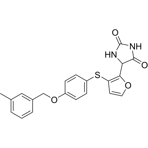
Aderamastat

Based on 1 Customer Validation
Aderamastat (FP-025) is an orally active, selective MMP-12 inhibitor. Aderamastat provides protection against house dust mite (HDM)-sensitized allergic asthma. Aderamastat can be used in the research of respiratory diseases, including chronic inflammatory airway diseases and pulmonary fibrosis.
Nos produits utilisent uniquement pour la recherche. Nous ne vendons pas aux patients.
- Pureté : 99.02%
- CAS No.: 877176-23-3
- Formule: C21H18N2O4S
- Masse moléculaire:394.44
-
Stockage:Powder -20°C, 3 years , 4°C, 2 years ; In solvent -80°C, 6 months , -20°C, 1 month

To place orders, for customer services and technical support, please contact: MedChemExpress USA
Tel: 609-228-6898 E-mail: [email protected] [email protected]


-
 Activité biologique
-
Chemical Information
-
Solvant et solubilité
- Protocole
- Pureté et documentation
- Références
-
 Help & FAQs




Activité biologique
Description
IC50 & Target
[3]|
MMP-12 |
In Vivo
MedChemExpress (MCE) has not independently confirmed the accuracy of these methods. They are for reference only.
-
Animal Model:C57BL/6J mice + HDM-sensitized allergic asthma model[3]
-
Dosage:10 mg/kg, 30 mg/kg, 100 mg/kg
-
Administration:oral administration, daily, 7 days
-
Result:Did not induce behavioral or physical signs of discomfort nor differences in body weight in mice even at the highest dose.
Abrogated AHR to a similar extent as 5 mg/kg Prednisone, significantly reduced the number of total BAL cells, eosinophils, neutrophils, macrophages, inflammatory-migratory dendritic cells (DCs), B lymphocytes and CD4+ T lymphocytes (but not CD8+ T lymphocytes).
Reduced peri-bronchial and peri-arterial cellular infiltrates in a dose-dependent manner.
Decreased lung fibrosis (assessed by α-SMA stain) and mucus production (assessed by PAS stain).
Highly reduced MMP-12 expression in bronchial epithelial cells, and dose-dependently reduced MMP-12 levels in BAL and lung parenchyma.
Did not affect the number of total inflammatory cells and various immune cells (eosinophils, neutrophils, macrophages, CD11b+ DCs, CD4+ lymphocytes, CD8+ lymphocytes, B lymphocytes) in lung parenchyma, nor the levels of mediators of innate, Th1- and Th2-responses, chemoattractant chemokines and growth factors.
Essai clinique
| NCT Number | Sponsor | Condition | Start Date |
Phase

|
|---|---|---|---|---|
| NCT01329991 | 2011-05 | PHASE1 |


Chemical Information
-
CAS No. 877176-23-3
-
Appearance Solid
-
Masse moléculaire 394.44
-
Formule C21H18N2O4S
-
Color White to light yellow
-
SMILES
O=C1NC(C(C2=C(SC3=CC=C(C=C3)OCC4=CC(C)=CC=C4)C=CO2)N1)=O
-
Synonyms
FP-025
-
Livraison
Room temperature in continental US; may vary elsewhere.
-
Stockage
Powder -20°C 3 years 4°C 2 years In solvent -80°C 6 months -20°C 1 month


Solvant et solubilité
In Vitro:
DMSO : 100 mg/mL (253.52 mM; Need ultrasonic; Hygroscopic DMSO has a significant impact on the solubility of product, please use newly opened DMSO)
Please refer to the solubility information to select the appropriate solvent. Once prepared, please aliquot and store the solution to prevent product inactivation from repeated freeze-thaw cycles.
Storage method and period of stock solution: -80°C, 6 months; -20°C, 1 month. When stored at -80°C, please use it within 6 months. When stored at -20°C, please use it within 1 month.
Please refer to the solubility information to select the appropriate solvent. Once prepared, please aliquot and store the solution to prevent product inactivation from repeated freeze-thaw cycles.
Storage method and period of stock solution: -80°C, 6 months; -20°C, 1 month. When stored at -80°C, please use it within 6 months. When stored at -20°C, please use it within 1 month.
Concentration (start) × Volume (start) = Concentration (final) × Volume (final)
In Vivo:
Select the appropriate dissolution method based on your experimental animal and administration route.
- For the following dissolution methods, please ensure to first prepare a clear stock solution using an In Vitro approach and then sequentially add co-solvents:
- To ensure reliable experimental results, the clarified stock solution can be appropriately stored based on storage conditions. As for the working solution for In Vivo experiments, it is recommended to prepare freshly and use it on the same day.
- The percentages shown for the solvents indicate their volumetric ratio in the final prepared solution. If precipitation or phase separation occurs during preparation, heat and/or sonication can be used to aid dissolution.
Add each solvent one by one: 10% DMSO 40% PEG300 5% Tween-80 45% Saline
Solubility: 2.5 mg/mL (6.34 mM); Clear solution; Need ultrasonic
This protocol yields a clear solution of 2.5 mg/mL.
Taking 1 mL working solution as an example, add 100 μL DMSO stock solution (25.0 mg/mL) to 400 μL PEG300, and mix evenly; then add 50 μL Tween-80 and mix evenly; then add 450 μL Saline to adjust the volume to 1 mL.
Preparation of Saline: Dissolve 0.9 g sodium chloride in ddH₂O and dilute to 100 mL to obtain a clear Saline solution.
Add each solvent one by one: 10% DMSO 90% (20% SBE-β-CD in Saline)
Solubility: 2.5 mg/mL (6.34 mM); Clear solution; Need ultrasonic
This protocol yields a clear solution of 2.5 mg/mL.
Taking 1 mL working solution as an example, add 100 μL DMSO stock solution (25.0 mg/mL) to 900 μL 20% SBE-β-CD in Saline, and mix evenly.
Preparation of 20% SBE-β-CD in Saline (4°C, storage for one week): 2 g SBE-β-CD powder is dissolved in 10 mL Saline, completely dissolve until clear.
In Vivo Dissolution Calculator
Please enter the basic information of animal experiments:
-
-
-
-
Recommended: Prepare an additional quantity of animals to account for potential losses during experiments.
Please enter your animal formula composition:
-
%DMSO +
Recommended: Keep the proportion of DMSO in working solution below 2% if your animal is weak.
-
%+
-
+%Tween-80 + +
-
%Saline +
The co-solvents required include: DMSO, . All of co-solvents are available by MedChemExpress (MCE). , Tween 80. All of co-solvents are available by MedChemExpress (MCE).
Working solution concentration: 0.22 mg/mL
Method for preparing stock solution: mg drug dissolved in μL DMSO. Stock solution concentration: mg/mL.
1. Take μL DMSO stock solution;
2. Add μL .
μL , mix evenly;
3. Then add μL Tween 80, mix evenly;
4. Then add μL
Please ensure that the stock solution in the first step is dissolved to a clear state, and add co-solvents in sequence. You can use ultrasonic heating (ultrasonic cleaner, recommended frequency 20-40 kHz), vortexing, etc. to assist dissolution.


Pureté et documentation
-
Fiche technique (275 KB)
-
SDS (254 KB)
- English - EN (254 KB)
- Français - FR (254 KB)
- Deutsch - DE (254 KB)
- Norwegian - NO (254 KB)
- Español - ES (254 KB)
- Swedish - SV (254 KB)
- Italian - IT (254 KB)
- Korean - KR (254 KB)
- Portuguese - PT (254 KB)
-
Instruction de manipulation (2659 KB)
Références
[1]. Abd-Elaziz KS, et al. Validation of a method for the determination of Aderamastat (FP-025) in K2EDTA human plasma by LC-MS/MS. J Chromatogr B Analyt Technol Biomed Life Sci. 2024 Sep 15;1245:124244. [Content Brief]
[2]. Abd-Elaziz K, et al. First-in-Man Safety, Tolerability, and Pharmacokinetics of a Novel and Highly Selective Inhibitor of Matrix Metalloproteinase-12, FP-025: Results from Two Randomized Studies in Healthy Subjects. Clin Drug Investig. 2021 Jan;41(1):65-76. [Content Brief]
[3]. Ravanetti L, et al. Efficacy of FP-025: A novel matrix metalloproteinase-12 (MMP-12) inhibitor in murine allergic asthma. Allergy. 2023 Feb;78(2):559-562. [Content Brief]


Complete Stock Solution Preparation Table
Please refer to the solubility information to select the appropriate solvent. Once prepared, please aliquot and store the solution to prevent product inactivation from repeated freeze-thaw cycles.
Storage method and period of stock solution: -80°C, 6 months; -20°C, 1 month. When stored at -80°C, please use it within 6 months. When stored at -20°C, please use it within 1 month.
| Optional Solvent | Concentration Solvent Mass | 1 mg | 5 mg | 10 mg | 25 mg |
|---|---|---|---|---|---|
| DMSO | 1 mM | 2.5352 mL | 12.6762 mL | 25.3524 mL | 63.3810 mL |
| 5 mM | 0.5070 mL | 2.5352 mL | 5.0705 mL | 12.6762 mL | |
| 10 mM | 0.2535 mL | 1.2676 mL | 2.5352 mL | 6.3381 mL | |
| 15 mM | 0.1690 mL | 0.8451 mL | 1.6902 mL | 4.2254 mL | |
| 20 mM | 0.1268 mL | 0.6338 mL | 1.2676 mL | 3.1690 mL | |
| 25 mM | 0.1014 mL | 0.5070 mL | 1.0141 mL | 2.5352 mL | |
| 30 mM | 0.0845 mL | 0.4225 mL | 0.8451 mL | 2.1127 mL | |
| 40 mM | 0.0634 mL | 0.3169 mL | 0.6338 mL | 1.5845 mL | |
| 50 mM | 0.0507 mL | 0.2535 mL | 0.5070 mL | 1.2676 mL | |
| 60 mM | 0.0423 mL | 0.2113 mL | 0.4225 mL | 1.0563 mL | |
| 80 mM | 0.0317 mL | 0.1585 mL | 0.3169 mL | 0.7923 mL | |
| 100 mM | 0.0254 mL | 0.1268 mL | 0.2535 mL | 0.6338 mL |


Aderamastat Related Classifications
HY-156963 Related Classifications
 Powered by Bioz