



Acyclovir sodium
Based on 26 publication(s) in Google Scholar
Acyclovir (Aciclovir) sodium is a potent, orally active antiviral agent. Acyclovir sodium has antiherpetic activity with IC50 values of 0.85 μM and 0.86 μM for HSV-1 and HSV-2, respectively. Acyclovir sodium induces cell cycle perturbation and apoptosis. Acyclovir sodium prevents bacterial infections during induction therapy for acute leukaemia.
Nos produits utilisent uniquement pour la recherche. Nous ne vendons pas aux patients.
- Pureté : 99.82%
- CAS No.: 69657-51-8
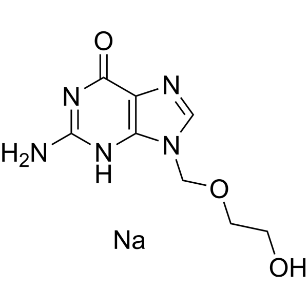
- Formule: C8H11N5NaO3
- Masse moléculaire:248.19
-
Stockage:Powder -20°C, 3 years , 4°C, 2 years ; In solvent -80°C, 6 months , -20°C, 1 month

To place orders, for customer services and technical support, please contact: MedChemExpress USA
Tel: 609-228-6898 E-mail: [email protected] [email protected]


-
 Activité biologique
-
Chemical Information
-
Solvant et solubilité
- Protocole
- Pureté et documentation
- Références
-
 Help & FAQs


Publications Citing Use of MedChemExpress (MCE) Acyclovir sodium
More- Signal Transduct Target Ther. 2025 Dec 15;10(1):406. [Abstract]
- Mol Cell. 2026 Feb 19;86(4):722-739.e8. [Abstract]
- Sci Adv. 2025 Nov 21;11(47):eadw4206. [Abstract]
- Phytomedicine. 2026 Feb:151:157802. [Abstract]
- Phytomedicine. 2024 Nov:134:156015. [Abstract]
- Acta Pharmacol Sin. 2024 Nov;45(11):2380-2393. [Abstract]
- EMBO J. 2025 Apr;44(7):2157-2182. [Abstract]
- Cell Rep. 2026 Mar 12;45(3):117099. [Abstract]
- Eur J Med Chem. 2023 Mar 15:250:115184. [Abstract]
- Ocul Surf. 2026 Jul 16:42:129-143.
- Drug Des Devel Ther. 2022 Dec 20:16:4311-4323. [Abstract]
- Front Microbiol. 2021 Jun 18:12:691008. [Abstract]
- Microchem J. 2021, 106587.
- Mol Med Rep. 2025 Jul;32(1):203. [Abstract]
- Antiviral Res. 2024 Aug:228:105936. [Abstract]
- Antiviral Res. 2024 Jan:221:105787. [Abstract]
- Virol J. 2025 Jul 19;22(1):249. [Abstract]
- Microb Pathog. 2026 Apr:213:108349. [Abstract]
- J Med Virol. 2023 Jan;95(1):e28226. [Abstract]
- Front Oncol. 2024 Dec 19:14:1501981. [Abstract]
- Vet Microbiol. 2026 Jun 17:320:111115. [Abstract]
- SSRN. 2026 Jun 24.
- Zoonoses. 2026 Jun 5.
- bioRxiv. 2026 May 4.
- University of Auckland. 2025.
- Biomed Pharmacother. 2023 Jun:162:114595. [Abstract]
 Customer Validation & Images

-
Cell Proliferation/Viability Assay
-
Cell Imaging/Staining
-
Bio/Physico-chemical Assay
-
Cell Proliferation/Viability Assay
-
WB
Voir tous les produits spécifiques à Isoform Antibiotic
More

Activité biologique
Description
IC50 & Target
[2]|
HSV-1 0.85 μM (IC50) |
HSV-2 0.86 μM (IC50) |
In Vitro
Acyclovir (Aciclovir) sodium (3-100 µM; 24-72 hours; Jurkat, U937, and K562 leukemia cells) reduces cell viability in a dose- and time-dependent[1].
Acyclovir (Aciclovir) sodium (10-100 µM; 24-72 hours; Jurkat cells) blocks DNA synthesis, thereby arresting the cell cycle in G2/M and S phases and increasing the sub-G1 hypodiploid peak in a dose-dependent manner[1].
Acyclovir (Aciclovir) sodium (10-100 µM; 24-72 hours; Jurkat cells) induces apoptosis through activates caspase-3 and presences nuclear DNA fragmentation[1].
MedChemExpress (MCE) has not independently confirmed the accuracy of these methods. They are for reference only. Further protocols information, click here.
-
Cell Line:Jurkat, U937 and K562 leukemia cells
-
Concentration:3, 10, 30 and 100 µM
-
Incubation Time:24, 48 and 72 hours
-
Result:Showed a dose- and time-dependent reduction of cell viability.
-
Cell Line:Jurkat cells
-
Concentration:10 and 100 µM
-
Incubation Time:24, 48 and 72 hours
-
Result:Increased of caspase-3 activity and cleavaged the internucleosomal DNA.
-
Cell Line:Jurkat cells
-
Concentration:10 and 100 µM
-
Incubation Time:24, 48 and 72 hours
-
Result:Revealed a dose-dependent accumulation of cells in S phase after 24 and 48 h. Showed a dose-dependent increase of the sub-G1 hypodiploid peak after 72 h.
In Vivo
MedChemExpress (MCE) has not independently confirmed the accuracy of these methods. They are for reference only.
-
Animal Model:Specific-pathogen-free BALB/c mice (7-week-old) infected with HSV-1[3]
-
Dosage:20 mg/kg
-
Administration:Oral administration; three times daily; for 10 days
-
Result:Suppressed the development of skin lesions and resulted in a dissociation between DTH response and antibody production.
Essai clinique
| NCT Number | Sponsor | Condition | Start Date |
Phase

|
|---|---|---|---|---|
| NCT01329991 | Plexxikon| | 2011-05 | PHASE1 |


Chemical Information
-
CAS No. 69657-51-8
-
Appearance Solid
-
Masse moléculaire 248.19
-
Formule C8H11N5NaO3
-
Color White to off-white
-
SMILES
O=C1N=C(N)NC2=C1N=CN2COCCO.[Na]
-
Synonyms
Aciclovir sodium; Acycloguanosine sodium
-
Livraison
Room temperature in continental US; may vary elsewhere.
-
Stockage
Powder -20°C 3 years 4°C 2 years In solvent -80°C 6 months -20°C 1 month


Publications (26)
-
Journal Impact Factor
-
Most Recent
-
Signal Transduct Target Ther
Selective depletion of tumor-associated SAMHD1 enhances chemotherapeutic efficacy and antitumor immune responses. [Abstract]2025 Dec 15;10(1):406. PMID: 41392286 -
Mol Cell
Asparagine sensing by TBK1 controls its phase separation to drive antiviral innate immune responses. [Abstract]2026 Feb 19;86(4):722-739.e8. PMID: 41653919 -
Sci Adv
Transition of pseudorabies virus from latency to reactivation state selectively triggered by pathogenic bacteria. [Abstract]2025 Nov 21;11(47):eadw4206. PMID: 41259522
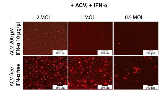
Acyclovir sodium purchased from MedChemExpress. Usage Cited in: Sci Adv. 2025 Nov 21;11(47):eadw4206. [Abstract]
Fluorescence microscopy images depicting N2a cells infected with different doses of HuBXY/2018-mCherry with or without Acyclovir (ACV: 200 μM) and IFN-α treatments. The images were acquired on 8 dpi.
-
Phytomedicine
Inhibition of herpes simplex virus infection by Caesappanin C targeting virus gB protein and host HSP90β. [Abstract]2026 Feb:151:157802. PMID: 41544463
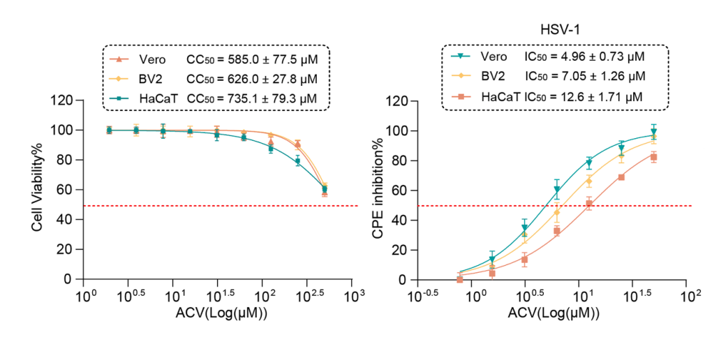
Acyclovir sodium purchased from MedChemExpress. Usage Cited in: Phytomedicine. 2026 Feb:151:157802. [Abstract]
The antiviral activity of Acyclovir (ACV) against HSV-1 in different cell cultures was evaluated using the CPE inhibition assay (MOI = 1).
-
Phytomedicine
Salvianolic acid A inhibits pseudorabies virus infection by directly inactivating the virus particle. [Abstract]2024 Nov:134:156015. PMID: 39244942 -
Acta Pharmacol Sin
A small molecule compound targeting hemagglutinin inhibits influenza A virus and exhibits broad-spectrum antiviral activity. [Abstract]2024 Nov;45(11):2380-2393. PMID: 38987389
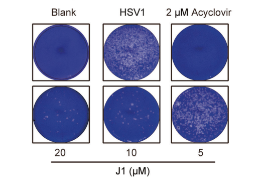
Acyclovir sodium purchased from MedChemExpress. Usage Cited in: Acta Pharmacol Sin. 2024 Nov;45(11):2380-2393. [Abstract]
Acyclovir (2 μM). The inhibitory effect of J1 on HSV1 (MOI = 0.1) infection in Vero cells was analyzed by plaque assay.
-
EMBO J
A novel biosensor for the spatiotemporal analysis of STING activation during innate immune responses to dsDNA. [Abstract]2025 Apr;44(7):2157-2182. PMID: 39984755
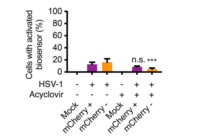
Acyclovir sodium purchased from MedChemExpress. Usage Cited in: EMBO J. 2025 Apr;44(7):2157-2182. [Abstract]
HeLa SIRF biosensor cells were analyzed 24 hours after being treated with a simulated method or infected with 0.5 MOI of HSV-1 virus expressing mCherry, with or without Acyclovir (50 µM).
-
Cell Rep
Enucleated cells with Nectin-1 overexpression capture HSV-1 and promote viral elimination for herpes simplex encephalitis therapy. [Abstract]2026 Mar 12;45(3):117099. PMID: 41824450 -
Eur J Med Chem
Design and syntheses of a bimolecular STING agonist based on the covalent STING antagonist. [Abstract]2023 Mar 15:250:115184. PMID: 36758305 -
-
Drug Des Devel Ther
2022 Dec 20:16:4311-4323. PMID: 36573068 -
Front Microbiol
In vitro Studies and Clinical Observations Imply a Synergistic Effect Between Epstein-Barr Virus and Dengue Virus Infection. [Abstract]2021 Jun 18:12:691008. PMID: 34220783 -
-
Mol Med Rep
Identification of metabolites associated with capecitabine‑induced hand‑foot syndrome using untargeted metabolomics in patients with cancer. [Abstract]2025 Jul;32(1):203. PMID: 40377002 -
Antiviral Res
Flexible nano-liposomes-encapsulated recombinant UL8-siRNA (r/si-UL8) based on bioengineering strategy inhibits herpes simplex virus-1 infection. [Abstract]2024 Aug:228:105936. PMID: 38908520 -
Antiviral Res
Pralatrexate inhibited the replication of varicella zoster virus and vesicular stomatitis virus: An old dog with new tricks. [Abstract]2024 Jan:221:105787. PMID: 38145756 -
Virol J
Small molecule UCM05 inhibits HSV-2 infection via targeting viral glycoproteins and fatty acid synthase with potentiating antiviral immunity. [Abstract]2025 Jul 19;22(1):249. PMID: 40684179 -
Microb Pathog
mTORC2-dependent autophagy inhibition regulates the replication of HSV-1 and adenovirus in viral keratitis & conjunctivitis. [Abstract]2026 Apr:213:108349. PMID: 41628839 -
J Med Virol
Small molecule RAF265 as an antiviral therapy acts against HSV-1 by regulating cytoskeleton rearrangement and cellular translation machinery. [Abstract]2023 Jan;95(1):e28226. PMID: 36251738 -
Front Oncol
Gut virome dysbiosis impairs antitumor immunity and reduces 5-fluorouracil treatment efficacy for colorectal cancer. [Abstract]2024 Dec 19:14:1501981. PMID: 39791120 -
Vet Microbiol
Pseudorabies virus EP0 recruits TECPR2 and CK2 to promote COPII accumulation and viral egress. [Abstract]2026 Jun 17:320:111115. PMID: 42322707 -
-
-
-
-
Biomed Pharmacother
A potential anti-HIV-1 compound, Q308, inhibits HSV-2 infection and replication in vitro and in vivo. [Abstract]2023 Jun:162:114595. PMID: 36989723
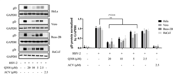
Acyclovir sodium purchased from MedChemExpress. Usage Cited in: Biomed Pharmacother. 2023 Jun:162:114595. [Abstract]
Acyclovir (ACV; 2.5 μM; 48 h) significantly inhibits the expression of gD in HSV-2-infected HeLa, Vero, Beas-2B and HaCaT cells.


Solvant et solubilité
In Vitro:
H2O : 6.25 mg/mL (25.18 mM; Need ultrasonic)
Please refer to the solubility information to select the appropriate solvent. Once prepared, please aliquot and store the solution to prevent product inactivation from repeated freeze-thaw cycles.
Storage method and period of stock solution: -80°C, 6 months; -20°C, 1 month. When stored at -80°C, please use it within 6 months. When stored at -20°C, please use it within 1 month.
* Note: If you choose water as the stock solution, please dilute it to the working solution, then filter and sterilize it with a 0.22 μm filter before use.
Please refer to the solubility information to select the appropriate solvent. Once prepared, please aliquot and store the solution to prevent product inactivation from repeated freeze-thaw cycles.
Storage method and period of stock solution: -80°C, 6 months; -20°C, 1 month. When stored at -80°C, please use it within 6 months. When stored at -20°C, please use it within 1 month.
* Note: If you choose water as the stock solution, please dilute it to the working solution, then filter and sterilize it with a 0.22 μm filter before use.
Concentration (start) × Volume (start) = Concentration (final) × Volume (final)

Protocole
-
Research Protocol for Infectious Diseases
Infectious-disease experiments test how pathogens interact with host barriers, innate immune receptors, inflammatory signaling, pathogen replication, and tissue injury; pattern-recognition receptors such as TLRs, RIG-I-like receptors, NOD-like receptors, and inflammasomes detect microbial molecules and activate NF-κB, interferon, and cytokine responses. The central hypothesis is that infection severity reflects the balance between pathogen burden and host response: protective inflammation restricts pathogen growth, whereas excessive or mislocalized inflammation contributes to tissue damage and disease phenotype. Unresolved questions include which host pathways are protective versus pathogenic, why some infection models fail to translate to human disease, and which combined readouts best predict clinically relevant infection outcomes.
-
Apoptosis
Apoptosis, also called programmed cell death, is generally characterized by distinct morphological characteristics.
-
TUNEL staining for apoptotic DNA fragmentation
TUNEL staining detects DNA strand breaks by using terminal deoxynucleotidyl transferase to add labeled nucleotides to exposed 3′-OH DNA termini, generating either microscopic staining in fixed cells or tissue sections, or fluorescence/cytometric signal in cell suspensions. TUNEL positivity reflects DNA fragmentation but should not be interpreted alone as definitive apoptosis, because TUNEL can also label necrotic, autolytic, mechanically damaged, or DNA-repair-associated DNA breaks.
-
Cell Cytotoxicity Assay
Cytotoxicity assays are usually based on the assessment of cell membrane damage, which can also be indirectly detected by measuring cell viability. Detection methods include MTT assay, CKK-8 assay, LDH assay and ATP assay, etc.
-
Flow cytometric DNA-content cell-cycle staining
Flow cytometric DNA-content cell-cycle staining measures the fluorescence intensity of DNA-bound fluorochromes in single cells or nuclei to estimate DNA content distributions, allowing assignment of populations to G0/G1, S, and G2/M phases by DNA histogram deconvolution. Propidium iodide (PI) intercalates into DNA, and PI fluorescence is proportional to cellular DNA content when staining is performed under conditions that make DNA accessible and minimize non-DNA signal. Cells with G2/M DNA content are expected to show approximately twice the fluorescence intensity of G0/G1 cells, while S-phase cells occupy intermediate fluorescence values. PI-based DNA-content analysis can also detect cells with fractional DNA content, often reported as sub-G1, when DNA fragmentation and extraction during staining reduce retained DNA signal in apoptotic cells. DAPI is an alternative DNA fluorochrome for univariate DNA-content analysis, while bivariate approaches combining DNA content with proliferation
-
Annexin V plus membrane-impermeant dye apoptosis staining
Annexin V-based apoptosis assays rely on the detection of phosphatidylserine (PS) externalization from the inner leaflet of the plasma membrane to the outer leaflet, an early biochemical hallmark of apoptosis. Fluorescently labeled Annexin V binds PS in a calcium-dependent manner, enabling identification of early apoptotic cells by flow cytometry or fluorescence microscopy. When combined with a membrane-impermeant DNA-binding dye (e. g. , propidium iodide), this approach allows discrimination between viable (Annexin V−/dye−), early apoptotic (Annexin V+/dye−), and late apoptotic or necrotic (Annexin V+/dye+) cell populations by assessing membrane integrity and PS exposure.
-
BrdU Incorporation Assay
Bromodeoxyuridine (BrdU) incorporation assay is based on the principle that BrdU, a thymidine analog, is incorporated into newly synthesized DNA during the S phase of the cell cycle, thereby serving as a marker of DNA replication and cellular proliferation. Incorporated BrdU can be detected using anti-BrdU antibodies following DNA denaturation, enabling visualization or quantification of proliferating cells through immunochemical detection methods such as immunofluorescence or immunohistochemistry.
-
Apoptosis Solutions
Apoptosis is a regulated, generally non-lytic cell-death pathway that removes unwanted, damaged, infected, or abnormal cells through coordinated morphological changes, caspase activation, DNA fragmentation, and membrane remodeling. The intrinsic apoptosis pathway is controlled mainly by mitochondrial outer membrane permeabilization, BCL-2 family proteins, cytochrome c release, apoptosome formation, caspase-9 activation, and downstream executioner caspase-3/7 activation. The extrinsic apoptosis pathway is initiated by death receptors such as Fas, TNFR, and TRAIL receptors, which recruit adaptor proteins and activate caspase-8 before engaging executioner caspases or mitochondrial amplification through BID cleavage. Apoptosis is linked to many phenotypes, including cancer cell killing, tissue homeostasis, immune regulation, neurodegeneration, infection response, and treatment-induced cytotoxicity; unresolved questions include how apoptosis interacts with necroptosis, pyroptosis, ferroptos
-
Protocol for Cell Cycle
Cell-cycle analysis by flow cytometry measures DNA content in single cells to estimate the fraction of cells in G0/G1, S, and G2/M phases. Propidium iodide intercalates into DNA, and after RNA removal with RNase, fluorescence intensity reflects cellular DNA content: 2N cells are assigned to G0/G1, cells between 2N and 4N to S phase, and 4N cells to G2/M. DNA-content analysis alone cannot reliably separate G0 from G1 or G2 from M. Ki-67 can distinguish quiescent G0 cells from cycling cells, EdU or BrdU incorporation marks active DNA synthesis in S phase, and phospho-histone H3 staining identifies mitotic cells within the 4N population.


Pureté et documentation
-
Fiche technique (278 KB)
-
SDS (476 KB)
- English - EN (476 KB)
- Français - FR (476 KB)
- Deutsch - DE (476 KB)
- Norwegian - NO (476 KB)
- Español - ES (476 KB)
- Swedish - SV (476 KB)
- Italian - IT (476 KB)
- Korean - KR (476 KB)
- Portuguese - PT (476 KB)
-
Instruction de manipulation (2659 KB)
Références
[1]. Benedetti S, et, al. Acyclovir induces cell cycle perturbation and apoptosis in Jurkat leukemia cells, and enhances chemotherapeutic drug cytotoxicity. Life Sci. 2018 Dec 15;215:80-85. [Content Brief]
[2]. Suzuki M, et, al. Synergistic antiviral activity of acyclovir and vidarabine against herpes simplex virus types 1 and 2 and varicella-zoster virus. Antiviral Res. 2006 Nov;72(2):157-61. [Content Brief]
[3]. Li Z, et, al. Acyclovir treatment of skin lesions results in immune deviation in mice infected cutaneously with herpes simplex virus. Antivir Chem Chemother. 1999 Sep;10(5):251-7. [Content Brief]
[4]. Lönnqvist B, et, al. Oral acyclovir as prophylaxis for bacterial infections during induction therapy for acute leukaemia in adults. The Leukemia Group of Middle Sweden. Support Care Cancer. 1993 May;1(3):139-44. [Content Brief]


Complete Stock Solution Preparation Table
Please refer to the solubility information to select the appropriate solvent. Once prepared, please aliquot and store the solution to prevent product inactivation from repeated freeze-thaw cycles.
Storage method and period of stock solution: -80°C, 6 months; -20°C, 1 month. When stored at -80°C, please use it within 6 months. When stored at -20°C, please use it within 1 month.
| Optional Solvent | Concentration Solvent Mass | 1 mg | 5 mg | 10 mg | 25 mg |
|---|---|---|---|---|---|
| H2O | 1 mM | 4.0292 mL | 20.1459 mL | 40.2917 mL | 100.7293 mL |
| 5 mM | 0.8058 mL | 4.0292 mL | 8.0583 mL | 20.1459 mL | |
| 10 mM | 0.4029 mL | 2.0146 mL | 4.0292 mL | 10.0729 mL | |
| 15 mM | 0.2686 mL | 1.3431 mL | 2.6861 mL | 6.7153 mL | |
| 20 mM | 0.2015 mL | 1.0073 mL | 2.0146 mL | 5.0365 mL | |
| 25 mM | 0.1612 mL | 0.8058 mL | 1.6117 mL | 4.0292 mL |
* Note: If you choose water as the stock solution, please dilute it to the working solution, then filter and sterilize it with a 0.22 μm filter before use.


Acyclovir sodium Related Classifications
HY-17422A Related Classifications
 Powered by Bioz