



Cdc7-IN-7

Based on 1 Customer Validation
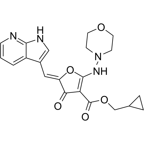
Cdc7-IN-7 (compound I-E) is a potent Cdc7 kinase inhibitor extracted from patent WO2019165473A1, compound I-E. Cdc7 is a serine-threonine protein kinase enzyme which is essential for the initiation of DNA replication in the cell cycle.
For research use only. We do not sell to patients.
- Purity : 98.03%
- CAS No.: 1402059-17-9
- Formula: C21H22N4O5
- Molecular Weight:410.42
-
Storage:Powder -20°C, 3 years ; In solvent -80°C, 6 months , -20°C, 1 month

To place orders, for customer services and technical support, please contact: MedChemExpress USA
Tel: 609-228-6898 E-mail: [email protected] [email protected]


-
 Biological Activity
-
Chemical Information
-
Solvent & Solubility
- Protocols
- Purity & Documentation
- References
-
 Help & FAQs




Biological Activity
Description


Chemical Information
-
CAS No. 1402059-17-9
-
Appearance Solid
-
Molecular Weight 410.42
-
Formula C21H22N4O5
-
Color White to off-white
-
SMILES
O=C(C(C/1=O)=C(NN2CCOCC2)OC1=C/C3=CNC4=NC=CC=C43)OCC5CC5
-
Shipping
Room temperature in continental US; may vary elsewhere.
-
Storage
Powder -20°C 3 years In solvent -80°C 6 months -20°C 1 month


Solvent & Solubility
In Vitro:
DMSO : 41.67 mg/mL (101.53 mM; Need ultrasonic; Hygroscopic DMSO has a significant impact on the solubility of product, please use newly opened DMSO)
Please refer to the solubility information to select the appropriate solvent. Once prepared, please aliquot and store the solution to prevent product inactivation from repeated freeze-thaw cycles.
Storage method and period of stock solution: -80°C, 6 months; -20°C, 1 month. When stored at -80°C, please use it within 6 months. When stored at -20°C, please use it within 1 month.
Please refer to the solubility information to select the appropriate solvent. Once prepared, please aliquot and store the solution to prevent product inactivation from repeated freeze-thaw cycles.
Storage method and period of stock solution: -80°C, 6 months; -20°C, 1 month. When stored at -80°C, please use it within 6 months. When stored at -20°C, please use it within 1 month.
Concentration (start) × Volume (start) = Concentration (final) × Volume (final)


Purity & Documentation
-
Data Sheet (273 KB)
-
SDS (252 KB)
- English - EN (252 KB)
- Français - FR (252 KB)
- Deutsch - DE (252 KB)
- Norwegian - NO (252 KB)
- Español - ES (252 KB)
- Swedish - SV (252 KB)
- Italian - IT (252 KB)
- Korean - KR (252 KB)
- Portuguese - PT (252 KB)
-
Handling Instructions (2659 KB)
References


Complete Stock Solution Preparation Table
Please refer to the solubility information to select the appropriate solvent. Once prepared, please aliquot and store the solution to prevent product inactivation from repeated freeze-thaw cycles.
Storage method and period of stock solution: -80°C, 6 months; -20°C, 1 month. When stored at -80°C, please use it within 6 months. When stored at -20°C, please use it within 1 month.
| Optional Solvent | Concentration Solvent Mass | 1 mg | 5 mg | 10 mg | 25 mg |
|---|---|---|---|---|---|
| DMSO | 1 mM | 2.4365 mL | 12.1826 mL | 24.3653 mL | 60.9132 mL |
| 5 mM | 0.4873 mL | 2.4365 mL | 4.8731 mL | 12.1826 mL | |
| 10 mM | 0.2437 mL | 1.2183 mL | 2.4365 mL | 6.0913 mL | |
| 15 mM | 0.1624 mL | 0.8122 mL | 1.6244 mL | 4.0609 mL | |
| 20 mM | 0.1218 mL | 0.6091 mL | 1.2183 mL | 3.0457 mL | |
| 25 mM | 0.0975 mL | 0.4873 mL | 0.9746 mL | 2.4365 mL | |
| 30 mM | 0.0812 mL | 0.4061 mL | 0.8122 mL | 2.0304 mL | |
| 40 mM | 0.0609 mL | 0.3046 mL | 0.6091 mL | 1.5228 mL | |
| 50 mM | 0.0487 mL | 0.2437 mL | 0.4873 mL | 1.2183 mL | |
| 60 mM | 0.0406 mL | 0.2030 mL | 0.4061 mL | 1.0152 mL | |
| 80 mM | 0.0305 mL | 0.1523 mL | 0.3046 mL | 0.7614 mL | |
| 100 mM | 0.0244 mL | 0.1218 mL | 0.2437 mL | 0.6091 mL |


Cdc7-IN-7 Related Classifications
HY-130519 Related Classifications
 Powered by Bioz