



Semaglutide
Based on 53 publication(s) in Google Scholar
Semaglutide is a long-acting, selective, competitive GLP-1R agonist that can penetrate the blood-brain barrier. After activating GLP-1R, Semaglutide promotes insulin secretion, inhibits gastric emptying and appetite, and at the same time enhances autophagy, inhibits oxidative stress and apoptosis. Semaglutide also regulates mitochondrial function and lipid metabolism (such as reducing de novo lipogenesis in the liver). Semaglutide has activities such as lowering blood sugar, reducing weight, neuroprotection (such as improving motor function in Parkinson's disease models, reducing α-synuclein aggregation) and improving hepatic steatosis. Semaglutide can be used for the study of neurodegenerative diseases and liver diseases such as type 2 diabetes, obesity, Parkinson's disease, metabolic associated fatty liver disease (MASLD), and cancer.
Nur für Forschungszwecke. Wir verkaufen nicht an Patienten.
- Reinheit : 99.74%
- CAS. Nr.: 910463-68-2
- Formel: C187H291N45O59
- Molecular Weight:4113.58
-
Speicherung:
Sealed storage, away from moisture.
Powder -80°C, 2 years , -20°C, 1 year* In solvent : -80°C, 6 months; -20°C, 1 month (sealed storage, away from moisture)


To place orders, for customer services and technical support, please contact: MedChemExpress USA
Tel: 609-228-6898 E-mail: [email protected] [email protected]


-
 Biologische Aktivität
-
Chemical Information
-
Lösungsmittel & Löslichkeit
- Protokoll
- Reinheit & Dokumentation
- Verweise
-
 Help & FAQs


-

HY-P0014
LiraglutideGLP-1R Agonist -

HY-13443
Exendin-4GLP-1 Receptor Agonist -

HY-P3506
RetatrutideGCGR/GIPR/GLP-1R Agonist -
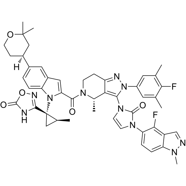
HY-112185
OrforglipronGLP-1R Agonist -

HY-P0264
Exendin(9-39) amideGLP-1R Antagonist -

HY-P0119
LixisenatideGLP-1 Agonist -

HY-P0120
DulaglutideGLP-1 Receptor Agonist -

HY-18739
Phorbol 12-myristate 13-acetatePKC Activator -
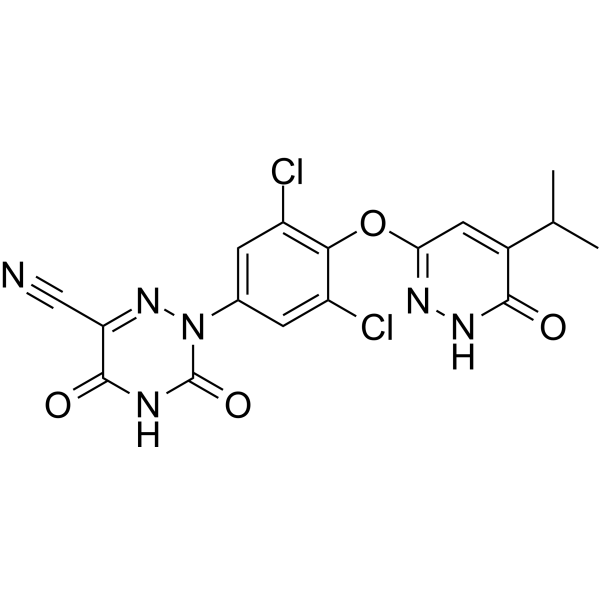
HY-12216
ResmetiromTHR-β Agonist -
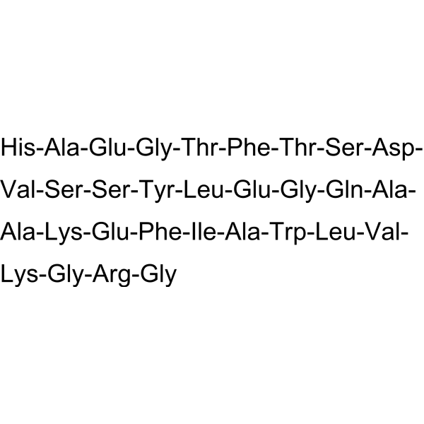
HY-P0055
GLP-1(7-37)Intestinal Insulinotropic Hormone -

HY-13259
MG-132Proteasome Inhibitor
Publications Citing Use of MedChemExpress (MCE) Semaglutide
More- Nat Metab. 2026 Sep 15.
- J Adv Res. 2026 Feb 12:S2090-1232(26)00137-2. [Abstract]
- Metabolism. 2025 Oct 13:156414. [Abstract]
- Adv Sci (Weinh). 2025 Aug 28:e17664. [Abstract]
- Cell Rep Med. 2026 Jul 7;7(7).
- Cell Rep Med. 2026 Jun 18:102875. [Abstract]
- Mater Today Bio. 2026 May 14:38:103231. [Abstract]
- J Orthop Translat. 2026 Jun 23;59:101166. [Abstract]
- Int J Mol Med. 2026 Jan;57(1):25. [Abstract]
- Int J Mol Med. 2021 Dec;48(6):219. [Abstract]
- Free Radic Biol Med. 2026 Jul:250:352-369. [Abstract]
- Free Radic Biol Med. 2026 Jun:249:138-151. [Abstract]
- ACS Appl Mater Interfaces. 2026 Jun 10;18(22):31046-31060. [Abstract]
- Cell Prolif. 2025 Aug 27:e70118. [Abstract]
- Drug Des Devel Ther. 2026 May 22:20:599539. [Abstract]
- Diabetes Obes Metab. 2026 Jan 20. [Abstract]
- Drug Des Devel Ther. 2024 Nov 30:18:5485-5500. [Abstract]
- Eur J Pharmacol. 2026 Jan 12:1011:178459. [Abstract]
- Acta Physiol. 2025 Dec 7;242(1):e70141.
- Int J Mol Sci. 2026 Sep 2;27(17):7855. [Abstract]
- Int J Mol Sci. 2026 Jun 26;27(13):5775.
- Int J Obes. 2026 Feb 21. [Abstract]
- Mol Med Rep. 2025 May;31(5):111. [Abstract]
- J Lipid Res. 2025 Feb;66(2):100736. [Abstract]
- J Physiol. 2026 Apr 3. [Abstract]
- iScience. 2026 Aug 13;29(9):117174.
- Bioconjug Chem. 2025 Dec 22. [Abstract]
- Curr Issues Mol Biol. 2026 Jun 30;48(7):675.
- J Chromatogr A. 2026 Sep 27:1785:467292.
- Diabetes Metab Syndr Obes. 2025 Apr 1:18:969-983. [Abstract]
- Mol Biol Rep. 2025 Nov 4;53(1):44. [Abstract]
- Surgery. 2025 Mar:179:108943. [Abstract]
- Microcirculation. 2026 Jul;33(5):e70070. [Abstract]
- Int J Gen Med. 2026 Feb 18:19:564902. [Abstract]
- Chem Pharm Bull. 2024;72(7):658-663. [Abstract]
- bioRxiv. 2026 Jul 23.
- bioRxiv. 2026 Jun 8:2026.06.03.729910. [Abstract]
- bioRxiv. 2026 Apr 8:2026.04.07.716956. [Abstract]
- bioRxiv. 2025 Dec 27:2025.12.26.696603. [Abstract]
- bioRxiv. 2025 Nov 18:2025.05.09.653092. [Abstract]
- bioRxiv. 2025 Nov 13.
- bioRxiv. 2025 Aug 31.
- Explor Endocr Metab Dis. 2025 Jun 24;2:101433.
- Res Sq. 2025 May 25.
- bioRxiv. 2025 April 15.
- NPJ Metab Health Dis. 2025;3(1):10. [Abstract]
- Patent. US12233110.
- Patent. US12233111.
- Patent. US12233112.
- bioRxiv. 2025 Jan 10.
- Patent. US20240366602A1.
- bioRxiv. 2024 Apr 3.
- bioRxiv. 2023 Jul 19.
 Customer Validation & Images

-
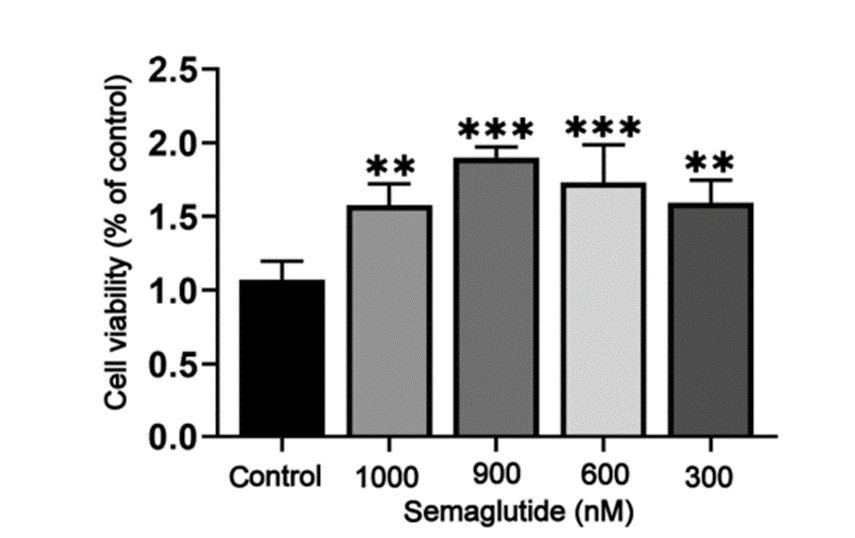
Cell Proliferation/Viability Assay
-
WB
-
IF
-
WB
-
RT-PCR
Alle α-synuclein Isoform-spezifische Produkte anzeigen
More

Biologische Aktivität
Beschreibung
In Vitro
1. Anti-Aβ25-35 injury experiment:
Semaglutide (1-100 nM; 24 h) significantly increases the survival rate of SH-SY5Y cells, increases the expression of autophagy-related proteins such as LC3II, Atg7, Beclin-1 and P62, inhibits Bax and upregulated Bcl-2, and protectes neurons by enhancing autophagy, inhibiting apoptosis[1][2].
2. Oral squamous cell carcinoma (OSCC) cell experiment:
Semaglutide (5-40 μM; 48 h) dose-dependently inhibits the proliferation, migration and invasion of Cal27 and HSC4 cells, upregulates E-cadherin and downregulates Vimentin, activates the P38 MAPK signaling pathway (increased p-P38 expression), and induces cell apoptosis[3].
MedChemExpress (MCE) has not independently confirmed the accuracy of these methods. They are for reference only.
-
Cell Line:SH-SY5Y human neuroblastoma cells
-
Concentration:0, 1, 10, 100 nM
-
Incubation Time:24 h
-
Result:Cell Viability: Significantly increased cell survival rate in a dose-dependent manner, reversing Aβ25-35-induced cytotoxicity.
WB (Western Blot): Upregulated autophagy markers (LC3II, Atg7, Beclin-1, P62) and anti-apoptotic protein Bcl-2, while downregulating pro-apoptotic protein Bax
In Vivo
Oral squamous cell carcinoma (OSCC) xenograft model
Semaglutide (3 μmol/kg; subcutaneous injection; 3 times a week; 3 weeks) significantly inhibits the growth of tumor volume in nude mouse oral squamous cell carcinoma (OSCC) xenograft model, downregulates proliferation markers Ki67 and PCNA, upregulates pro-apoptotic protein Bax and downregulates anti-apoptotic protein Bcl-xL, and induces tumor cell apoptosis by activating the P38 MAPK pathway[3].
Chronic MPTP-induced Parkinson's disease model
Semaglutide (25 nmol/kg; intraperitoneal injection; once every 2 days; 30 days) improves the chronic MPTP-induced Parkinson's disease model in mice and its motor dysfunction, increases the number of nigral tyrosine (TH)-positive neurons, reduces α-synuclein aggregation and glial activation, and reduces the level of oxidative stress marker 4-HNE[4].
Metabolic dysfunction-associated fatty liver disease (MASLD) model
Semaglutide (25 μg/kg/week + 100 μg/kg/week; subcutaneous injection; once a week; 11 weeks) reduces body weight, blood glucose and serum liver enzymes (ALT, AST, AP), reduces hepatic triglyceride deposition, improves hepatic steatosis and hepatocyte ballooning, and downregulates the de novo lipogenesis markers Acaca and Scd1 in the mouse metabolic dysfunction-associated fatty liver disease (MASLD) model[5].
MedChemExpress (MCE) has not independently confirmed the accuracy of these methods. They are for reference only.
-
Animal Model:BALB/c nude mice (male, 4-6 weeks old) with oral squamous cell carcinoma (OSCC) xenograft model[3]
-
Dosage:3 μmol/kg
-
Administration:Subcutaneous injection, 3 times weekly, for 3 weeks
-
Result:Significantly reduced tumor volume and weight compared to the control group.
Decreased expression of proliferation markers (Ki67, PCNA) and mesenchymal marker Vimentin, while increasing epithelial marker E-cadherin.
- Mitochondrial membrane-potential fluorescent assay
- Annexin V plus membrane-impermeant dye apoptosis staining
- ROS/oxidative-stress fluorescent staining
- Human Islet Cell Culture
- Research Protocol for Metabolic Diseases
- Liver Histomorphometry
- Detection of Apoptosis
- TUNEL staining for apoptotic DNA fragmentation
- Research Protocol for Endocrine Diseases
- Human pluripotent stem cell midbrain dopaminergic neuron differentiation
- Autophagy
- Mitochondrial membrane-potential and mitochondrial mass staining
- Apoptosis Solutions
- Lysosome and acidic-vesicle live-cell staining
- Macroautophagy Solutions


Chemical Information
-
CAS. Nr. 910463-68-2
-
Appearance Solid
-
Molecular Weight 4113.58
-
Formel C187H291N45O59
-
Color White to off-white
-
Sequence
His-{Aib}-Glu-Gly-Thr-Phe-Thr-Ser-Asp-Val-Ser-Ser-Tyr-Leu-Glu-Gly-Gln-Ala-Ala-{C18 diacid-γ-Glu-(AEEA)2-Lys}-Glu-Phe-Ile-Ala-Trp-Leu-Val-Arg-Gly-Arg-Gly
-
Sequence Shortening
H-{Aib}-EGTFTSDVSSYLEGQAA-{C18 diacid-γ-Glu-(AEEA)2-Lys}-EFIAWLVRGRG
-
Versand
Room temperature in continental US; may vary elsewhere.
-
Speicherung
Sealed storage, away from moisture
Powder -80°C 2 years -20°C 1 year * In solvent : -80°C, 6 months; -20°C, 1 month (sealed storage, away from moisture)


Publications (53)
-
Journal Impact Factor
-
Most Recent
-
-
J Adv Res
A novel GIPR/GLP-1R dual agonist improves systemic metabolism through differentially regulating inflammation and lipid metabolism in obesity. [Abstract]2026 Feb 12:S2090-1232(26)00137-2. PMID: 41690462 -
Metabolism
A melanocortin 4- and glucagon-like peptide 1 receptor multiple agonist for the treatment of diabetes and obesity. [Abstract]2025 Oct 13:156414. PMID: 41093057 -
Adv Sci (Weinh)
The GLP1R Agonist Semaglutide Inhibits Reactive Astrocytes and Enhances the Efficacy of Neural Stem Cell Transplantation Therapy in Parkinson's Disease Mice. [Abstract]2025 Aug 28:e17664. PMID: 40874950 -
-
Cell Rep Med
2026 Jun 18:102875. PMID: 42314683 -
Mater Today Bio
Met@MPDA rejuvenates BMSC energy metabolism to promote bone regeneration in semaglutide-treated obese periodontitis. [Abstract]2026 May 14:38:103231. PMID: 42182879 -
J Orthop Translat
Semaglutide alleviates osteoarthritis independent of weight loss via GLP-1R-mediated activation of autophagy through AKT/mTOR inhibition. [Abstract]2026 Jun 23;59:101166. PMID: 42381999 -
Int J Mol Med
Synergistic effects of Akebia saponin D and Semaglutide on diabetic nephropathy and osteoporosis via the Klotho‑p53 signaling axis. [Abstract]2026 Jan;57(1):25. PMID: 41268608 -
Int J Mol Med
Semaglutide attenuates seizure severity and ameliorates cognitive dysfunction by blocking the NLR family pyrin domain containing 3 inflammasome in pentylenetetrazole‑kindled mice. [Abstract]2021 Dec;48(6):219. PMID: 34676876
Semaglutide purchased from MedChemExpress. Usage Cited in: Int J Mol Med. 2021 Dec;48(6):219. [Abstract]
Proliferation of BV2 cells treated with Semaglutide at different concentrations (300, 600, 900 and 1,000 nM) was assessed using Cell Counting Kit‑8 analysis.
Semaglutide purchased from MedChemExpress. Usage Cited in: Int J Mol Med. 2021 Dec;48(6):219. [Abstract]
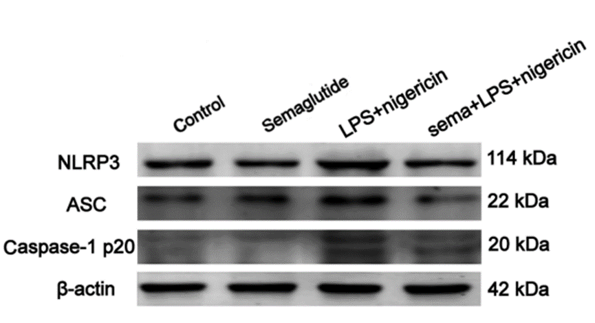
Representative WB images in different BV2 cell groups with Semaglutide (900 nM,20 h).
Semaglutide purchased from MedChemExpress. Usage Cited in: Int J Mol Med. 2021 Dec;48(6):219. [Abstract]
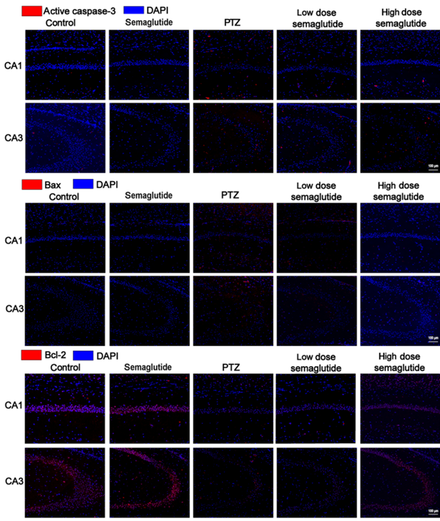
Semaglutide (10 and 25 nM/kg; ip.; single dose) decreased the fluorescence intensity of caspase‑3, Bax and Bcl-2 in the CA1 and CA3 regions.
Semaglutide purchased from MedChemExpress. Usage Cited in: Int J Mol Med. 2021 Dec;48(6):219. [Abstract]
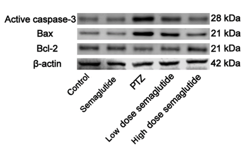
Western blot showing that 10 and 25 nM/kg Semaglutide, i.p, reduced the band intensity of active caspase‑3 and increased the Bcl‑2/Bax ratio.
Semaglutide purchased from MedChemExpress. Usage Cited in: Int J Mol Med. 2021 Dec;48(6):219. [Abstract]
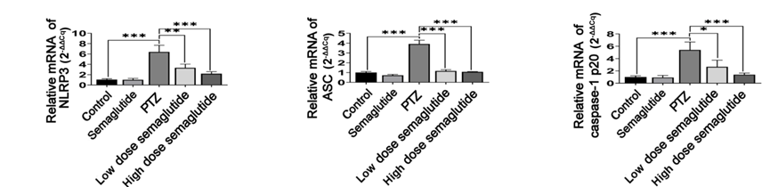
Semaglutide (10-25 nM/kg; i.p.; single dose) reduced the mRNA levels of NLRP3, ASC and caspase-1 p20.
-
Free Radic Biol Med
Semaglutide ameliorates aortic endothelial cell dysfunction in sarcopenia through the SIRT1/cGAS-STING signaling axis. [Abstract]2026 Jul:250:352-369. PMID: 41951015 -
Free Radic Biol Med
Semaglutide alleviates age-related dry eye disease by restoring lacrimal gland structure and function. [Abstract]2026 Jun:249:138-151. PMID: 41812831 -
ACS Appl Mater Interfaces
2026 Jun 10;18(22):31046-31060. PMID: 42198918 -
Cell Prolif
Development of NAFLD-Specific Human Liver Organoid Models on a Microengineered Array Chip for Semaglutide Efficacy Evaluation. [Abstract]2025 Aug 27:e70118. PMID: 40873114 -
Drug Des Devel Ther
Semaglutide Inhibits Osteoblast Ferroptosis Induced by Diabetic Periodontitis via Modulating the Wnt5a/Ror2/p38 MAPK Signaling Pathway. [Abstract]2026 May 22:20:599539. PMID: 42206237 -
Diabetes Obes Metab
Efsubaglutide Alfa attenuates metabolic dysfunction-associated steatohepatitis in mice with improvements in second harmonic generation-derived fibrosis features. [Abstract]2026 Jan 20. PMID: 41555840 -
Drug Des Devel Ther
Cardiometabolic Modulation by Semaglutide Contributes to Cardioprotection in Rats with Myocardial Infarction. [Abstract]2024 Nov 30:18:5485-5500. PMID: 39640291 -
Eur J Pharmacol
GLP-1RA partially alleviates obesity-induced reproductive dysfunction driven by the interplay mechanisms of inflammation and metabolic dysregulation via the SIRT-associated pathway. [Abstract]2026 Jan 12:1011:178459. PMID: 41380824 -
-
Int J Mol Sci
Combination GLP-1RA and Low-Dose IL-2 Modulates Peripheral Immune Activation and Attenuates CNS Inflammatory Transcript Signatures In Vivo. [Abstract]2026 Sep 2;27(17):7855. PMID: 42737751 -
-
Int J Obes
Efficacy of GLP-1 analog peptides, semaglutide, tirzepatide, and retatrutide on MC4R deficient obesity and their comparison. [Abstract]2026 Feb 21. PMID: 41723268 -
Mol Med Rep
Semaglutide enhances PINK1/Parkin‑dependent mitophagy in hypoxia/reoxygenation‑induced cardiomyocyte injury. [Abstract]2025 May;31(5):111. PMID: 40017118 -
J Lipid Res
Microglial activation and hypothalamic structural plasticity in HFD obesity: insights from semaglutide and minocycline. [Abstract]2025 Feb;66(2):100736. PMID: 39724960 -
J Physiol
Sex-specific metabolic responses to glucagon receptor agonism and modulation of the FGF21-glucagon axis in female mice. [Abstract]2026 Apr 3. PMID: 41931110 -
-
Bioconjug Chem
Development of a Long-Acting and Stapled Dual Amylin and Calcitonin Receptor Agonist as Monotherapy and Combination with GLP-1R Agonists for the Treatment of Obesity. [Abstract]2025 Dec 22. PMID: 41429154 -
-
-
Diabetes Metab Syndr Obes
Opaganib Promotes Weight Loss and Suppresses High-Fat Diet-Induced Obesity and Glucose Intolerance. [Abstract]2025 Apr 1:18:969-983. PMID: 40191829 -
Mol Biol Rep
Targeting UBE2B-mediated U2AF1 degradation to alleviate endothelial dysfunction in renal ischemia-reperfusion injury: therapeutic potential of semaglutide. [Abstract]2025 Nov 4;53(1):44. PMID: 41186784 -
Surgery
GLP-1R agonist promotes proliferation of neuroendocrine neoplasm cells expressing GLP-1 receptors. [Abstract]2025 Mar:179:108943. PMID: 39665969 -
Microcirculation
GLP-1 Receptors Are Enriched in the Lymphatic Endothelium and Their Pharmacological Activation With Semaglutide Improves the Pumping Capacity of Lymphatic Vessels. [Abstract]2026 Jul;33(5):e70070. PMID: 42231627 -
Int J Gen Med
Evaluating Semaglutide's Protection in H/R - Injured AC16 Cardiomyocytes: Oxidative Stress, Inflammation, Apoptosis, and Autophagy Insights. [Abstract]2026 Feb 18:19:564902. PMID: 41737537 -
Chem Pharm Bull
The Application of Microsampling Disks in Circular Dichroism Spectroscopy for Peptide and Nucleic Acid Drugs. [Abstract]2024;72(7):658-663. PMID: 38987173 -
-
bioRxiv
Intestinal fructose metabolism drives unsaturated fat absorption and synergizes with GLP-1 receptor agonism to promote weight loss. [Abstract]2026 Jun 8:2026.06.03.729910. PMID: 42327235 -
bioRxiv
Engineered Lactate Catabolizing Probiotics Reveal Timescale Dependent Microbiome-Host Metabolic Coupling. [Abstract]2026 Apr 8:2026.04.07.716956. PMID: 41993390 -
bioRxiv
GLP-1R Agonism Directly Improves the Pumping Capacity of Murine Collecting Lymphatic Vessels. [Abstract]2025 Dec 27:2025.12.26.696603. PMID: 41509291 -
bioRxiv
Protease-Resistant Azapeptide GLP-1 Analogue Improves Metabolic Control in Diet-Induced Obesity. [Abstract]2025 Nov 18:2025.05.09.653092. PMID: 41278761 -
-
-
-
-
-
NPJ Metab Health Dis
Incretin triple agonist retatrutide (LY3437943) alleviates obesity-associated cancer progression. [Abstract]2025;3(1):10. PMID: 40094000 -
-
-
-
-
-
-


Lösungsmittel & Löslichkeit
In Vitro:
0.5 M NH4OH : ≥ 25 mg/mL (6.08 mM)
* "≥" means soluble, but saturation unknown.
Please refer to the solubility information to select the appropriate solvent. Once prepared, please aliquot and store the solution to prevent product inactivation from repeated freeze-thaw cycles.
Storage method and period of stock solution: -80°C, 6 months; -20°C, 1 month (sealed storage, away from moisture). When stored at -80°C, please use it within 6 months. When stored at -20°C, please use it within 1 month.
Please refer to the solubility information to select the appropriate solvent. Once prepared, please aliquot and store the solution to prevent product inactivation from repeated freeze-thaw cycles.
Storage method and period of stock solution: -80°C, 6 months; -20°C, 1 month (sealed storage, away from moisture). When stored at -80°C, please use it within 6 months. When stored at -20°C, please use it within 1 month.
Konzentration (Stammlösung) × Volumen (Stammlösung) = Konzentration (Ziellösung) × Volumen (Ziellösung)


Reinheit & Dokumentation
-
Data Sheet (295 KB)
-
SDS (419 KB)
- English - EN (419 KB)
- Français - FR (419 KB)
- Deutsch - DE (419 KB)
- Norwegian - NO (419 KB)
- Español - ES (419 KB)
- Swedish - SV (419 KB)
- Italian - IT (419 KB)
- Korean - KR (419 KB)
- Portuguese - PT (419 KB)
-
Handling Instructions (2659 KB)
Verweise
[1]. Chang YF, et al. Semaglutide-mediated protection against Aβ correlated with enhancement of autophagy and inhibition of apotosis. J Clin Neurosci. 2020 Nov;81:234-239. [Content Brief]
[2]. Liu DX, et al. Semaglutide Protects against 6-OHDA Toxicity by Enhancing Autophagy and Inhibiting Oxidative Stress. Parkinsons Dis. 2022 Jul 13;2022:6813017. [Content Brief]
[3]. Wang C, et al. Semaglutide, a glucagon-like peptide-1 receptor agonist, inhibits oral squamous cell carcinoma growth through P38 MAPK signaling pathway. J Cancer Res Clin Oncol. 2025 Mar 7;151(3):103. [Content Brief]
[4]. Zhang L, et al. Semaglutide is Neuroprotective and Reduces α-Synuclein Levels in the Chronic MPTP Mouse Model of Parkinson's Disease. J Parkinsons Dis. 2019;9(1):157-171. [Content Brief]
[5]. Soto-Catalán M, et al. Semaglutide Improves Liver Steatosis and De Novo Lipogenesis Markers in Obese and Type-2-Diabetic Mice with Metabolic-Dysfunction-Associated Steatotic Liver Disease. Int J Mol Sci. 2024 Mar 4;25(5):2961. [Content Brief]
[6]. Stephen T Buckley, et al. Transcellular stomach absorption of a derivatized glucagon-like peptide-1 receptor agonist. Sci Transl Med. 2018 Nov 14;10(467):eaar7047. [Content Brief]


Complete Stock Solution Preparation Table
Please refer to the solubility information to select the appropriate solvent. Once prepared, please aliquot and store the solution to prevent product inactivation from repeated freeze-thaw cycles.
Storage method and period of stock solution: -80°C, 6 months; -20°C, 1 month (sealed storage, away from moisture). When stored at -80°C, please use it within 6 months. When stored at -20°C, please use it within 1 month.
| Optional Solvent | Concentration Solvent Mass | 1 mg | 5 mg | 10 mg | 25 mg |
|---|---|---|---|---|---|
| 0.5 M NH4OH | 1 mM | 0.2431 mL | 1.2155 mL | 2.4310 mL | 6.0774 mL |
| 5 mM | 0.0486 mL | 0.2431 mL | 0.4862 mL | 1.2155 mL |


Semaglutide Related Classifications
HY-114118 Related Classifications
 Powered by Bioz