



CP-471474
Based on 1 publication(s) in Google Scholar
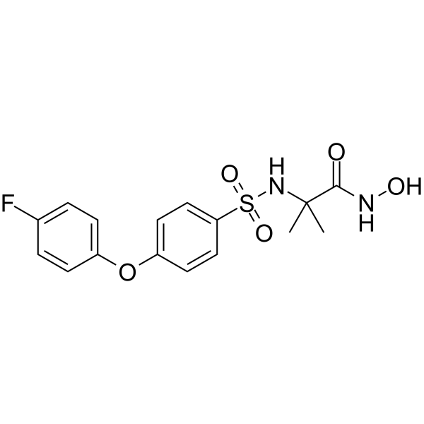
CP-471474 is an orally active and pan MMP inhibitor, with IC50 values of 1170 nM (MMP-1), 0.7 nM (MMP-2), 16 nM (MMP-3), 13 nM (MMP-9) and 0.9 nM (MMP-13), respectively.
For research use only. We do not sell to patients.
- Purity : 99.2%
- CAS No.: 210755-45-6
- Formula: C16H17FN2O5S
- Molecular Weight:368.38
-
Storage:Powder -20°C, 3 years , 4°C, 2 years ; In solvent -80°C, 6 months , -20°C, 1 month

To place orders, for customer services and technical support, please contact: MedChemExpress USA
Tel: 609-228-6898 E-mail: [email protected] [email protected]


-
 Biological Activity
-
Chemical Information
- Protocols
- Purity & Documentation
- References
-
 Help & FAQs


-
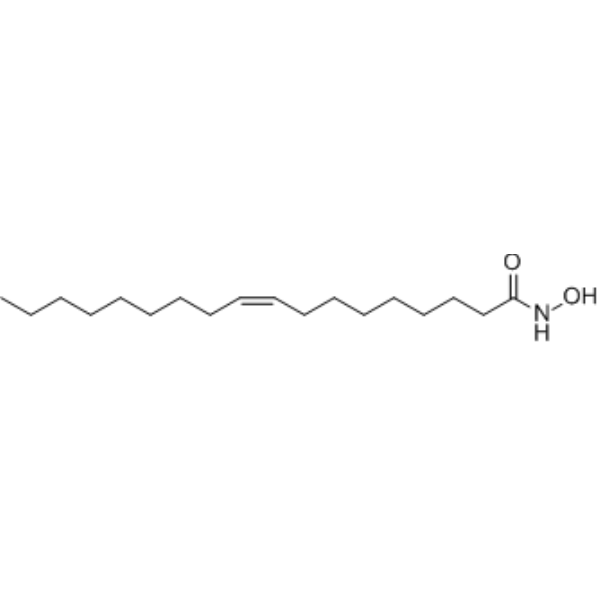
HY-112546
MMP-2 Inhibitor IMMP-2 Inhibitor -
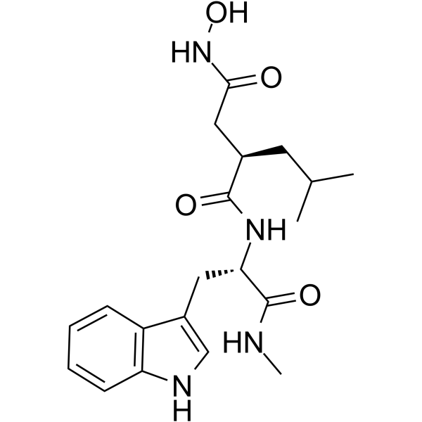
HY-15768
IlomastatMMP Inhibitor -
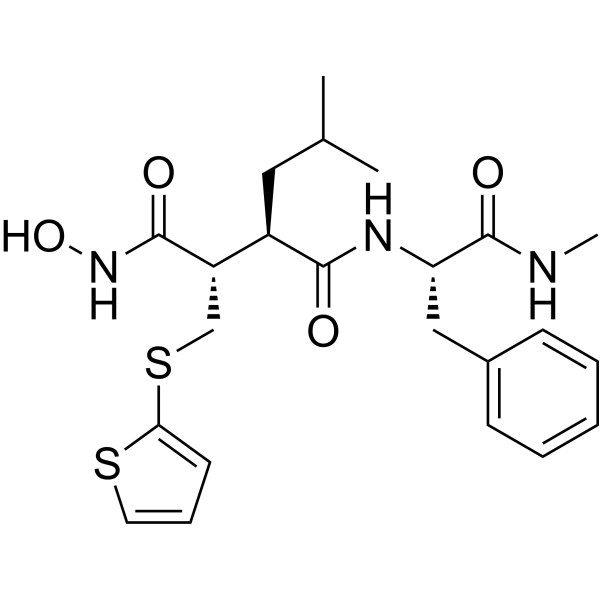
HY-13564
BatimastatMMP Inhibitor -
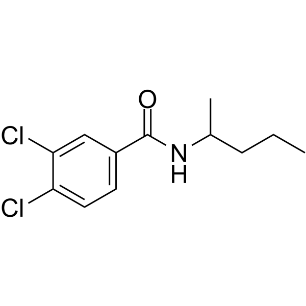
HY-15827
NSC 405020MMP14 Inhibitor -
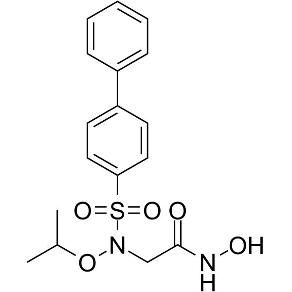
HY-103444
ARP-100MMP-2 Inhibitor -
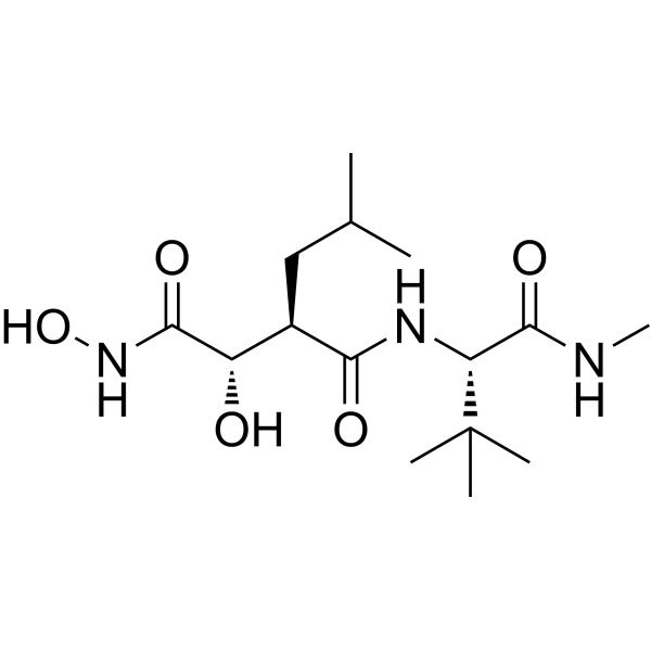
HY-12169
MarimastatMMP Inhibitor -
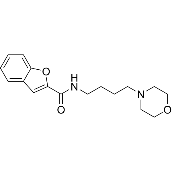
HY-100359
CL-82198MMP-13 Inhibitor -
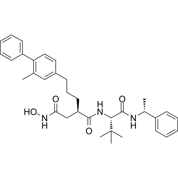
HY-107394
UK 356618MMP-3 Inhibitor -
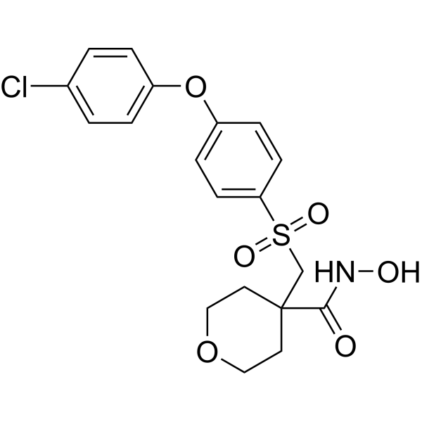
HY-10398
CTS-1027MMP Inhibitor -
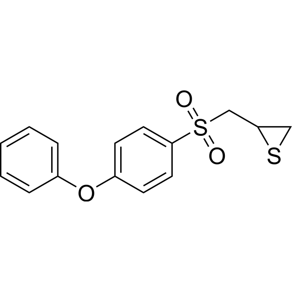
HY-12354
SB-3CTMMP2/9 Inhibitor -
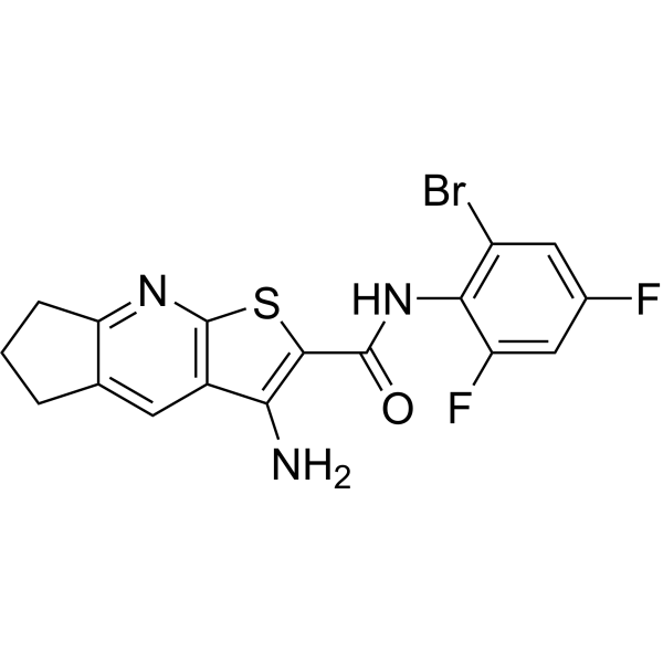
HY-107636
DC_AC50Atox1/CCS Inhibitor -
HY-101535
ARP101MMP-2 Inhibitor
Publications Citing Use of MedChemExpress (MCE) CP-471474
More

Biological Activity
Description
IC50 & Target
[1]|
MMP-2 0.7 nM (IC50) |
MMP-13 0.9 nM (IC50) |
MMP-9 13 nM (IC50) |
MMP-3 16 nM (IC50) |
MMP-1 1170 nM (IC50) |
In Vivo
CP-471,474 significantly reduces both the extent and severity of inflammation at 2 months. At 4 months, a spontaneous reduction of the inflammatory response is observed in both treated and untreated animals, and consequently no difference is observed between both[2].
CP-471474 significantly decreases the destructive lesions mainly at 2 months and also at 4 months[2].
MedChemExpress (MCE) has not independently confirmed the accuracy of these methods. They are for reference only.
-
Animal Model:Guinea pigs weighing 400 to 450 g (COPD)[2].
-
Dosage:20 mg/kg (also supplemented daily by the diet (200 mg/200 g powdered chow)).
-
Administration:Subcutaneously (in 20% ethanol/80% polyethylene glycol), once a day during the entire course.
-
Result:Lungs derived from smoking animals treated with the compound demonstrated a statistically significant reduction in the destructive lesions.
Showed an increase of both pro–MMP-9 and its active form (lanes 5 to 8) as compared with control animals.


Chemical Information
-
CAS No. 210755-45-6
-
Appearance Solid
-
Molecular Weight 368.38
-
Formula C16H17FN2O5S
-
Color White to off-white
-
SMILES
CC(C)(NS(=O)(C1=CC=C(OC2=CC=C(F)C=C2)C=C1)=O)C(NO)=O
-
Shipping
Room temperature in continental US; may vary elsewhere.
-
Storage
Powder -20°C 3 years 4°C 2 years In solvent -80°C 6 months -20°C 1 month


Publications (1)
-
Journal Impact Factor
-
Most Recent
-
Virulence
Monkeypox virus replication and host response in vaginal and ectocervical epithelial cells. [Abstract]2026 Mar 25:2649986. PMID: 41880196


Purity & Documentation
-
Data Sheet (270 KB)
-
SDS (392 KB)
- English - EN (392 KB)
- Français - FR (392 KB)
- Deutsch - DE (392 KB)
- Norwegian - NO (392 KB)
- Español - ES (392 KB)
- Swedish - SV (392 KB)
- Italian - IT (392 KB)
- Korean - KR (392 KB)
- Portuguese - PT (392 KB)
-
Handling Instructions (2659 KB)
References
[1]. L E Rohde, et al. Matrix metalloproteinase inhibition attenuates early left ventricular enlargement after experimental myocardial infarction in mice. Circulation. 1999 Jun 15;99(23):3063-70. [Content Brief]
[2]. Moisés Selman, et al. Matrix metalloproteinases inhibition attenuates tobacco smoke-induced emphysema in Guinea pigs. Chest. 2003 May;123(5):1633-41. [Content Brief]

Calculators
Concentration (start) × Volume (start) = Concentration (final) × Volume (final)


CP-471474 Related Classifications
HY-W011085 Related Classifications
 Powered by Bioz