



Neomycin sulfate
Based on 82 publication(s) in Google Scholar
Neomycin sulfate, an aminoglycoside antibiotic, exerts antibacterial activity through irreversible binding of the nuclear 30S ribosomal subunit, thereby blocking bacterial protein synthesis. Neomycin sulfate is a known phospholipase C (PLC) inhibitor. Neomycin sulfate potently inhibits both the nuclear translocation of angiogenin and angiogenin-induced cell proliferation and angiogenesis. Neomycin sulfate inhibits IP3-mediated Ca2+ release, MgATP-dependent Ca2+ uptake, and electrical excitation-evoked skeletal muscle Ca2+ transients. Neomycin sulfate depletes gut microbiota in specific mouse models, causes hearing impairment, and kidney damage with prolonged exposure. Neomycin sulfate can be used for the research of cancer.
For research use only. We do not sell to patients.
- Purity : 99.92%
- CAS No.: 1405-10-3
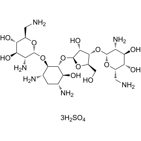
- Formula: C23H52N6O25S3
- Molecular Weight:908.88
-
Storage:
4°C, sealed storage, away from moisture
* In solvent : -80°C, 6 months; -20°C, 1 month (sealed storage, away from moisture)

To place orders, for customer services and technical support, please contact: MedChemExpress USA
Tel: 609-228-6898 E-mail: [email protected] [email protected]


-
 Biological Activity
-
Chemical Information
-
Solvent & Solubility
- Protocols
- Purity & Documentation
- References
-
 Help & FAQs


-
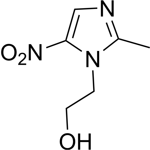
HY-B0318
MetronidazoleAntibiotic -
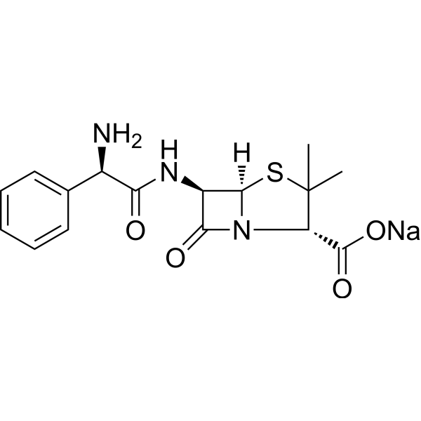
HY-B0522A
Ampicillin sodiumBacterial Inhibitor -
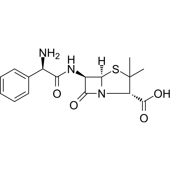
HY-B0522
AmpicillinBacterial Inhibitor -
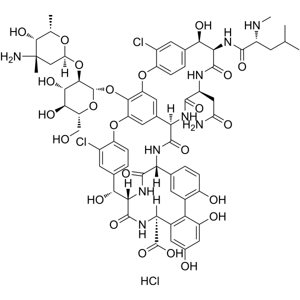
HY-17362
Vancomycin hydrochlorideAntibiotic -
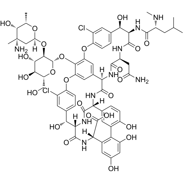
HY-B0671
VancomycinAntibiotic -
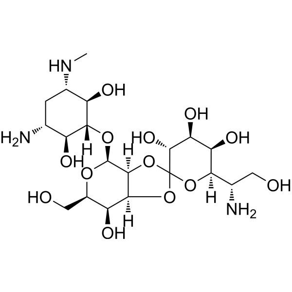
HY-B0490
Hygromycin BAntibiotic -
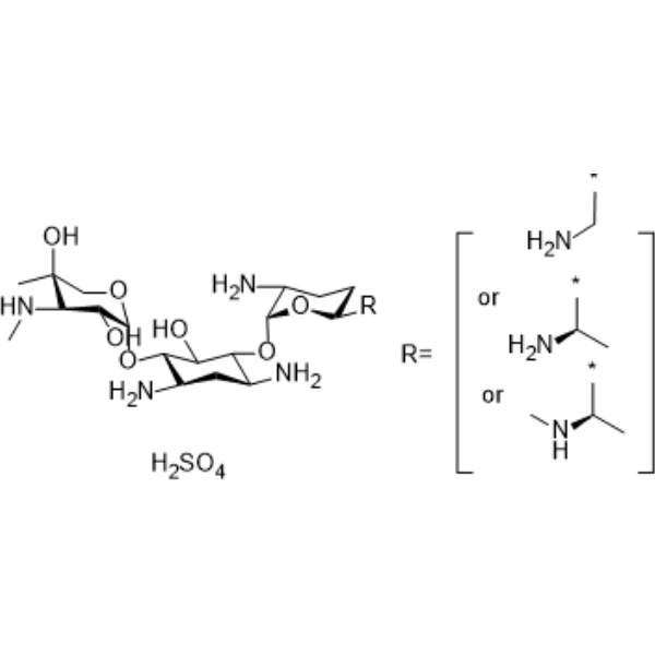
HY-A0276
Gentamicin sulfateAminoglycoside Antibiotic -
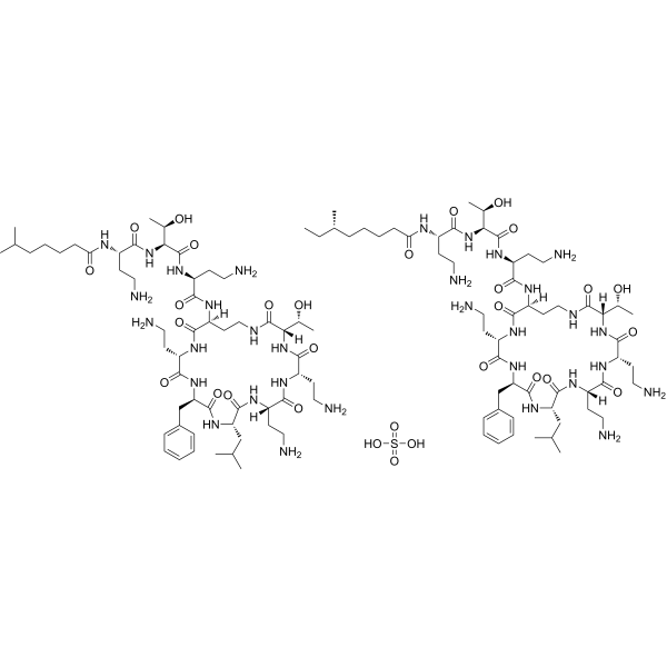
HY-A0248
Polymyxin B SulfateAntibiotic -
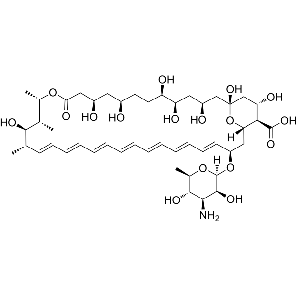
HY-B0221
Amphotericin BAntifungal Agent -
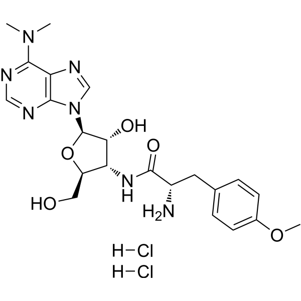
HY-B1743A
Puromycin dihydrochlorideProtein Synthesis Inhibitor -
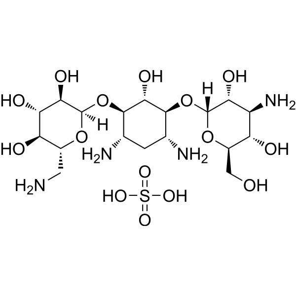
HY-16566A
Kanamycin sulfateAntibacterial Agent -
HY-B0472
Streptomycin sulfateBacterial Inhibitor
Publications Citing Use of MedChemExpress (MCE) Neomycin sulfate
More- Cancer Cell. 2025 Sep 25:S1535-6108(25)00394-0. [Abstract]
- Science. 2026 Mar 19;391(6791):eadu7686. [Abstract]
- Cell Metab. 2025 Jan 7;37(1):87-103.e10. [Abstract]
- Cell Metab. 2023 Nov 7;35(11):1961-1975.e6. [Abstract]
- Immunity. 2026 Mar 27:S1074-7613(26)00086-5. [Abstract]
- Immunity. 2026 Mar 10;59(3):598-617.e11. [Abstract]
- Cell Host Microbe. 2026 Jul 8;34(7):1350-1366.e8. [Abstract]
- Cancer Res. 2026 May 15. [Abstract]
- Cancer Res. 2025 Apr 17. [Abstract]
- Gut Microbes. 2026 Dec 31;18(1):2620125. [Abstract]
- Gut Microbes. 2024 Jan-Dec;16(1):2390164. [Abstract]
- Gut Microbes. 2023 Dec;15(2):2249143. [Abstract]
- Gut Microbes. 2023 Dec;15(2):2282790. [Abstract]
- J Nanobiotechnology. 2025 Jun 13;23(1):443. [Abstract]
- J Nanobiotechnology. 2024 Apr 8;22(1):157. [Abstract]
- Adv Sci (Weinh). 2025 Oct 13:e09383. [Abstract]
- Adv Sci (Weinh). 2025 Jun;12(23):e2416348. [Abstract]
- Adv Sci (Weinh). 2025 May;12(20):e2409837. [Abstract]
- Adv Sci (Weinh). 2025 Apr;12(16):e2415041. [Abstract]
- Cell Rep Med. 2024 Oct 15;5(10):101753. [Abstract]
- Sci Adv. 2023 Feb 17;9(7):eade4770. [Abstract]
- Research (Wash D C). 2024 Jul 16:7:0415. [Abstract]
- Pharmacol Res. 2024 Jul:205:107229. [Abstract]
- Cell Death Dis. 2021 Jun 7;12(6):582. [Abstract]
- Cancer Lett. 2026 May 1:645:218392. [Abstract]
- Int J Biol Sci. 2026 Apr 23;22(9):4784-4805. [Abstract]
- NPJ Biofilms Microbiomes. 2024 Oct 28;10(1):111. [Abstract]
- Phytomedicine. 2026 Jun:155:158086. [Abstract]
- EBioMedicine. 2026 Apr:126:106213. [Abstract]
- Cell Death Discov. 2022 Feb 22;8(1):77. [Abstract]
- Ultrason Sonochem. 2026 Sep:132:107985.
- NPJ Precis Oncol. 2025 Dec 23. [Abstract]
- Sci China Life Sci. 2026 Apr;69(4):1271-1283. [Abstract]
- Genome Biol. 2023 Apr 30;24(1):98. [Abstract]
- Int J Biol Macromol. 2024 Nov;281(Pt 4):135835. [Abstract]
- Microbiol Res. 2026 Jun:307:128477. [Abstract]
- Neural Regen Res. 2024 Sep 1;19(9):2081-2088. [Abstract]
- Clin Transl Med. 2026 Aug;16(8):e70754.
- Clin Transl Med. 2024 Oct;14(10):e70038. [Abstract]
- Cancer Immunol Res. 2022 Dec 2;10(12):1542-1558. [Abstract]
- Am J Chin Med. 2024;52(5):1527-1554. [Abstract]
- Cell Rep. 2026 Jun 25;45(7):117613. [Abstract]
- Cell Rep. 2023 Dec 27;43(1):113591. [Abstract]
- Cell Rep. 2021 Sep 7;36(10):109660. [Abstract]
- J Pineal Res. 2024 Apr;76(3):e12954. [Abstract]
- Brain Behav Immun. 2021 Feb:92:102-114. [Abstract]
- Neurotherapeutics. 2023 Sep;20(5):1405-1426. [Abstract]
- Emerg Microbes Infect. 2022 Dec;11(1):815-828. [Abstract]
- J Ethnopharmacol. 2026 Mar 1:358:120992. [Abstract]
- J Agric Food Chem. 2024 Apr 10. [Abstract]
- CNS Neurosci Ther. 2026 May;32(5):e70933. [Abstract]
- CNS Neurosci Ther. 2025 Feb;31(2):e70253. [Abstract]
- Biochem Pharmacol. 2024 Dec;230(Pt 2):116593. [Abstract]
- Life Sci. 2020 Nov 15:261:118473. [Abstract]
- Life Metab. 2025 Oct 8.
- Food Funct. 2025 Jul 14;16(14):5885-5899. [Abstract]
- EMBO Rep. 2024 Nov;25(11):4827-4845. [Abstract]
- Nutrients. 2023 Oct 18;15(20):4427. [Abstract]
- Int J Mol Sci. 2026 Feb 13;27(4):1807. [Abstract]
- Front Aging Neurosci. 2022 Jul 28;14:858130. [Abstract]
- Exp Neurol. 2025 Mar:385:115142. [Abstract]
- Neoplasia. 2024 Jan:47:100963. [Abstract]
- Mol Cell Biochem. 2026 Jul 30.
- Mol Cell Biochem. 2021 Jun;476(6):2503-2512. [Abstract]
- iScience. 2025 May 27;28(6):112763. [Abstract]
- iScience. 2025 May 13;28(7):112658. [Abstract]
- Aquaculture. 2021, 736248.
- Naunyn Schmiedebergs Arch Pharmacol. 2025 Apr;398(4):3681-3695. [Abstract]
- J Cell Physiol. 2022 Mar;237(3):1845-1856. [Abstract]
- Microb Pathog. 2026 Oct:219:108723.
- AMB Express. 2026 May 20. [Abstract]
- Mol Immunol. 2022 Sep:149:94-106. [Abstract]
- Microbiologyopen. 2019 Oct;8(10):e873. [Abstract]
- Water. 2025 Jun 6.
- Cell Biochem Biophys. 2025 Oct 31. [Abstract]
- Cancer Genomics Proteomics. 2026 May-Jun;23(3):407-429. [Abstract]
- Biochem Biophys Res Commun. 2025 Aug 30:776:152161. [Abstract]
- STAR Protoc. 2025 Jan 23;6(1):103585. [Abstract]
- medRxiv. 2026 Aug 10.
- SSRN. 2026 Aug 7.
- Res Sq. 2024 Oct 02.
- SSRN. 2023 Sep 11.
 Customer Validation & Images

-
In Vivo Efficacy Study
-
Others
-
PK/PD Analysis
-
Bio/Physico-chemical Assay
All Phospholipase Isoforms
MoreAll Antibiotic Isoforms
More

Biological Activity
Description
IC50 & Target
|
Aminoglycoside |
In Vitro
Neomycin sulfate (10-500 μM; 30 min) inhibits nuclear translocation of angiogenin in HUVE cells with 60% inhibition at 100 μM and residual 14% translocation at 500 μM[1].
Neomycin sulfate (5-50 μM; 48 h) completely abolishes angiogenin-induced proliferation of HUVE cells at 50 μM, with 49% inhibition at 5 μM and 69% inhibition at 25 μM[1].
Neomycin sulfate (5-50 μM; 2 h) does not inhibit the ribonucleolytic activity of angiogenin against yeast tRNA at concentrations up to 50 μM[1].
Neomycin sulfate (0.25-1.25 mM; 6 minutes after cell permeabilization) inhibits IP3-mediated Ca2+ release in saponin-permeabilized RINmSF insulinoma cells in a dose-dependent manner and also inhibits the initial rate of net Ca2+ uptake into non-mitochondrial pools[2].
Neomycin sulfate (1.25 mM) inhibits IP₃-mediated 45Ca2+ release from saponin-permeabilized rat hepatocytes, with greater inhibition at low IP₃ concentrations[2].
Neomycin sulfate covalently linked to glass beads avidly binds IP3 (93% retention) and ATP (97-98% retention) and also binds PIP2 (70% retention), but binds IP1 and myo-inositol poorly[2].
MedChemExpress (MCE) has not independently confirmed the accuracy of these methods. They are for reference only.
-
Cell Line:human umbilical vein endothelial (HUVE) cells
-
Concentration:5, 10, 25, 50 μM
-
Incubation Time:48 h
-
Result:Inhibited angiogenin-induced proliferation in a dose-dependent, non-linear manner; reduced proliferation by 49% at 5 μM, by 69% at 25 μM, and completely abolished angiogenin-induced proliferation at 50 μM; did not affect basal proliferation or cell viability when used alone.
In Vivo
Neomycin sulfate (1 g/L; p.o.; daily; 14 days) as part of a broad-spectrum antibiotic cocktail depletes over 90% of gut microbiota in 7.5-month-old APP/PS1 transgenic mice[3].
MedChemExpress (MCE) has not independently confirmed the accuracy of these methods. They are for reference only.
-
Animal Model:SPAFAS (fertilized eggs)[1]
-
Dosage:4 ng; 20 ng; 200 ng
-
Administration:applied to CAM; single dose
-
Result:Decreased the percentage of angiogenin-induced positive angiogenesis from 55% to 40% with 4 ng; decreased it to 20% (equivalent to water control background) with 20 ng; decreased it to 25% with 200 ng.
-
Animal Model:
-
Dosage:1 g/L (as part of broad-spectrum antibiotic cocktail)
-
Administration:p.o.; daily; 14 days (in drinking water)
-
Result:Achieved >90% depletion of gut microbiota.
- Research Protocol for Infectious Diseases
- Human pluripotent stem cell endothelial-cell differentiation
- Vascular/Branching Fractal Analysis
- BrdU Incorporation Assay
- Somatic Cell Culture
- CFSE Dye Dilution Proliferation Assay
- Dye-dilution cell tracking and proliferation staining
- Research Protocol for Microbiome Analysis
- CCK-8/WST-8 Cell Proliferation Assay
- Protocol for Cell Cycle
- Cell Counting-Based Growth Curve Assay
- Ca2+ Staining Technique
- Colony Formation (Clonogenic) Assay
- Bacterial live/dead nucleic-acid viability staining
- Cell Viability Determination by MTT Colorimetric Assay
- Cytoplasmic-Nuclear Fractionated Protein Extraction
- EdU Incorporation Assay (Click Chemistry-Based DNA Synthesis Measurement)
- Ki-67 Immunostaining Proliferation Assay
- PCNA Immunodetection Proliferation Assay
- Protocol for Cell Counting and Cell Density Analysis
- Endothelial Tube Formation Assay
- MTT Cell Proliferation Assay
- Endothelial Cell Migration/Angiogenic Sprouting Assay


Chemical Information
-
CAS No. 1405-10-3
-
Appearance Solid
-
Molecular Weight 908.88
-
Formula C23H52N6O25S3
-
Color White to light yellow
-
SMILES
O[C@H]1[C@H](O)[C@@H](N)[C@@H](O[C@@H]2[C@@H](N)C[C@@H](N)[C@H](O)[C@H]2O[C@@H]3O[C@H](CO)[C@@H](O[C@H]4O[C@@H](CN)[C@@H](O)[C@H](O)[C@H]4N)[C@H]3O)O[C@@H]1CN.[3H2SO4]
-
Initial Source
Micromonospora species
-
Shipping
Room temperature in continental US; may vary elsewhere.
-
Storage
4°C, sealed storage, away from moisture
* In solvent : -80°C, 6 months; -20°C, 1 month (sealed storage, away from moisture)


Publications (82)
-
Journal Impact Factor
-
Most Recent
-
Cancer Cell
Ketogenic diet inhibits glioma progression by promoting gut microbiota-derived butyrate production. [Abstract]2025 Sep 25:S1535-6108(25)00394-0. PMID: 41005305 -
Science
Commensal-driven serotonin production modulates in vivo delivery of synthetic and viral vectors. [Abstract]2026 Mar 19;391(6791):eadu7686. PMID: 41855345 -
Cell Metab
Stress triggers irritable bowel syndrome with diarrhea through a spermidine-mediated decline in type I interferon. [Abstract]2025 Jan 7;37(1):87-103.e10. PMID: 39366386 -
Cell Metab
High dietary fructose promotes hepatocellular carcinoma progression by enhancing O-GlcNAcylation via microbiota-derived acetate. [Abstract]2023 Nov 7;35(11):1961-1975.e6. PMID: 37797623 -
Immunity
NF-κB-activated fibroblasts orchestrate inflammaging and emergence of pro-inflammatory granzyme K+ T cells. [Abstract]2026 Mar 27:S1074-7613(26)00086-5. PMID: 41903549 -
Immunity
2026 Mar 10;59(3):598-617.e11. PMID: 41722568 -
Cell Host Microbe
Gut bacteria that produce fatty acid ethanolamides alleviate diarrhea-predominant IBS with insulin resistance. [Abstract]2026 Jul 8;34(7):1350-1366.e8. PMID: 42320473 -
Cancer Res
Distinct Inflammatory Cytotoxic T Lymphocyte Populations Mediate PD-1 Blockade Induced Immune-Related Adverse Events in Multiple Organs. [Abstract]2026 May 15. PMID: 42139517 -
Cancer Res
Mannose Enhances Immunotherapy Efficacy in Ovarian Cancer by Modulating Gut Microbial Metabolites. [Abstract]2025 Apr 17. PMID: 40245117 -
Gut Microbes
Gut microbial ammonia as a mediator of PFOS neurotoxicity and its remediation by the flavonoid Icaritin. [Abstract]2026 Dec 31;18(1):2620125. PMID: 41629743 -
Gut Microbes
Time-restricted feeding ameliorates non-alcoholic fatty liver disease through modulating hepatic nicotinamide metabolism via gut microbiota remodeling. [Abstract]2024 Jan-Dec;16(1):2390164. PMID: 39154362 -
Gut Microbes
Microbial metabolite butyrate promotes anti-PD-1 antitumor efficacy by modulating T cell receptor signaling of cytotoxic CD8 T cell. [Abstract]2023 Dec;15(2):2249143. PMID: 37635362
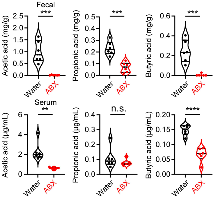
Neomycin sulfate purchased from MedChemExpress. Usage Cited in: Gut Microbes. 2023 Dec;15(2):2249143. [Abstract]
Targeted metabolomics analysis of gut microbial metabolites (AA, PA, and BA) in colon contents (feces) and serum from mice with or without ABX treatment (n = 6 per group). ABX: an antibiotic cocktail of 0.5 g/L Vancomycin, 1 g/L Metronidazole,1 g/L Neomycin and 1 g/L Ampicillin
-
Gut Microbes
Multi-omics data reveals aberrant gut microbiota-host glycerophospholipid metabolism in association with neuroinflammation in APP/PS1 mice. [Abstract]2023 Dec;15(2):2282790. PMID: 37992400
Neomycin sulfate purchased from MedChemExpress. Usage Cited in: Gut Microbes. 2023 Dec;15(2):2282790. [Abstract]
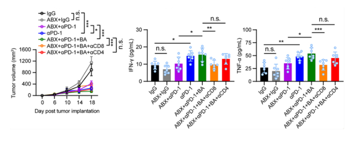
Depletion of CD8+ T cells in ABX-treated B16-F0 tumor-bearing mice dampened the antitumor effect of butyrate combined with anti-PD-1 (Figure 4b–e). Decreased serum IFN-γ and TNF-α production during the depletion of CD8+ T cells was also observed. ABX:an antibiotic cocktail of 0.5 g/L Vancomycin, 1 g/L Metronidazole, 1 g/L Neomycin and 1 g/L Ampicillin.
-
J Nanobiotechnology
Overcoming acquired immunotherapy resistance in non-small cell lung cancer using ginsenoside Rb1-loaded, peptide-enhanced exosome delivery systems. [Abstract]2025 Jun 13;23(1):443. PMID: 40514658 -
J Nanobiotechnology
Gold nanoparticles exhibit anti-osteoarthritic effects via modulating interaction of the "microbiota-gut-joint" axis. [Abstract]2024 Apr 8;22(1):157. PMID: 38589904 -
Adv Sci (Weinh)
Faecalibaculum rodentium Alleviates Ionizing Radiation-Induced Damage in Mice by Improving Intestinal Integrity and Hematopoiesis via Its Metabolite Butyrate. [Abstract]2025 Oct 13:e09383. PMID: 41082369 -
Adv Sci (Weinh)
The Lung Microbiome Modulates Pain-Like Behavior Via the Lung-Brain Axis in a Nitroglycerin-Induced Chronic Migraine Mouse Model. [Abstract]2025 Jun;12(23):e2416348. PMID: 40162625 -
Adv Sci (Weinh)
Microbiota-Derived Inosine Suppresses Systemic Autoimmunity via Restriction of B Cell Differentiation and Migration. [Abstract]2025 May;12(20):e2409837. PMID: 40289872 -
Adv Sci (Weinh)
Metabolomic and Cellular Mechanisms of Drug-Induced Ototoxicity and Nephrotoxicity: Therapeutic Implications of Uric Acid Modulation. [Abstract]2025 Apr;12(16):e2415041. PMID: 40041973
Neomycin sulfate purchased from MedChemExpress. Usage Cited in: Adv Sci (Weinh). 2025 Apr;12(16):e2415041. [Abstract]
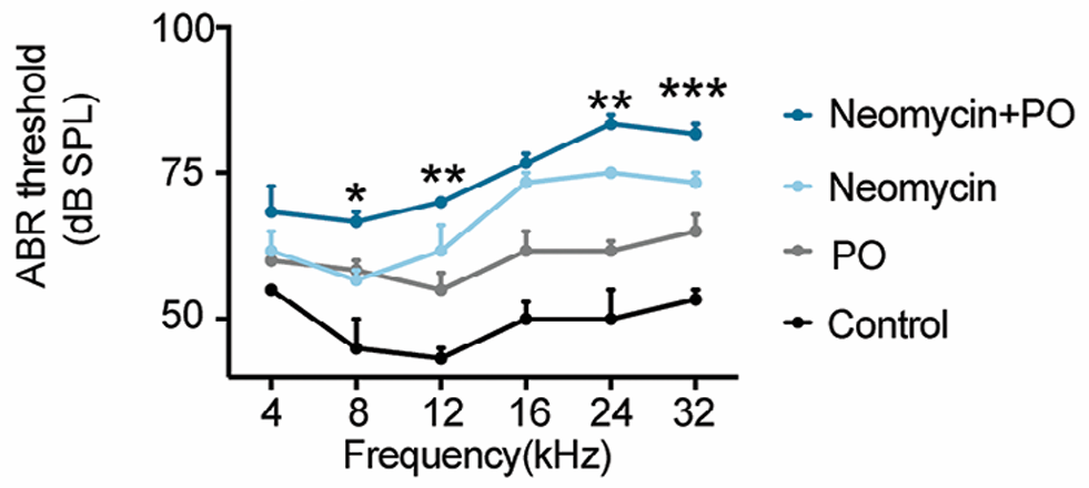
ABR thresholds in each group. The statistical differences were analyzed between Neomycin (100 mg/kg once daily for 14 days, ip) + PO group and Neomycin group.
Neomycin sulfate purchased from MedChemExpress. Usage Cited in: Adv Sci (Weinh). 2025 Apr;12(16):e2415041. [Abstract]
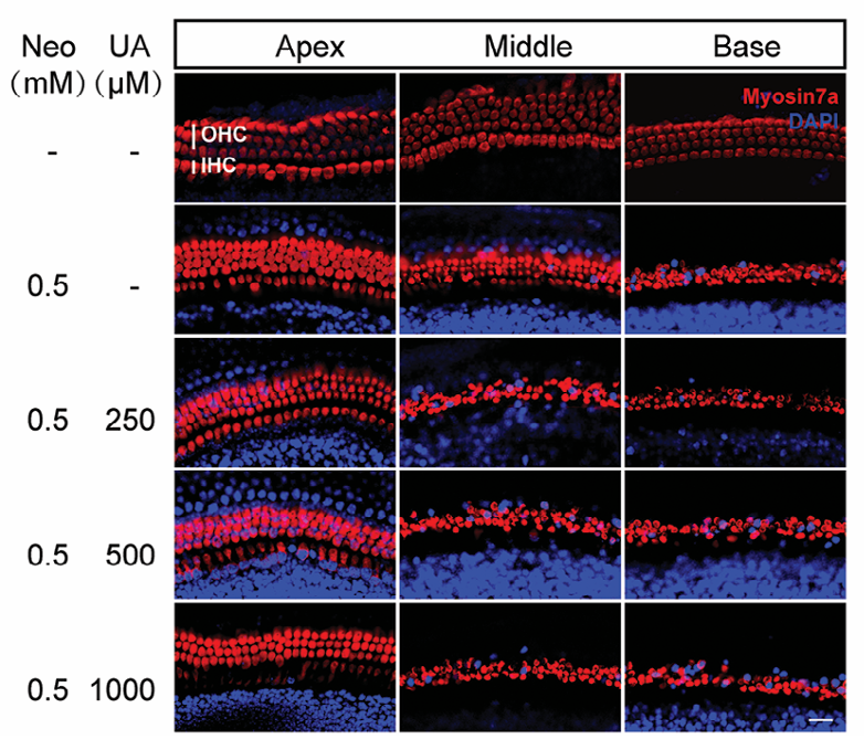
Effects of different levels of UA on HCs in the apical, middle, and basal turns after Neomycin (0.5 mM) treatment for 24 h.
-
Cell Rep Med
Leucine restriction ameliorates Fusobacterium nucleatum-driven malignant progression and radioresistance in nasopharyngeal carcinoma. [Abstract]2024 Oct 15;5(10):101753. PMID: 39357525 -
Sci Adv
Imaging of Escherichia coli K5 and glycosaminoglycan precursors via targeted metabolic labeling of capsular polysaccharides in bacteria. [Abstract]2023 Feb 17;9(7):eade4770. PMID: 36800421 -
Research (Wash D C)
Bacillus siamensis Targeted Screening from Highly Colitis-Resistant Pigs Can Alleviate Ulcerative Colitis in Mice. [Abstract]2024 Jul 16:7:0415. PMID: 39015206 -
Pharmacol Res
Multi-omics approaches for the understanding of therapeutic mechanism for Huang-Qi-Long-Dan Granule against ischemic stroke. [Abstract]2024 Jul:205:107229. PMID: 38782148 -
Cell Death Dis
Effects of short-chain fatty acids in inhibiting HDAC and activating p38 MAPK are critical for promoting B10 cell generation and function. [Abstract]2021 Jun 7;12(6):582. PMID: 34099635 -
Cancer Lett
Hypoxia-driven phase separation of the PABP1/eIF4B complex forms stress granules and activates ChaC2 translation to promote polyunsaturated lipids-supported peritoneal metastasis in gastric cancer. [Abstract]2026 May 1:645:218392. PMID: 41780839 -
Int J Biol Sci
Tryptophan-Restricted Intermittent Diet Alleviates Estrogen Deficiency-Induced Osteoporosis via Regulating Coupling Effects of "Gut-Bone" Axis. [Abstract]2026 Apr 23;22(9):4784-4805. PMID: 42157925 -
NPJ Biofilms Microbiomes
Gut microbiota dysbiosis-mediated ceramides elevation contributes to corticosterone-induced depression by impairing mitochondrial function. [Abstract]2024 Oct 28;10(1):111. PMID: 39468065 -
Phytomedicine
Usenamine a potentiates anti-CRC activity of sorafenib by inducing autophagy and inhibiting YAP pathway through targeting SOD2. [Abstract]2026 Jun:155:158086. PMID: 41861687 -
EBioMedicine
2026 Apr:126:106213. PMID: 41861519 -
Cell Death Discov
Long noncoding RNA SNHG1 silencing accelerates hepatocyte-like cell differentiation of bone marrow-derived mesenchymal stem cells to alleviate cirrhosis via the microRNA-15a/SMURF1/UVRAG axis. [Abstract]2022 Feb 22;8(1):77. PMID: 35194023 -
-
NPJ Precis Oncol
RICH1 enhances pro-inflammatory TAM infiltration in breast cancer via promoting TRIM21-mediated ubiquitination of RhoA and inhibiting STAT3 phosphorylation. [Abstract]2025 Dec 23. PMID: 41436617 -
Sci China Life Sci
Endometrial microbiota-dimethylglycine-Treg cell axis affects endometrial receptivity in recurrent implantation failure. [Abstract]2026 Apr;69(4):1271-1283. PMID: 41627665 -
Genome Biol
Caloric restriction remodels the hepatic chromatin landscape and bile acid metabolism by modulating the gut microbiota. [Abstract]2023 Apr 30;24(1):98. PMID: 37122023 -
Int J Biol Macromol
Tremella aurantialba polysaccharides alleviate ulcerative colitis in mice by improving intestinal barrier via modulating gut microbiota and inhibiting ferroptosis. [Abstract]2024 Nov;281(Pt 4):135835. PMID: 39306158 -
Microbiol Res
Gut commensal Clostridium sporogenes-derived 5-aminovaleric acid attenuates liver injury by suppressing M1 macrophage activation in mice. [Abstract]2026 Jun:307:128477. PMID: 41793891 -
Neural Regen Res
Gut microbiota dysbiosis contributes to α-synuclein-related pathology associated with C/EBPβ/AEP signaling activation in a mouse model of Parkinson's disease. [Abstract]2024 Sep 1;19(9):2081-2088. PMID: 38227539 -
-
Clin Transl Med
Deubiquitination of RIPK2 by OTUB2 augments NOD2 signalling and protective effects in intestinal inflammation. [Abstract]2024 Oct;14(10):e70038. PMID: 39358938 -
Cancer Immunol Res
NET-Triggered NLRP3 Activation and IL18 Release Drive Oxaliplatin-Induced Peripheral Neuropathy. [Abstract]2022 Dec 2;10(12):1542-1558. PMID: 36255412 -
Am J Chin Med
Astragaloside IV Ameliorates Colonic Adenomatous Polyps Development by Orchestrating Gut Bifidobacterium and Serum Metabolome. [Abstract]2024;52(5):1527-1554. PMID: 39164214 -
Cell Rep
Type I unconventional protein secretion of the SARS-CoV-2 nucleocapsid protein promotes inflammatory cytokine release. [Abstract]2026 Jun 25;45(7):117613. PMID: 42360880 -
Cell Rep
Oral fecal transplantation enriches Lachnospiraceae and butyrate to mitigate acute liver injury. [Abstract]2023 Dec 27;43(1):113591. PMID: 38153838 -
Cell Rep
Multiplatform discovery and regulatory function analysis of structural variations in non-small cell lung carcinoma. [Abstract]2021 Sep 7;36(10):109660. PMID: 34496260 -
J Pineal Res
Gut microbially produced tryptophan metabolite melatonin ameliorates osteoporosis via modulating SCFA and TMAO metabolism. [Abstract]2024 Apr;76(3):e12954. PMID: 38618998 -
Brain Behav Immun
Antibiotic-induced microbiome depletion in adult mice disrupts blood-brain barrier and facilitates brain infiltration of monocytes after bone-marrow transplantation. [Abstract]2021 Feb:92:102-114. PMID: 33242652 -
Neurotherapeutics
Fecal Microbiota Transplantation from Aged Mice Render Recipient Mice Resistant to MPTP-Induced Nigrostriatal Degeneration Via a Neurogenesis-Dependent but Inflammation-Independent Manner. [Abstract]2023 Sep;20(5):1405-1426. PMID: 37596429 -
Emerg Microbes Infect
2022 Dec;11(1):815-828. PMID: 35191819 -
J Ethnopharmacol
Quyushengxin formula restores the integrity of intestinal barrier by regulating the gut microbiota to ameliorate DSS-induced ulcerative colitis in mice. [Abstract]2026 Mar 1:358:120992. PMID: 41344523 -
J Agric Food Chem
Lactiplantibacillus plantarum-Derived Indole-3-lactic Acid Ameliorates Intestinal Barrier Integrity through the AhR/Nrf2/NF-κB Axis. [Abstract]2024 Apr 10. PMID: 38597152 -
CNS Neurosci Ther
Sleep Deprivation Exacerbates Ischemic Stroke Outcomes via Akkermansia Depletion and Metabolic Dysregulation. [Abstract]2026 May;32(5):e70933. PMID: 42157727 -
CNS Neurosci Ther
Berberine Alleviates Kainic Acid-Induced Acute Epileptic Seizures in Mice via Reshaping Gut Microbiota-Associated Lipid Metabolism. [Abstract]2025 Feb;31(2):e70253. PMID: 39915895 -
Biochem Pharmacol
MNAM enhances Blautia abundance and modulates Th17/Treg balance to alleviate diabetes in T2DM mice. [Abstract]2024 Dec;230(Pt 2):116593. PMID: 39454734 -
Life Sci
Electroacupuncture preserves intestinal barrier integrity through modulating the gut microbiota in DSS-induced chronic colitis. [Abstract]2020 Nov 15:261:118473. PMID: 32971101 -
-
Food Funct
Bifidobacterium breve M-16V alleviates cow's milk allergy by regulating the gut microbiota and metabolites in human microbiota-associated mice. [Abstract]2025 Jul 14;16(14):5885-5899. PMID: 40600295 -
EMBO Rep
2024 Nov;25(11):4827-4845. PMID: 39333628 -
Nutrients
Ketogenic Diet Exacerbates L-Arginine-Induced Acute Pancreatitis and Reveals the Therapeutic Potential of Butyrate. [Abstract]2023 Oct 18;15(20):4427. PMID: 37892502 -
Int J Mol Sci
KIF18B Is Essential for Lung Adenocarcinoma Progression Through the E2F Transcriptional Network. [Abstract]2026 Feb 13;27(4):1807. PMID: 41751942 -
Front Aging Neurosci
Transplantation of fecal microbiota from APP/PS1 mice and Alzheimer's disease patients enhanced endoplasmic reticulum stress in the cerebral cortex of wild-type mice. [Abstract]2022 Jul 28;14:858130. PMID: 35966768 -
Exp Neurol
Indoleamine 2, 3-dioxygenase 1 inhibition mediates the therapeutic effects in Parkinson's disease mice by modulating inflammation and neurogenesis in a gut microbiota dependent manner. [Abstract]2025 Mar:385:115142. PMID: 39793693 -
Neoplasia
Hsa_circ_0001583 fuels bladder cancer metastasis by promoting staphylococcal nuclease and tudor domain containing 1-mediated MicroRNA decay. [Abstract]2024 Jan:47:100963. PMID: 38176295 -
-
Mol Cell Biochem
Long non-coding RNA DANCR modulates osteogenic differentiation by regulating the miR-1301-3p/PROX1 axis. [Abstract]2021 Jun;476(6):2503-2512. PMID: 33629241 -
iScience
Gut microbiota from voluntary exercised mice protects the intestinal barrier by inhibiting neutrophil extracellular trap formation. [Abstract]2025 May 27;28(6):112763. PMID: 40585508 -
iScience
AF6 regulates intestinal IgA via crosstalk between intestinal epithelial cells and immune cells in inflammatory bowel disease. [Abstract]2025 May 13;28(7):112658. PMID: 40687779 -
-
Naunyn Schmiedebergs Arch Pharmacol
5-aminosalicylic acid alleviates colitis and protects intestinal barrier function by modulating gut microbiota in mice. [Abstract]2025 Apr;398(4):3681-3695. PMID: 39352537 -
J Cell Physiol
Lactobacillus plantarum alleviates irradiation-induced intestinal injury by activation of FXR-FGF15 signaling in intestinal epithelia. [Abstract]2022 Mar;237(3):1845-1856. PMID: 34881818 -
-
AMB Express
Odoribacter laneus protects intestinal barrier by bile acid-FXR axis in acute pancreatitis. [Abstract]2026 May 20. PMID: 42162499 -
Mol Immunol
Alteration of intestinal microecology by oral antibiotics promotes oral squamous cell carcinoma development. [Abstract]2022 Sep:149:94-106. PMID: 35803000 -
Microbiologyopen
Hepatic progenitor cell activation is induced by the depletion of the gut microbiome in mice. [Abstract]2019 Oct;8(10):e873. PMID: 31094067 -
-
Cell Biochem Biophys
Histone Deacetylase Inhibitor Panobinostat Augments the Antitumor Efficacy of Bromodomain Inhibitor JQ1 by Hijacking Mechanisms of Apoptosis in Head Neck Squamous Cell Carcinoma. [Abstract]2025 Oct 31. PMID: 41171581 -
Cancer Genomics Proteomics
Integrated Proteomics Reveal ALDH1A1 as an Oncogenic Driver and Regulatory Hub in Hepatocellular Carcinoma. [Abstract]2026 May-Jun;23(3):407-429. PMID: 42055623 -
Biochem Biophys Res Commun
Multi-omics approaches to explore the therapeutic mechanism for ginsenoside Rg1 against MASLD. [Abstract]2025 Aug 30:776:152161. PMID: 40527176 -
STAR Protoc
2025 Jan 23;6(1):103585. PMID: 39854205 -
-
-
-


Solvent & Solubility
In Vitro:
H2O : 250 mg/mL (275.06 mM; Need ultrasonic)
DMSO : < 1 mg/mL (insoluble or slightly soluble)
Please refer to the solubility information to select the appropriate solvent. Once prepared, please aliquot and store the solution to prevent product inactivation from repeated freeze-thaw cycles.
Storage method and period of stock solution: -80°C, 6 months; -20°C, 1 month (sealed storage, away from moisture). When stored at -80°C, please use it within 6 months. When stored at -20°C, please use it within 1 month.
* Note: If you choose water as the stock solution, please dilute it to the working solution, then filter and sterilize it with a 0.22 μm filter before use.
Please refer to the solubility information to select the appropriate solvent. Once prepared, please aliquot and store the solution to prevent product inactivation from repeated freeze-thaw cycles.
Storage method and period of stock solution: -80°C, 6 months; -20°C, 1 month (sealed storage, away from moisture). When stored at -80°C, please use it within 6 months. When stored at -20°C, please use it within 1 month.
* Note: If you choose water as the stock solution, please dilute it to the working solution, then filter and sterilize it with a 0.22 μm filter before use.
Concentration (start) × Volume (start) = Concentration (final) × Volume (final)
In Vivo:
For the following dissolution methods, please prepare the working solution directly:
It is recommended to prepare fresh solutions and use them promptly within a short period of time.
The percentages shown for the solvents indicate their volumetric ratio in the final prepared solution. If precipitation or phase separation occurs during preparation, heat and/or sonication can be used to aid dissolution.
Add each solvent one by one: PBS
Solubility: 50 mg/mL (55.01 mM); Clear solution; Need ultrasonic


Purity & Documentation
-
Data Sheet (285 KB)
-
SDS (791 KB)
- English - EN (791 KB)
- Français - FR (791 KB)
- Deutsch - DE (791 KB)
- Norwegian - NO (791 KB)
- Español - ES (791 KB)
- Swedish - SV (791 KB)
- Italian - IT (791 KB)
- Korean - KR (791 KB)
- Portuguese - PT (791 KB)
-
Handling Instructions (2659 KB)
References
[1]. Hu GF. Neomycin inhibits angiogenin-induced angiogenesis. Proc Natl Acad Sci U S A. 1998;95(17):9791-9795. [Content Brief]
[2]. Prentki M, et al. Neomycin: a specific drug to study the inositol-phospholipid signalling system. FEBS Lett. 1986;197(1-2):285-288. [Content Brief]
[3]. Qian X, et al. Multi-omics data reveals aberrant gut microbiota-host glycerophospholipid metabolism in association with neuroinflammation in APP/PS1 mice. Gut Microbes. 2023;15(2):2282790. [Content Brief]


Complete Stock Solution Preparation Table
Please refer to the solubility information to select the appropriate solvent. Once prepared, please aliquot and store the solution to prevent product inactivation from repeated freeze-thaw cycles.
Storage method and period of stock solution: -80°C, 6 months; -20°C, 1 month (sealed storage, away from moisture). When stored at -80°C, please use it within 6 months. When stored at -20°C, please use it within 1 month.
| Optional Solvent | Concentration Solvent Mass | 1 mg | 5 mg | 10 mg | 25 mg |
|---|---|---|---|---|---|
| H2O | 1 mM | 1.1003 mL | 5.5013 mL | 11.0026 mL | 27.5064 mL |
| 5 mM | 0.2201 mL | 1.1003 mL | 2.2005 mL | 5.5013 mL | |
| 10 mM | 0.1100 mL | 0.5501 mL | 1.1003 mL | 2.7506 mL | |
| 15 mM | 0.0734 mL | 0.3668 mL | 0.7335 mL | 1.8338 mL | |
| 20 mM | 0.0550 mL | 0.2751 mL | 0.5501 mL | 1.3753 mL | |
| 25 mM | 0.0440 mL | 0.2201 mL | 0.4401 mL | 1.1003 mL | |
| 30 mM | 0.0367 mL | 0.1834 mL | 0.3668 mL | 0.9169 mL | |
| 40 mM | 0.0275 mL | 0.1375 mL | 0.2751 mL | 0.6877 mL | |
| 50 mM | 0.0220 mL | 0.1100 mL | 0.2201 mL | 0.5501 mL | |
| 60 mM | 0.0183 mL | 0.0917 mL | 0.1834 mL | 0.4584 mL | |
| 80 mM | 0.0138 mL | 0.0688 mL | 0.1375 mL | 0.3438 mL | |
| 100 mM | 0.0110 mL | 0.0550 mL | 0.1100 mL | 0.2751 mL |
* Note: If you choose water as the stock solution, please dilute it to the working solution, then filter and sterilize it with a 0.22 μm filter before use.


Neomycin sulfate Related Classifications
HY-B0470 Related Classifications
 Powered by Bioz