



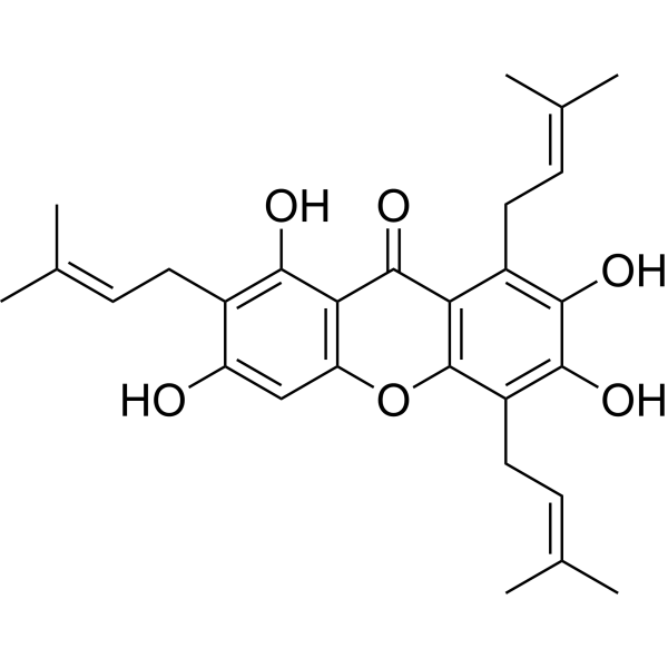
Garcinone E

Based on 1 Customer Validation
Garcinone E is a xanthone natural product that exerts antioxidant, anti-inflammatory, antitumor, and immunomodulatory activities by inhibiting JNK, TNF-α, NF-κB, and FAS, and enhancing Nrf2 signaling pathways. Garcinone E can be used in research on autoimmune hepatitis, obesity, and various cancers (including breast cancer, colorectal cancer, cervical cancer, liver cancer, lung cancer, and gastric cancer).
Nos produits utilisent uniquement pour la recherche. Nous ne vendons pas aux patients.
- Pureté : 98.19%
- CAS No.: 112649-21-5
- Formule: C28H32O6
- Masse moléculaire:464.55
-
Stockage:
4°C, sealed storage, away from moisture and light
* In solvent : -80°C, 6 months; -20°C, 1 month (sealed storage, away from moisture and light)

To place orders, for customer services and technical support, please contact: MedChemExpress USA
Tel: 609-228-6898 E-mail: [email protected] [email protected]


-
 Activité biologique
-
Chemical Information
- Protocole
- Pureté et documentation
- Références
-
 Help & FAQs


Voir tous les produits spécifiques à Isoform TNF Receptor
More

Activité biologique
Description
|
FAS 3.3 μM (IC50) |
NF-κB |
JNK |
Cellular Effect
|
Cell Line

|
Type | Value | Description | References |
|---|---|---|---|---|
| HeLa | IC50 |
128 μM
|
Antiproliferative activity against human HeLa cervical carcinoma cells assessed as reduction in cell viability after 72 hrs by MTT assay.
Antiproliferative activity against human HeLa cervical carcinoma cells assessed as reduction in cell viability after 72 hrs by MTT assay.
|
32676834 |
| MDA-MB-231 | IC50 |
0.68 μM
|
Cytotoxicity against human MDA-MB-231 breast cancer cells assessed as reduction in cell viability incubated for 48 hrs by CCK-8 assay.
Cytotoxicity against human MDA-MB-231 breast cancer cells assessed as reduction in cell viability incubated for 48 hrs by CCK-8 assay.
|
37259475 |
| MCF7 | IC50 |
2.27 μM
|
Cytotoxicity against human MCF-7 breast cancer cells assessed as reduction in cell viability incubated for 48 hrs by CCK-8 assay.
Cytotoxicity against human MCF-7 breast cancer cells assessed as reduction in cell viability incubated for 48 hrs by CCK-8 assay.
|
37259475 |
| 4T1 | IC50 |
1.21 μM
|
Cytotoxicity against mouse 4T1 breast cancer cells assessed as reduction in cell viability incubated for 48 hrs by CCK-8 assay.
Cytotoxicity against mouse 4T1 breast cancer cells assessed as reduction in cell viability incubated for 48 hrs by CCK-8 assay.
|
37259475 |
| HONE1 cell line | IC50 |
8.83 μM
|
Antiproliferative activity against human HONE1 nasopharyngeal carcinoma cells assessed as reduction in cell viability incubated for 72 hrs by MTS assay.
Antiproliferative activity against human HONE1 nasopharyngeal carcinoma cells assessed as reduction in cell viability incubated for 72 hrs by MTS assay.
|
37203204 |
In Vitro
Garcinone E (16-128 μM; 24-72 h) selectively decreases the proliferation of human HeLa cervical cancer cells in a concentration- and time-dependent manner, restricting cell viability to approximately 5% after treatment with 128 μM for 72 h[2].
Garcinone E (16-128 μM; 48 h) significantly inhibits the clonogenic ability of human HeLa cervical cancer cells, reducing colony formation from 450 to nearly 50 at 128 μM[2].
Garcinone E (16-128 μM; 12 h) reduces the migration and invasion abilities of human HeLa cervical cancer cells in a dose-dependent manner[2].
Garcinone E (GE) exhibits cytotoxic activity against MDA-MB-231, MCF-7, and 4T1 breast cancer cells, with IC50 values of 0.68, 2.27, and 1.21 μM, respectively[4].
Garcinone E (25-100 nM) dose-dependently inhibits M2 macrophage-induced proliferation of MCF-7, MDA-MB-231, 4T1, and HUVEC cells, as evidenced by reduced BrdU incorporation[4].
Garcinone E (GAR E) (0.5-2 μM; 24 h) inhibits colony formation and migration in HT-29 and Caco-2 human colorectal cancer cells in a concentration-dependent manner[5].
Garcinone E (0.5-2 μM; 24 h) reduces the cell viability of HT-29 and Caco-2 human colorectal cancer cells in a dose-dependent manner, an effect that is partially reversed by Acetylcysteine (NAC) (HY-B0215) and attenuated by the JNK inhibitor SP600125 (HY-12041)[5].
Garcinone E (1-2 μM; 2 h) induces the production of intracellular reactive oxygen species in HT-29 and Caco-2 human colorectal cancer cells[5].
Garcinone E (2 μM; 24 h) induces loss of mitochondrial membrane potential in HT-29 and Caco-2 human colorectal cancer cells, an effect that is reversed by the antioxidant NAC and attenuated by the JNK inhibitor SP600125[5].
Garcinone E (2 μM; 24 h) induces apoptosis in HT-29 and Caco-2 human colorectal cancer cells, an effect that is abolished by the antioxidant Acetylcysteine or attenuated by the JNK inhibitor SP600125[5].
Garcinone E (1 μM; 48 h) induces cell cycle arrest at the Sub G1 phase in HT-29 and Caco-2 human colorectal cancer cells, an effect that is abolished by N-acetylcysteine pretreatment and attenuated by the JNK inhibitor SP600125[5].
Garcinone E (2 μM; 24 h) activates the JNK signaling pathway in HT-29 human colorectal cancer cells and induces the expression of apoptosis-related proteins, an effect that is abolished by the antioxidant NAC[5].
Garcinone E (2 μM; 24 h) activates the JNK signaling pathway and induces the expression of apoptosis-related proteins in Caco-2 human colorectal cancer cells, an effect that is abolished by the antioxidant NAC[5].
Garcinone E (2 μM; 24 h) activates the JNK signaling pathway and induces the expression of apoptosis-related proteins in HT-29 and Caco-2 human colorectal cancer cells, and this effect is abolished by the JNK inhibitor SP600125[5].
Garcinone E (16-128 μM) induces apoptotic cell death in human HeLa cervical cancer cells in a dose-dependent manner[2].
Garcinone E (16-128 μM; 24 h) induces caspase-dependent apoptotic cell death in human HeLa cervical cancer cells, manifested by upregulation of Bax and caspases and downregulation of Bcl-2[2].
Garcinone E (16-128 μM; 24 h) induces G2/M phase cell cycle arrest in human HeLa cervical cancer cells[2].
Garcinone E acts as a potent, rapidly binding inhibitor of the overall FAS reaction, with an IC50 of 3.3 μM[3].
Garcinone E inhibits the KR domain activity of FAS with an IC50 of 14.6 μM, which is weaker than its effect on the overall reaction[3].
Garcinone E (20.0 μM) acts as a slow-binding irreversible inhibitor of the overall FAS reaction (kobs = 0.009 min-1 at 20 μM), but exhibits only weak irreversible inhibition of the KR domain (kobs = 0.0004 min-1)[3].
Garcinone E (50 and 100 nM) significantly downregulates the mRNA expression of M2-specific markers (CD206, Arg-1, Fizz-1, Ym1) in IL-4 + IL-13-treated RAW 264.7 macrophages[4].
Garcinone E (25-100 nM) inhibits IL-4/IL-13-induced STAT6 phosphorylation, Arg-1 expression, and nuclear translocation in RAW 264.7 macrophages[4].
Garcinone E (25-100 nM) inhibits M2 macrophage-induced angiogenesis in HUVECs[4].
Garcinone E (50 and 100 nM) reduces the secretion of pro-angiogenic and pro-metastatic factors, including VEGF, MMP9, MMP2, and TGF-β, from M2-polarized RAW 264.7 macrophages[4].
Garcinone E (2.5-20 μmol/L; 24-72 h) significantly inhibits the viability of HK1, HONE1, and S18 cells, with 72 h IC50 values of 7.64, 8.83, and 4.65 μmol/L, respectively[6].
Garcinone E (1.25-5 μmol/L; 9-12 days) reduces the colony-forming ability of HK1, HONE1, and S18 cells and statistically confirms the decrease in relative colony area of HK1 and HONE1 cells[6].
Garcinone E (7.5-12.5 μmol/L; 24 h) induces cell cycle arrest at the G0/G1 and S phases in HK1 and HONE1 cells[6].
Garcinone E (5-10 μmol/L; 48 h) increases the number of autophagosomes in HK1 and HONE1 cells[6].
Garcinone E (5-10 μmol/L; 48 h) blocks autophagic flux in HK1, HONE1, and S18 cells, manifested as accumulation of yellow puncta[6].
Garcinone E (5-10 μmol/L; 48 h) induces lysosomal damage and blocks autophagosome-lysosome fusion in HK1 and S18 cells[6].
Garcinone E (5-10 μmol/L; 48 h) blocks autophagic flux in HK1 and HONE1 cells by inhibiting autophagosome-lysosome fusion[6].
Garcinone E (5-10 μmol/L; 24 h) enhances the expression of cell cycle-related proteins, autophagy-related proteins, and signaling pathway proteins in HK1 and HONE1 cells[6].
Treatment with Garcinone E (5-10 μmol/L; 24 h) significantly alters the mRNA expression profiles in HK1 and HONE1 cells and enriches autophagy-related pathways[6].
Garcinone E (5-10 μmol/L; 24 h) upregulates BECN1, SQSTM1, and MAP1LC3B mRNA and downregulates AKT1 mRNA in HK1 and HONE1 cells[6].
MedChemExpress (MCE) has not independently confirmed the accuracy of these methods. They are for reference only. Further protocols information, click here.
-
Cell Line:human HeLa cervical carcinoma cells
-
Concentration:8, 16, 32, 64, 128 μM
-
Incubation Time:24, 48, and 72 h
-
Result:Reduced cell viability from 100% to almost 10% over the concentration range of 0-128 μM.
Limited viability to approximately 35%, 20%, and 5% after 24 h, 48 h, and 72 h, respectively.
-
Cell Line:human HeLa cervical carcinoma cells
-
Concentration:16, 64, 128 μM
-
Incubation Time:24 h
-
Result:Increased accumulation of cells in the G2/M-phase as the concentration increased from 0 to 128 μM.
-
Cell Line:human HeLa cervical carcinoma cells
-
Concentration:16, 64, 128 μM
-
Incubation Time:12 h
-
Result:Declined the number of migrated cells with increasing doses in contrast to controls.
Suppressed the invasion potency of cells in a dose-dependent manner.
-
Cell Line:human HeLa cervical carcinoma cells
-
Concentration:16, 64, 128 μM
-
Incubation Time:24 h
-
Result:Increased the expression of proapoptotic protein Bax and Caspases (-3, -8, and -9) while decreasing the expression of antiapoptotic protein Bcl-2 with increasing doses.
-
Cell Line:RAW 264.7 macrophages and differentiated THP-1 macrophages
-
Concentration:0.06, 0.125, 0.25, 0.5 μM
-
Incubation Time:48 h
-
Result:Showed no effect on cell viability at 0.06-0.125 μM in either cell type.
Significantly reduced cell viability in both RAW 264.7 and THP-1 cells at 0.25-0.5 μM.
Had no effect on LDH release at 0.06-0.125 μM in either cell type.
Significantly increased LDH release in both RAW 264.7 and THP-1 cells at 0.25-0.5 μM.
-
Cell Line:RAW 264.7 macrophages
-
Concentration:25, 50, 100 nM
-
Incubation Time:48 h
-
Result:Significantly downregulated the mRNA expression of M2-specific markers (CD206, Arg-1, Fizz-1, Ym1) in IL-4 + IL-13-treated RAW 264.7 macrophages at 50 and 100 nM.
-
Cell Line:RAW 264.7 macrophages
-
Concentration:25, 50, 100 nM
-
Incubation Time:48 h
-
Result:Inhibited IL-4/IL-13-induced STAT6 phosphorylation and Arg-1 expression in RAW 264.7 macrophages at concentrations of 25-100 nM.
-
Cell Line:RAW 264.7 macrophages
-
Concentration:25, 50, 100 nM
-
Incubation Time:48 h
-
Result:Inhibited IL-4/IL-13-induced STAT6 nuclear translocation in RAW 264.7 macrophages at concentrations of 25-100 nM.
-
Cell Line:MCF-7, MDA-MB-231, 4T1 cells, and HUVECs
-
Concentration:25, 50, 100 nM
-
Incubation Time:48 h
-
Result:Dose-dependently suppressed M2 macrophage-induced proliferation of MCF-7, MDA-MB-231, 4T1, and HUVEC cells, as evidenced by a reduced BrdU incorporation.
-
Cell Line:HT-29 and Caco-2 human colon cancer cells
-
Concentration:0.5, 1, 2 μM
-
Incubation Time:24 h
-
Result:Suppressed the viability of HT-29 and Caco-2 cells dose-dependently after 24 h treatment.
Increased the viability of HT-29 cells preincubated with 5 mM NAC from 60.65% to 78.57%, 39.27% to 63.34%, and 27.14% to 45.90% after 24 h treatment with 0.5, 1, and 2 μM GAR E, respectively.
Increased the viability of Caco-2 cells preincubated with 3 mM NAC from 53.44% to 68.04%, 31.12% to 56.64%, and 13.25% to 34.27% after treatment with 0.5, 1, and 2 μM GAR E, respectively.
-
Cell Line:HT-29 and Caco-2 human colon cancer cells
-
Concentration:0.5, 1, 2 μM
-
Incubation Time:24 h
-
Result:Improved the viability of HT-29 cells treated with 0.5, 1, and 2 μM GAR E from 66.20% to 75.75%, 48.94% to 63.99%, and 30.60% to 46.39%, respectively, after pretreatment with 10 μM JNKI.
Increased the viability of 0.5, 1, and 2 μM GAR E-treated Caco-2 cells from 44.16% to 64.50%, 21.76% to 47.95%, and 8.86% to 21.54%, respectively, after JNKI pretreatment.
-
Cell Line:HT-29 and Caco-2 human colon cancer cells
-
Concentration:2 μM
-
Incubation Time:24 h
-
Result:Increased the apoptotic rates of HT-29 and Caco-2 cells to 323.2% and 2800%, respectively, after treatment with 2 μM GAR E.
Abolished the GAR E-induced increase in the apoptotic rates of these two cancer cells by N-acetylcysteine treatment.
-
Cell Line:HT-29 and Caco-2 human colon cancer cells
-
Concentration:2 μM
-
Incubation Time:24 h
-
Result:Decreased the percentage of apoptotic HT-29 and Caco-2 cells from 340.4% to 257% and from 2808% to 1595%, respectively, after preincubation with JNKI.
-
Cell Line:HT-29 human colon cancer cells
-
Concentration:1 μM
-
Incubation Time:48 h
-
Result:Increased the proportion of HT-29 cells in the Sub G1 phase from 2.25% to 9.81% after 48 h incubation with 1 μM GAR E.
Decreased the fraction of cells at the G1 phase from 77% to 67.07%.
-
Cell Line:Caco-2 human colon cancer cells
-
Concentration:1 μM
-
Incubation Time:48 h
-
Result:Stimulated an increase in the proportion of cells at the Sub G1 phase from 1.94% to 21.67% after 48 h treatment with GAR E.
Decreased the fraction of cells in the G1 phase from 59.17% to 48.6% after treatment with GAR E.
-
Cell Line:HT-29 and Caco-2 human colon cancer cells
-
Concentration:1 μM
-
Incubation Time:48 h
-
Result:Abolished the GAR E-induced fraction of cells in the Sub G1 phase in both HT-29 and Caco-2 cells by N-acetylcysteine pretreatment.
-
Cell Line:HT-29 and Caco-2 human colon cancer cells
-
Concentration:1 μM
-
Incubation Time:48 h
-
Result:Attenuated the GAR E-induced increase in the fraction of cells in the Sub G1 phase from 7.75% to 4.37% in HT-29 cells and from 21.67% to 12.31% in Caco-2 cancer cells by JNKI pretreatment.
-
Cell Line:HT-29 human colon cancer cells
-
Concentration:2 μM
-
Incubation Time:24 h
-
Result:Stimulated the cleavage of PARP, caspase 3, and 9, and promoted the phosphorylation of JNK1/2.
Decreased the level of expression of Bcl-2, while the level of Bax was unaltered.
Abrogated the GAR E-induced changes in the expression of phospho-JNK1/2, cleaved caspase 3 and 9, and cleaved PARP, and reversed the down-regulation of Bcl-2 by NAC pretreatment.
-
Cell Line:Caco-2 human colon cancer cells
-
Concentration:2 μM
-
Incubation Time:24 h
-
Result:Stimulated the cleavage of PARP, caspase 3, and 9, and promoted the phosphorylation of JNK1/2.
Decreased the level of expression of Bcl-2, while the level of Bax was unaltered.
Abrogated the GAR E-induced changes in the expression of phospho-JNK1/2, cleaved caspase 3 and 9, and cleaved PARP, and reversed the down-regulation of Bcl-2 by NAC pretreatment.
-
Cell Line:HT-29 and Caco-2 human colon cancer cells
-
Concentration:2 μM
-
Incubation Time:24 h
-
Result:Decreased the GAR E-induced increases in the levels of cleaved-PARP and phosphor-JNK1/2 by JNKI.
Almost abolished the cleavage of caspase 3 and caspase 9 by JNKI pretreatment.
Elevated the production of Bcl-2 by JNKI, whereas the level of expression of Bax was unaffected in both cell lines.
-
Cell Line:HK1, HONE1, and S18
-
Concentration:2.5, 3.75, 5, 7.5, 10, 15, 20 μmol/L
-
Incubation Time:24, 48, and 72 h
-
Result:Significantly inhibited cell viability.
Achieved IC50 values of 7.64 μmol/L for HK1, 8.83 μmol/L for HONE1, and 4.65 μmol/L for S18 cells at 72 h.
-
Cell Line:HK1 and HONE1
-
Concentration:7.5, 10, 12.5 μmol/L
-
Incubation Time:24 h
-
Result:Induced cell cycle arrest at the G0/G1 phase in HK1 cells.
Induced arrest at the G0/G1 phase with 7.5 and 10 μmol/L and at both G0/G1 and S phases with 12.5 μmol/L in HONE1 cells.
-
Cell Line:HK1 and HONE1
-
Concentration:5, 10 μmol/L
-
Incubation Time:48 h
-
Result:Increased MDC fluorescence intensity and the number of autophagosomes compared to the control group.
-
Cell Line:HK1, HONE1, and S18
-
Concentration:5, 7.5, 10 μmol/L
-
Incubation Time:48 h
-
Result:Induced the appearance of both green and red puncta, appearing as yellow puncta, indicating impaired autophagic flux and inhibition of lysosomal degradation.
-
Cell Line:HK1 and S18
-
Concentration:5, 10 μmol/L
-
Incubation Time:48 h
-
Result:Increased the number of Gal-3-labeled damaged lysosomes in both cell lines.\nReduced the co-localization of LC3-labeled autophagosomes and Gal-3-labeled damaged lysosomes.
-
Cell Line:HK1-stubRFP-sensGFP-LC3 and HONE1-stubRFP-sensGFP-LC3
-
Concentration:5, 10 μmol/L
-
Incubation Time:48 h
-
Result:Blocked autophagosome-lysosome fusion, as green and blue puncta did not completely overlap.
-
Cell Line:HK1 and HONE1
-
Concentration:5, 7.5, 10 μmol/L
-
Incubation Time:24 h
-
Result:Increased the expression of p21 and p27.
Decreased the expression of CDK2, CDK4, CDK6, CDK7, Cyclin A2, Cyclin D3, Cyclin E1, and Cyclin E2.\nEnhanced the expression of LC3B-I, LC3B-II, SQSTM1/p62, ULK1, PIK3R4, Beclin-1, and Atg16L1.
Did not affect the expression of Atg3, Atg5, Atg7, and Atg12.
-
Cell Line:HK1 and HONE1
-
Concentration:5, 7.5, 10 μmol/L
-
Incubation Time:24 h
-
Result:Decreased the expression of Akt, p70 S6K, p-p70 S6K, DNA Polymerase, Met, and Optineurin.
Increased the expression of p-GSK-3β.
Dysregulated the expression of 4E-BP1, p-4E-BP1(Thr37/46), p-4E-BP1(Thr70), and Non-p-4E-BP1(Thr46).
-
Cell Line:HK1 and HONE1
-
Concentration:5, 10 μmol/L
-
Incubation Time:24 h
-
Result:Upregulated the mRNA expression of Beclin-1, SQSTM1/p62, and LC3B.
Downregulated Akt mRNA expression.
In Vivo
Garcinone E (2 mg/kg; i.p.; once every other day) effectively inhibits tumor growth and lung metastasis in the 4T1 mouse breast cancer model, with a 52% reduction in tumor volume and a 50% reduction in lung metastatic nodules[4].
Garcinone E (2 mg/kg; i.p.; every other day) effectively inhibits breast cancer lung metastasis in a tail vein injection model, significantly reducing the number of metastatic nodules at 20 days (105 vs. 47)[4].
Garcinone E (35 mg/kg; i.p.; once every three days; for 18 days) inhibits the growth of S18 nasopharyngeal carcinoma xenografts in nude mice without severe side effects[6].
Garcinone E (2.5-5 mg/kg; i.p.; 2 days) significantly inhibits tumor growth in the HT-29 xenograft mouse model, reducing tumor volume to as low as 129 mm3 at a dose of 5 mg/kg[5].
MedChemExpress (MCE) has not independently confirmed the accuracy of these methods. They are for reference only.
-
Animal Model:BALB/c mice (Male; 20-22 g; Concanavalin A-induced)[1]
-
Dosage:25 mg/kg; 50 mg/kg
-
Administration:once daily; 5 days
-
Result:Reduced elevated serum indices of hepatic damage (ALT, AST, ALP, LDH, and γ-GT).
Suppressed the elevation of hepatic myeloperoxidase (MPO) level.
Minimized the immunostaining of CD4+ and PCNA in the liver.
Enhanced hepatic Nrf2 signaling and binding activity.
Increased downstream antioxidants (SOD, GSH, TAC, HO-1).
Reduced lipid peroxidative markers (MDA, 4-HNE, PC).
Attenuated elevated levels of NF-κB, IL-6, IL-1β, TNF-α, JNK, Bax, and caspase-3.
Elevated the anti-apoptotic Bcl2 level.
-
Animal Model:BALB/c nude mice (male, 4-6 weeks old, 20-22 g; subcutaneous injection of HT-29 human colon cancer cells)[5]
-
Dosage:2.5, 5 mg/kg
-
Administration:i.p.; 2 days
-
Result:Reduced tumor volumes to 139 mm3 at 2.5 mg/kg and 129 mm3 at 5 mg/kg, compared to ~485 mm3 in the saline-treated group.
Decreased tumor weights to 0.18 g and 0.15 g, respectively, versus 0.45 g in the saline group.
Increased the necrotic area of tumor tissues by approximately 6-fold compared with the saline group.
Elevated levels of cleaved PARP and phosphorylated JNK by approximately 75%, increased the Bax/Bcl-2 ratio by 67%, and increased expression of cleaved-caspase 3 and 9 by 14% and 30%, respectively, at 5 mg/kg.
-
Animal Model:BALB/c nude mice (Female, 4-week-old, subcutaneous inoculation of 3 × 10⁵ S18 cells)[6]
-
Dosage:35 mg/kg
-
Administration:i.p.; every three days; up to 18 days
-
Result:Significantly reduced tumour weight and volume compared to the vehicle group.
Did not significantly alter body weight compared to the vehicle group.


Chemical Information
-
CAS No. 112649-21-5
-
Appearance Solid
-
Masse moléculaire 464.55
-
Formule C28H32O6
-
Color Light yellow to yellow
-
SMILES
O=C1C2=C(OC3=C1C(O)=C(C/C=C(C)\C)C(O)=C3)C(C/C=C(C)\C)=C(O)C(O)=C2C/C=C(C)\C
-
Structure Classification
-
Initial Source
-
Livraison
Room temperature in continental US; may vary elsewhere.
-
Stockage
4°C, sealed storage, away from moisture and light
* In solvent : -80°C, 6 months; -20°C, 1 month (sealed storage, away from moisture and light)

Protocole
-
Research Protocol for Inflammation-related Diseases
The NLRP3 inflammasome is a cytosolic innate immune signaling platform that integrates priming signals and danger-signal activation to promote caspase-1 activation, maturation of IL-1β and IL-18, and gasdermin D-mediated pyroptotic cell death. The core experimental logic is to determine whether inflammatory disease phenotypes are driven by increased NLRP3 expression, ASC-containing inflammasome assembly, caspase-1 cleavage, GSDMD cleavage, and extracellular release of IL-1β/IL-18 rather than by nonspecific cell injury alone. The pathway is strongly linked to inflammation-related disease phenotypes because monosodium urate crystals activate NALP3/NLRP3 inflammasome signaling in gout-like crystal inflammation, cholesterol crystals activate NLRP3 inflammasomes in atherogenesis models, and DSS-induced intestinal inflammation has been reported to involve NLRP3 inflammasome activity. However, experimental colitis studies also show context-dependent protective effects of NLRP3 inflammasome co
-
Liver Cancer Modeling
Liver cancer can be classified into primary liver cancer and secondary liver cancer. Secondary liver cancer is the metastatic liver cancer. Primary liver cancer includes hepatocellular carcinoma (HCC), intrahepatic cholangiocarcinoma (ICC) and fibrolamellar HCC, of which HCC is the most common form, accounting for approximately 90% of primary liver cancers[1]. HCC mouse models include chemical agent-induced models, transplanted tumor models, and genetic engineered models.
-
Research Protocol for Metabolic Diseases
AMP-activated protein kinase, AMPK, is a conserved cellular energy sensor that responds to reduced cellular energy status and coordinates metabolism by increasing ATP-generating catabolic pathways while suppressing ATP-consuming anabolic processes. In metabolic disease research, the AMPK pathway is experimentally relevant because it regulates hepatic lipid synthesis, fatty acid oxidation, glucose production, skeletal-muscle glucose disposal, mTORC1-linked biosynthesis, autophagy, mitochondrial homeostasis, and whole-body energy balance. The central pathway logic is that energy stress, metformin, exercise-like stimulation, or direct AMPK activators increase AMPKα Thr172 phosphorylation and downstream substrate phosphorylation, including ACC and RAPTOR. Phosphorylation of ACC suppresses lipogenesis and supports fatty acid oxidation, whereas phosphorylation of RAPTOR suppresses mTORC1 signaling and links cellular energy status to growth and protein synthesis control. The pathway is linked
-
Breast Cancer Modeling
Breast cancer is a heterogeneous cancer, and it has been distinguished into four subtypes: luminal A, luminal B, HER2-positive and basal-like. Molecular mutations, epigenetic alterations, hormone exposure and immune microenvironment are related to the progression of breast cancer.


Pureté et documentation
-
Fiche technique (324 KB)
-
SDS (251 KB)
- English - EN (251 KB)
- Français - FR (251 KB)
- Deutsch - DE (251 KB)
- Norwegian - NO (251 KB)
- Español - ES (251 KB)
- Swedish - SV (251 KB)
- Italian - IT (251 KB)
- Korean - KR (251 KB)
- Portuguese - PT (251 KB)
-
Instruction de manipulation (2659 KB)
Références

Calculators
Concentration (start) × Volume (start) = Concentration (final) × Volume (final)


Garcinone E Related Classifications
HY-123901 Related Classifications
 Powered by Bioz
Keywords