



Grosvenorine

Based on 1 Customer Validation
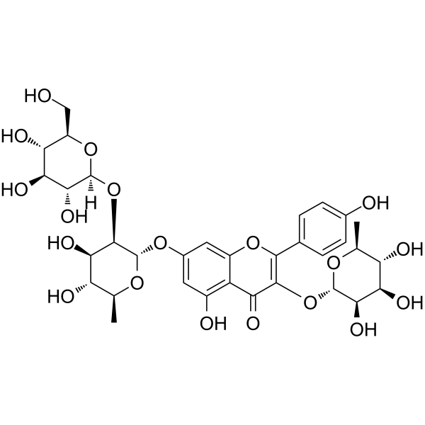
Grosvenorine is an orally active flavonoid glycoside found in S. grosvenorii. Grosvenorine exhibits antibacterial, antioxidant and antiinflammation activities. Grosvenorine can induce apoptosis and increases anti-apoptotic Bcl-2 protein expression and reduces pro-apoptotic P53 protein expression in gastric tissues. Grosvenorine enhances mucin/glycoprotein secretion, regulates gastric pH, and reduces gastric lesion incidence.Grosvenorine increases glutathione peroxidase, catalase, and SOD levels, reduces lipid peroxidation (MDA), and lowers TNF-α and IL-6 levels. Grosvenorine can be used for the researches of bacterial infection and Gastric ulcer.
For research use only. We do not sell to patients.
- Purity : 99.64%
- CAS No.: 156980-60-8
- Formula: C33H40O19
- Molecular Weight:740.66
-
Storage:
4°C, sealed storage, away from moisture and light
* In solvent : -80°C, 6 months; -20°C, 1 month (sealed storage, away from moisture and light)

To place orders, for customer services and technical support, please contact: MedChemExpress USA
Tel: 609-228-6898 E-mail: [email protected] [email protected]


-
 Biological Activity
-
Chemical Information
-
Solvent & Solubility
- Protocols
- Purity & Documentation
- References
-
 Help & FAQs




Biological Activity
Description
IC50 & Target
[1]|
Bcl-2 |
IL-6 |
In Vitro
Grosvenorine exhibits in vitro inhibitory activity against Gram-positive bacteria with a MIC of less than 70 μg/mL[2].
MedChemExpress (MCE) has not independently confirmed the accuracy of these methods. They are for reference only.
In Vivo
Grosvenorine (250-500 mg/kg; p.o.; single dose) administered orally up to 500 mg/kg is non-toxic in SD rats, with no observed adverse effects on behavior, organ function, or tissue architecture over a 2-week period[1].
MedChemExpress (MCE) has not independently confirmed the accuracy of these methods. They are for reference only.
-
Animal Model:Sprague-Dawley (male and female, 7-8 weeks old, 190-200 g; indomethacin-induced gastropathy)[1]
-
Dosage:30 mg/kg; 60 mg/kg
-
Administration:I.g.; single pretreatment dose
-
Result:Inhibited gastric lesion incidence by 69.03% (30 mg/kg) and 74.89% (60 mg/kg).
Reduced ulcer area and increased gastric mucus weigh.
Lowered total stomach acidity and increased Alcian blue binding capacity.
Elevated gastric tissue superoxide dismutase (SOD), catalase (CAT), and glutathione peroxidase (GPx) and reduced malondialdehyde (MDA.
Reduced serum tumor necrosis factor-alpha (TNF-α), interleukin-6 (IL-6), and increased interleukin-10 (IL-10).
Reduced mucosal injury, edema, and inflammatory cell infiltration.
Upregulated anti-apoptotic Bcl-2 protein and downregulated pro-apoptotic P53 protein in gastric tissues.
- Research Protocol for Infectious Diseases
- RNA extraction experimental
- Western Blot
- Detection of Apoptosis
- TUNEL staining for apoptotic DNA fragmentation
- Annexin V plus membrane-impermeant dye apoptosis staining
- Apoptosis Solutions
- Ferroptosis Solutions
- Protocol For Protein Expression And Purification


Chemical Information
-
CAS No. 156980-60-8
-
Appearance Solid
-
Molecular Weight 740.66
-
Formula C33H40O19
-
Color Off-white to light yellow
-
SMILES
O=C1C(O[C@H]2[C@@H]([C@@H]([C@@H](O)[C@H](C)O2)O)O)=C(C3=CC=C(O)C=C3)OC4=CC(O[C@H]5[C@@H]([C@@H]([C@@H](O)[C@H](C)O5)O)O[C@]6([H])O[C@@H]([C@@H](O)[C@H](O)[C@H]6O)CO)=CC(O)=C41
-
Structure Classification
-
Shipping
Room temperature in continental US; may vary elsewhere.
-
Storage
4°C, sealed storage, away from moisture and light
* In solvent : -80°C, 6 months; -20°C, 1 month (sealed storage, away from moisture and light)


Solvent & Solubility
In Vitro:
DMSO : 100 mg/mL (135.01 mM; Need ultrasonic; Hygroscopic DMSO has a significant impact on the solubility of product, please use newly opened DMSO)
Please refer to the solubility information to select the appropriate solvent. Once prepared, please aliquot and store the solution to prevent product inactivation from repeated freeze-thaw cycles.
Storage method and period of stock solution: -80°C, 6 months; -20°C, 1 month (sealed storage, away from moisture and light). When stored at -80°C, please use it within 6 months. When stored at -20°C, please use it within 1 month.
Please refer to the solubility information to select the appropriate solvent. Once prepared, please aliquot and store the solution to prevent product inactivation from repeated freeze-thaw cycles.
Storage method and period of stock solution: -80°C, 6 months; -20°C, 1 month (sealed storage, away from moisture and light). When stored at -80°C, please use it within 6 months. When stored at -20°C, please use it within 1 month.
Concentration (start) × Volume (start) = Concentration (final) × Volume (final)
In Vivo:
Select the appropriate dissolution method based on your experimental animal and administration route.
- For the following dissolution methods, please ensure to first prepare a clear stock solution using an In Vitro approach and then sequentially add co-solvents:
- To ensure reliable experimental results, the clarified stock solution can be appropriately stored based on storage conditions. As for the working solution for In Vivo experiments, it is recommended to prepare freshly and use it on the same day.
- The percentages shown for the solvents indicate their volumetric ratio in the final prepared solution. If precipitation or phase separation occurs during preparation, heat and/or sonication can be used to aid dissolution.
Add each solvent one by one: 10% DMSO 40% PEG300 5% Tween-80 45% Saline
Solubility: ≥ 2.5 mg/mL (3.38 mM); Clear solution
This protocol yields a clear solution of ≥ 2.5 mg/mL (saturation unknown).
Taking 1 mL working solution as an example, add 100 μL DMSO stock solution (25.0 mg/mL) to 400 μL PEG300, and mix evenly; then add 50 μL Tween-80 and mix evenly; then add 450 μL Saline to adjust the volume to 1 mL.
Preparation of Saline: Dissolve 0.9 g sodium chloride in ddH₂O and dilute to 100 mL to obtain a clear Saline solution.
Add each solvent one by one: 10% DMSO 90% (20% SBE-β-CD in Saline)
Solubility: ≥ 2.5 mg/mL (3.38 mM); Clear solution
This protocol yields a clear solution of ≥ 2.5 mg/mL (saturation unknown).
Taking 1 mL working solution as an example, add 100 μL DMSO stock solution (25.0 mg/mL) to 900 μL 20% SBE-β-CD in Saline, and mix evenly.
Preparation of 20% SBE-β-CD in Saline (4°C, storage for one week): 2 g SBE-β-CD powder is dissolved in 10 mL Saline, completely dissolve until clear.
In Vivo Dissolution Calculator
Please enter the basic information of animal experiments:
-
-
-
-
Recommended: Prepare an additional quantity of animals to account for potential losses during experiments.
Please enter your animal formula composition:
-
%DMSO +
Recommended: Keep the proportion of DMSO in working solution below 2% if your animal is weak.
-
%+
-
+%Tween-80 + +
-
%Saline +
The co-solvents required include: DMSO, . All of co-solvents are available by MedChemExpress (MCE). , Tween 80. All of co-solvents are available by MedChemExpress (MCE).
Working solution concentration: 0.22 mg/mL
Method for preparing stock solution: mg drug dissolved in μL DMSO. Stock solution concentration: mg/mL. * In solvent : -80°C, 6 months; -20°C, 1 month (sealed storage, away from moisture and light)
1. Take μL DMSO stock solution;
2. Add μL .
μL , mix evenly;
3. Then add μL Tween 80, mix evenly;
4. Then add μL
Please ensure that the stock solution in the first step is dissolved to a clear state, and add co-solvents in sequence. You can use ultrasonic heating (ultrasonic cleaner, recommended frequency 20-40 kHz), vortexing, etc. to assist dissolution.


Purity & Documentation
-
Data Sheet (292 KB)
-
SDS (393 KB)
- English - EN (393 KB)
- Français - FR (393 KB)
- Deutsch - DE (393 KB)
- Norwegian - NO (393 KB)
- Español - ES (393 KB)
- Swedish - SV (393 KB)
- Italian - IT (393 KB)
- Korean - KR (393 KB)
- Portuguese - PT (393 KB)
-
Handling Instructions (2659 KB)
References


Complete Stock Solution Preparation Table
Please refer to the solubility information to select the appropriate solvent. Once prepared, please aliquot and store the solution to prevent product inactivation from repeated freeze-thaw cycles.
Storage method and period of stock solution: -80°C, 6 months; -20°C, 1 month (sealed storage, away from moisture and light). When stored at -80°C, please use it within 6 months. When stored at -20°C, please use it within 1 month.
| Optional Solvent | Concentration Solvent Mass | 1 mg | 5 mg | 10 mg | 25 mg |
|---|---|---|---|---|---|
| DMSO | 1 mM | 1.3501 mL | 6.7507 mL | 13.5015 mL | 33.7537 mL |
| 5 mM | 0.2700 mL | 1.3501 mL | 2.7003 mL | 6.7507 mL | |
| 10 mM | 0.1350 mL | 0.6751 mL | 1.3501 mL | 3.3754 mL | |
| 15 mM | 0.0900 mL | 0.4500 mL | 0.9001 mL | 2.2502 mL | |
| 20 mM | 0.0675 mL | 0.3375 mL | 0.6751 mL | 1.6877 mL | |
| 25 mM | 0.0540 mL | 0.2700 mL | 0.5401 mL | 1.3501 mL | |
| 30 mM | 0.0450 mL | 0.2250 mL | 0.4500 mL | 1.1251 mL | |
| 40 mM | 0.0338 mL | 0.1688 mL | 0.3375 mL | 0.8438 mL | |
| 50 mM | 0.0270 mL | 0.1350 mL | 0.2700 mL | 0.6751 mL | |
| 60 mM | 0.0225 mL | 0.1125 mL | 0.2250 mL | 0.5626 mL | |
| 80 mM | 0.0169 mL | 0.0844 mL | 0.1688 mL | 0.4219 mL | |
| 100 mM | 0.0135 mL | 0.0675 mL | 0.1350 mL | 0.3375 mL |


Grosvenorine Related Classifications
HY-N3031 Related Classifications
 Powered by Bioz
Keywords