



HQ461
Based on 2 publication(s) in Google Scholar
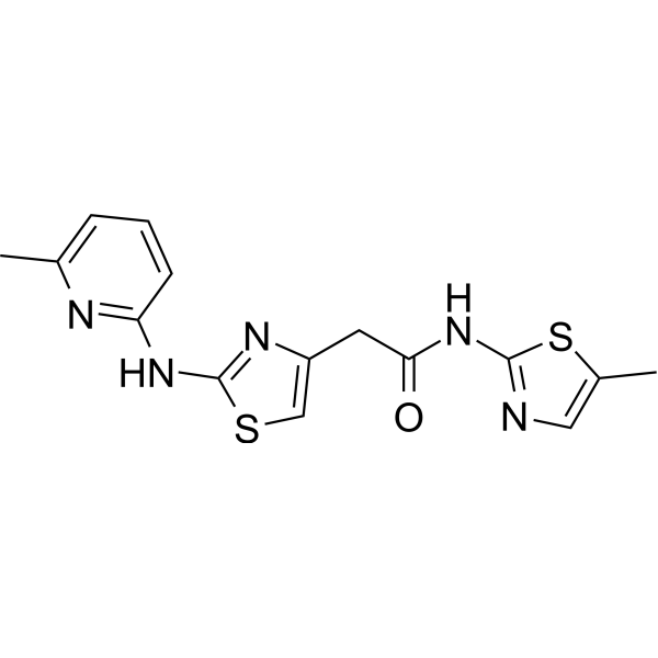
HQ461 is a molecular glue that promotes CDK12-DDB1 interaction to trigger cyclin K degradation. HQ461-mediated degradation of cyclin K impairs CDK12 function, resulting in decreased CDK12 substrate phosphorylation, downregulation of DNA damage response genes, and cell death.
商品は「研究用試薬」です。人や動物の医療用・臨床診断用・食品用の製品ではありません。
研究用途以外に使用した場合、当社は一切の責任を負いかねます。
- 純度 : 98.10%
- CAS 番号: 1226443-41-9
- 分子式: C15H15N5OS2
- 分子量:345.44
-
保管条件:Powder -20°C, 3 years ; In solvent -80°C, 6 months , -20°C, 1 month

To place orders, for customer services and technical support, please contact: MedChemExpress USA
Tel: 609-228-6898 E-mail: [email protected] [email protected]


-
 生物活性
生物活性
-
化学情報
-
溶剤 & 溶解度
- プロトコル
- 純度とドキュメンテーション
- 参考文献
-
 Help & FAQs


-
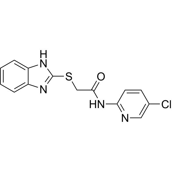
HY-144976
dCeMM3Molecular Glue Degrader -
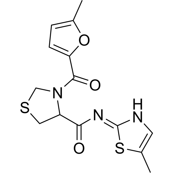
HY-144977
dCeMM4Cyclin K Degrader -
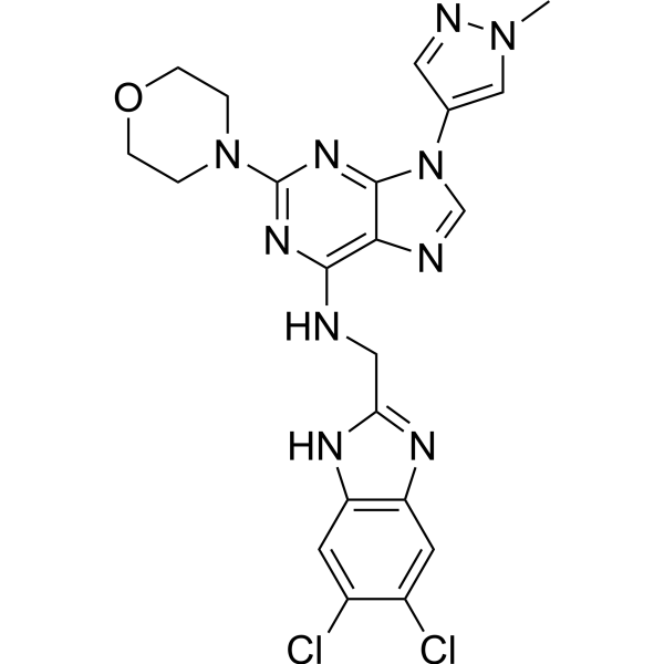
HY-130250
SR-4835CDK12/13 Inhibitor -
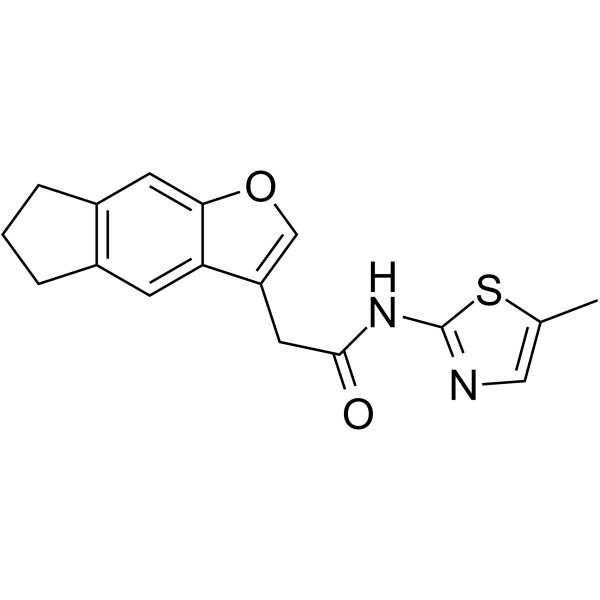
HY-W181530
NCT02CDK12/CCNK Molecular Glue -
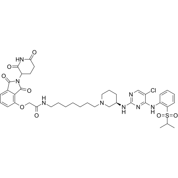
HY-139039
BSJ-4-116PROTAC CDK12 Degrader -
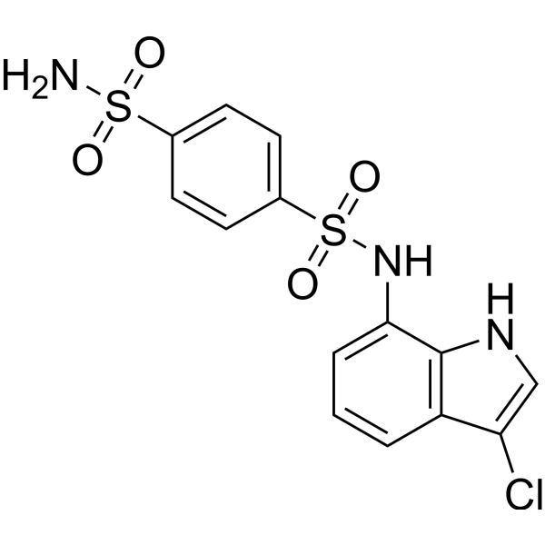
HY-13650
IndisulamCarbonic Anhydrase Inhibitor/RBM39 Molecular Glue Degrader -
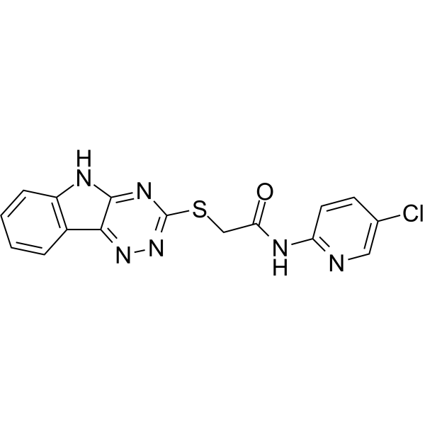
HY-144971
dCeMM2Glue Degrader -
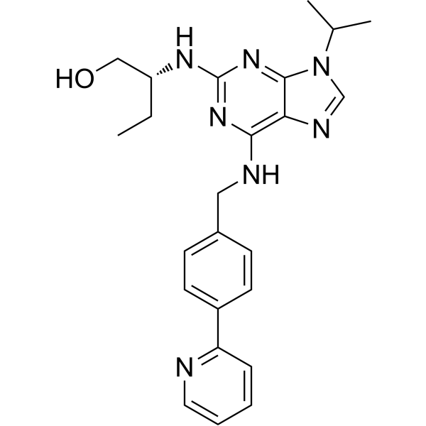
HY-18340
(R)-CR8CDK Inhibitor -
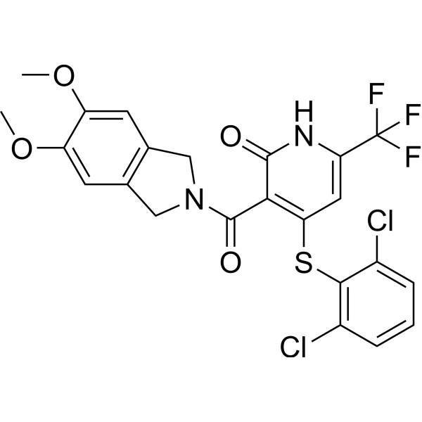
HY-111760
NRX-252262β-catenin Inhibitor -
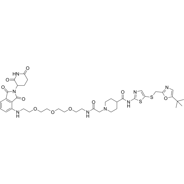
HY-123937
THAL-SNS-032CDK9 PROTAC Degrader -
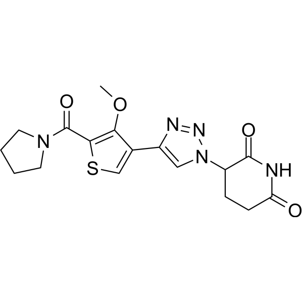
HY-145322
TMX-4116CK1α Degrader -
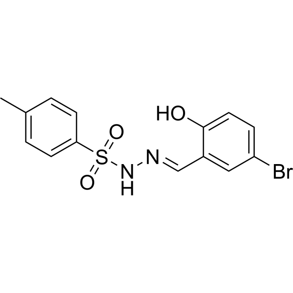
HY-W133627
dCeMM1RBM39 Inhibitor
MedChemExpress(MCE)の使用を引用している文献 HQ461
More

生物活性
製品説明
Cellular Effect
|
Cell Line

|
Type | Value | Description | References |
|---|---|---|---|---|
| A549 | IC50 |
1.3 μM
Compound: HQ461
|
Cytotoxicity against human A549 cells assessed as reduction in cell viability incubated for 72 hrs by Cell-titer glo luminescent assay
Cytotoxicity against human A549 cells assessed as reduction in cell viability incubated for 72 hrs by Cell-titer glo luminescent assay
|
[PMID: 37984298] |
| HCT-116 | IC50 |
1.3 μM
Compound: HQ461
|
Cytotoxicity against human HCT-116 cells assessed a s reduction in cell growth incubated for 72 hrs by CellTiter-Glo luminescent cell viability assay
Cytotoxicity against human HCT-116 cells assessed a s reduction in cell growth incubated for 72 hrs by CellTiter-Glo luminescent cell viability assay
|
[PMID: 37984298] |
体外実験
HQ461 inhibits A549 viabilbity with an IC50 of 1.3 µM[1].
HQ461 (10 μM, 8 h) reduces CDK12 protein level in A549 cells[1].
HQ461 (0-10 μM) mediates recruitment of CDK12/CCNK to DDB1 in A549 cells[1].
MedChemExpress (MCE) has not independently confirmed the accuracy of these methods. They are for reference only. Further protocols information, click here.
-
Cell Line:A549 cells
-
Concentration:10 μM
-
Incubation Time:0-8h
-
Result:Induces 50% reduction of CDK12 protein level at 8 h, and >8 fold reduction of the CCNK protein level at 4 h.


化学情報
-
CAS 番号 1226443-41-9
-
性状 Solid
-
分子量 345.44
-
分子式 C15H15N5OS2
-
Color Gray to light brown
-
SMILES
O=C(NC1=NC=C(C)S1)CC2=CSC(NC3=NC(C)=CC=C3)=N2
-
輸送条件
Room temperature in continental US; may vary elsewhere.
-
保管条件
Powder -20°C 3 years In solvent -80°C 6 months -20°C 1 month


Publications (2)
-
Journal Impact Factor
-
Most Recent
-
Sci Adv
Cyclin K condensates bridge CDK12 to phosphorylate and drive oncogenic YAP activation in hepatocellular carcinoma. [Abstract]2026 Jul 3;12(27):eaec6492. PMID: 42397909 -
bioRxiv
2025 Nov 3:2025.11.02.685764. PMID: 41279360


溶剤 & 溶解度
体外:
DMSO : 62.5 mg/mL (180.93 mM; ultrasonic and warming and heat to 60°C; Hygroscopic DMSO has a significant impact on the solubility of product, please use newly opened DMSO)
H2O : < 0.1 mg/mL (insoluble)
Ethanol : < 1 mg/mL (insoluble)
Please refer to the solubility information to select the appropriate solvent. Once prepared, please aliquot and store the solution to prevent product inactivation from repeated freeze-thaw cycles.
Storage method and period of stock solution: -80°C, 6 months; -20°C, 1 month. When stored at -80°C, please use it within 6 months. When stored at -20°C, please use it within 1 month.
Please refer to the solubility information to select the appropriate solvent. Once prepared, please aliquot and store the solution to prevent product inactivation from repeated freeze-thaw cycles.
Storage method and period of stock solution: -80°C, 6 months; -20°C, 1 month. When stored at -80°C, please use it within 6 months. When stored at -20°C, please use it within 1 month.
濃度 (開始) × 体積 (開始) = 濃度 (終了) × 体積 (終了)

プロトコル
-
Kinase activity and phosphorylation assays
Kinase activity assays measure the ability of kinases to transfer phosphate groups from ATP to specific substrates, while phosphorylation assays detect the presence and levels of phosphorylated proteins. Common methods include radiolabeled ATP incorporation (e. g. ,), ADP release detection via bioluminescence (e. g. ,[3]), enzyme-linked immunosorbent assays (ELISA) for phospho-specific epitopes (e. g. ,[6]), and microtiter-based formats for high-throughput screening (e. g. ,[8]). The ADP-Glo assay quantifies kinase activity by measuring ADP produced during phosphorylation using a luciferase-based system. Radiometric assays involve autoradiography or scintillation counting after incorporation of 32P-labeled ATP into substrate proteins. ELISA-based approaches rely on phospho-specific antibodies to detect activated kinases in cell lysates or purified samples.
-
Western Blot
Western blotting (WB) is a commonly used experimental method in molecular biology, biochemistry, and immunogenetics for identifying and quantifying target proteins. It combines gel electrophoresis with immunoassay, enabling researchers to analyze protein expression, post-translational modifications, and molecular weight.
-
Cell Cytotoxicity Assay
Cytotoxicity assays are usually based on the assessment of cell membrane damage, which can also be indirectly detected by measuring cell viability. Detection methods include MTT assay, CKK-8 assay, LDH assay and ATP assay, etc.
-
Mammalian live/dead viability and cytotoxicity staining
Live/dead viability and cytotoxicity staining assays are based on the simultaneous detection of intracellular esterase activity in metabolically active (viable) cells and membrane integrity loss in non-viable cells. In commonly used dual-staining approaches, membrane-permeant fluorogenic substrates are converted by intracellular esterases into fluorescent products in live cells, while impermeant DNA-binding dyes selectively enter cells with compromised plasma membranes and label nucleic acids in dead or dying cells, enabling discrimination between viable and non-viable populations by fluorescence microscopy or flow cytometry.
-
Apoptosis Solutions
Apoptosis is a regulated, generally non-lytic cell-death pathway that removes unwanted, damaged, infected, or abnormal cells through coordinated morphological changes, caspase activation, DNA fragmentation, and membrane remodeling. The intrinsic apoptosis pathway is controlled mainly by mitochondrial outer membrane permeabilization, BCL-2 family proteins, cytochrome c release, apoptosome formation, caspase-9 activation, and downstream executioner caspase-3/7 activation. The extrinsic apoptosis pathway is initiated by death receptors such as Fas, TNFR, and TRAIL receptors, which recruit adaptor proteins and activate caspase-8 before engaging executioner caspases or mitochondrial amplification through BID cleavage. Apoptosis is linked to many phenotypes, including cancer cell killing, tissue homeostasis, immune regulation, neurodegeneration, infection response, and treatment-induced cytotoxicity; unresolved questions include how apoptosis interacts with necroptosis, pyroptosis, ferroptos
-
Protocol for Kinase activity and phosphorylation assays
Kinase activity assays measure transfer of phosphate from ATP to a protein or peptide substrate, generating phosphorylated substrate, ADP, or incorporated radiolabeled phosphate as the readout; phosphorylation assays measure site-specific phosphorylation in cells or tissues as a proxy for kinase-pathway activation, inhibition, or substrate regulation. Phosphorylation can be detected by phospho-specific Western blot, immunoprecipitation kinase assay, phospho-immunofluorescence, phospho-flow cytometry, luminescent ADP detection, radiolabeled ATP incorporation, or reporter-based pathway assays, and these readouts can be applied to cancer cells, primary neurons, mouse tumors, organoids, inflammatory macrophages, ferroptosis studies, and mitophagy studies when the kinase target is biologically relevant.
-
Genotoxicity/Mutagenicity Study
The bacterial reverse mutation assay detects point mutations that restore amino-acid prototrophy in auxotrophic Salmonella typhimurium or Escherichia coli tester strains; after exposure to a test article, mutagenic activity is read out as an increased number of revertant colonies on minimal agar compared with the vehicle control. The assay uses tester strains with different mutation targets so that base-substitution and frameshift mutagens can be detected, and testing is performed with and without exogenous mammalian metabolic activation because some chemicals require biotransformation to become mutagenic.


純度とドキュメンテーション
-
データシート (272 KB)
-
SDS (393 KB)
- English - EN (393 KB)
- Français - FR (393 KB)
- Deutsch - DE (393 KB)
- Norwegian - NO (393 KB)
- Español - ES (393 KB)
- Swedish - SV (393 KB)
- Italian - IT (393 KB)
- Korean - KR (393 KB)
- Portuguese - PT (393 KB)
-
取扱説明書 (2659 KB)
参考文献


Complete Stock Solution Preparation Table
Please refer to the solubility information to select the appropriate solvent. Once prepared, please aliquot and store the solution to prevent product inactivation from repeated freeze-thaw cycles.
Storage method and period of stock solution: -80°C, 6 months; -20°C, 1 month. When stored at -80°C, please use it within 6 months. When stored at -20°C, please use it within 1 month.
| Optional Solvent | Concentration Solvent Mass | 1 mg | 5 mg | 10 mg | 25 mg |
|---|---|---|---|---|---|
| DMSO | 1 mM | 2.8949 mL | 14.4743 mL | 28.9486 mL | 72.3715 mL |
| 5 mM | 0.5790 mL | 2.8949 mL | 5.7897 mL | 14.4743 mL | |
| 10 mM | 0.2895 mL | 1.4474 mL | 2.8949 mL | 7.2371 mL | |
| 15 mM | 0.1930 mL | 0.9650 mL | 1.9299 mL | 4.8248 mL | |
| 20 mM | 0.1447 mL | 0.7237 mL | 1.4474 mL | 3.6186 mL | |
| 25 mM | 0.1158 mL | 0.5790 mL | 1.1579 mL | 2.8949 mL | |
| 30 mM | 0.0965 mL | 0.4825 mL | 0.9650 mL | 2.4124 mL | |
| 40 mM | 0.0724 mL | 0.3619 mL | 0.7237 mL | 1.8093 mL | |
| 50 mM | 0.0579 mL | 0.2895 mL | 0.5790 mL | 1.4474 mL | |
| 60 mM | 0.0482 mL | 0.2412 mL | 0.4825 mL | 1.2062 mL | |
| 80 mM | 0.0362 mL | 0.1809 mL | 0.3619 mL | 0.9046 mL | |
| 100 mM | 0.0289 mL | 0.1447 mL | 0.2895 mL | 0.7237 mL |


HQ461 関連分類
HY-144981 関連分類
 Powered by Bioz