



SCAL-255

Based on 1 Customer Validation
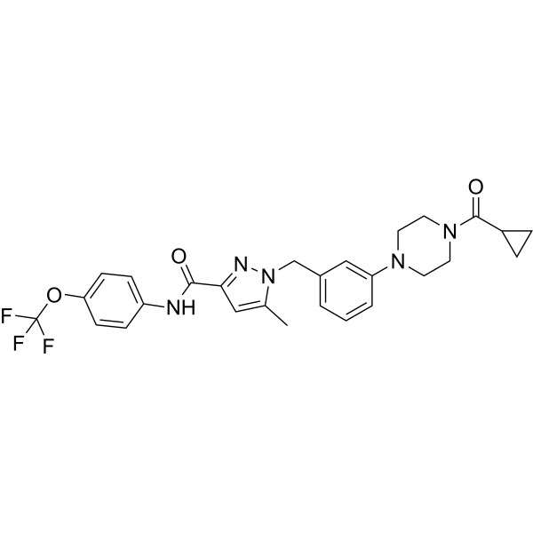
SCAL-255 is a potent mitochondrial complex I (CI) inhibitor with an IC50 of 1.14 μM. SCAL-255 blocks mitochondrial function, inhibits oxygen consumption rate (OCR), induces reactive oxygen species (ROS) production, and reduces MMP. SCAL-255 can be used in the research of oxidative phosphorylation (OXPHOS)-dependent cancers, such as colorectal cancer (CRC) and acute myeloid leukemia (AML), etc.
For research use only. We do not sell to patients.
- Purity : 99.15%
- CAS No.: 2798953-61-2
- Formula: C27H28F3N5O3
- Molecular Weight:527.54
-
Storage:Powder -20°C, 3 years , 4°C, 2 years ; In solvent -80°C, 6 months , -20°C, 1 month

To place orders, for customer services and technical support, please contact: MedChemExpress USA
Tel: 609-228-6898 E-mail: [email protected] [email protected]


-
 Biological Activity
-
Chemical Information
-
Solvent & Solubility
- Protocols
- Purity & Documentation
- References
-
 Help & FAQs




Biological Activity
Description
IC50 & Target
IC50: 1.14 μM (Mitochondrial complex I)[1]
Cellular Effect
|
Cell Line

|
Type | Value | Description | References |
|---|---|---|---|---|
| HCT-116 | IC50 |
1.02 nM
Compound: 5q; SCAL-255
|
Antiproliferative activity against human HCT-116 cells assessed as inhibition of cell growth incubated for 96 hrs by MTT assay
Antiproliferative activity against human HCT-116 cells assessed as inhibition of cell growth incubated for 96 hrs by MTT assay
|
[PMID: 37392582] |
| KG-1 | IC50 |
5.81 nM
Compound: 5q; SCAL-255
|
Antiproliferative activity against human KG-1 cells assessed as inhibition of cell growth incubated for 96 hrs by MTT assay
Antiproliferative activity against human KG-1 cells assessed as inhibition of cell growth incubated for 96 hrs by MTT assay
|
[PMID: 37392582] |
| NCM460 | IC50 |
>1000 nM
Compound: 5q; SCAL-255
|
Antiproliferative activity against human NCM460 cells assessed as inhibition of cell growth incubated for 96 hrs by MTT assay
Antiproliferative activity against human NCM460 cells assessed as inhibition of cell growth incubated for 96 hrs by MTT assay
|
[PMID: 37392582] |
In Vitro
SCAL-255 (0.01-10 nM; 96 h) significantly inhibits the proliferation of human colorectal cancer cell line HCT116 and acute myeloid leukemia cell line KG-1. SCAL-255 has little inhibitory effect on NCM460 cells[1].
SCAL-255 (2 nM, 10 nM; 24 h) inhibits the oxygen consumption rate (OCR), activates glycolysis, and changes the energy metabolism of HCT116 cells[1].
SCAL-255 (0.13-2 nM) induces the production of ROS and reduces the MMP in HCT116 and KG-1 cells[1].
MedChemExpress (MCE) has not independently confirmed the accuracy of these methods. They are for reference only.
-
Cell Line:HCT116, KG-1 cells
-
Concentration:0.01 nM, 0.1 nM, 1 nM, 10 nM
-
Incubation Time:96 h
-
Result:Ssignificantly inhibited the proliferation of HCT116 and KG-1 cells.
In Vivo
The maximum tolerated dose (MTD) values of SCAL-255 is 68 mg/kg in mice[1].
MedChemExpress (MCE) has not independently confirmed the accuracy of these methods. They are for reference only.
-
Animal Model:Female NOD-SCID mice (18-20 g, 5 weeks) inoculated with KG-1 cells[1].
-
Dosage:5 mg/kg
-
Administration:Intraperitoneal injection, five consecutive days per week for 24 days
-
Result:Exhibited adequate antitumor activity with a tumor growth inhibitory (TGI) rate of 61.5%.
No significant change of body weight, blood biochemical parameters, routine blood indexes, and histology of major organs were observed.


Chemical Information
-
CAS No. 2798953-61-2
-
Appearance Solid
-
Molecular Weight 527.54
-
Formula C27H28F3N5O3
-
Color White to off-white
-
SMILES
O=C(C1=NN(CC2=CC=CC(N3CCN(C(C4CC4)=O)CC3)=C2)C(C)=C1)NC5=CC=C(OC(F)(F)F)C=C5
-
Shipping
Room temperature in continental US; may vary elsewhere.
-
Storage
Powder -20°C 3 years 4°C 2 years In solvent -80°C 6 months -20°C 1 month


Solvent & Solubility
In Vitro:
DMSO : 50 mg/mL (94.78 mM; ultrasonic and warming and heat to 60°C; Hygroscopic DMSO has a significant impact on the solubility of product, please use newly opened DMSO)
Please refer to the solubility information to select the appropriate solvent. Once prepared, please aliquot and store the solution to prevent product inactivation from repeated freeze-thaw cycles.
Storage method and period of stock solution: -80°C, 6 months; -20°C, 1 month. When stored at -80°C, please use it within 6 months. When stored at -20°C, please use it within 1 month.
Please refer to the solubility information to select the appropriate solvent. Once prepared, please aliquot and store the solution to prevent product inactivation from repeated freeze-thaw cycles.
Storage method and period of stock solution: -80°C, 6 months; -20°C, 1 month. When stored at -80°C, please use it within 6 months. When stored at -20°C, please use it within 1 month.
Concentration (start) × Volume (start) = Concentration (final) × Volume (final)


Purity & Documentation
-
Data Sheet (271 KB)
-
SDS (251 KB)
- English - EN (251 KB)
- Français - FR (251 KB)
- Deutsch - DE (251 KB)
- Norwegian - NO (251 KB)
- Español - ES (251 KB)
- Swedish - SV (251 KB)
- Italian - IT (251 KB)
- Korean - KR (251 KB)
- Portuguese - PT (251 KB)
-
Handling Instructions (2659 KB)
References


Complete Stock Solution Preparation Table
Please refer to the solubility information to select the appropriate solvent. Once prepared, please aliquot and store the solution to prevent product inactivation from repeated freeze-thaw cycles.
Storage method and period of stock solution: -80°C, 6 months; -20°C, 1 month. When stored at -80°C, please use it within 6 months. When stored at -20°C, please use it within 1 month.
| Optional Solvent | Concentration Solvent Mass | 1 mg | 5 mg | 10 mg | 25 mg |
|---|---|---|---|---|---|
| DMSO | 1 mM | 1.8956 mL | 9.4780 mL | 18.9559 mL | 47.3898 mL |
| 5 mM | 0.3791 mL | 1.8956 mL | 3.7912 mL | 9.4780 mL | |
| 10 mM | 0.1896 mL | 0.9478 mL | 1.8956 mL | 4.7390 mL | |
| 15 mM | 0.1264 mL | 0.6319 mL | 1.2637 mL | 3.1593 mL | |
| 20 mM | 0.0948 mL | 0.4739 mL | 0.9478 mL | 2.3695 mL | |
| 25 mM | 0.0758 mL | 0.3791 mL | 0.7582 mL | 1.8956 mL | |
| 30 mM | 0.0632 mL | 0.3159 mL | 0.6319 mL | 1.5797 mL | |
| 40 mM | 0.0474 mL | 0.2369 mL | 0.4739 mL | 1.1847 mL | |
| 50 mM | 0.0379 mL | 0.1896 mL | 0.3791 mL | 0.9478 mL | |
| 60 mM | 0.0316 mL | 0.1580 mL | 0.3159 mL | 0.7898 mL | |
| 80 mM | 0.0237 mL | 0.1185 mL | 0.2369 mL | 0.5924 mL |


SCAL-255 Related Classifications
HY-155554 Related Classifications
 Powered by Bioz