



FC131 TFA

Based on 1 Customer Validation
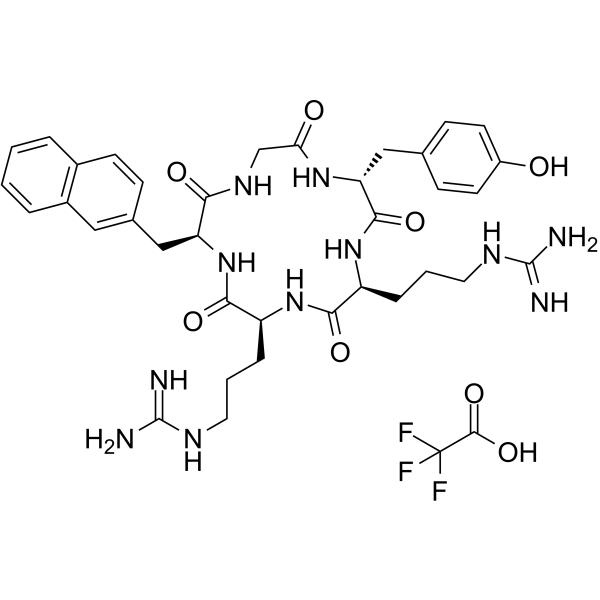
FC131 TFA is a CXCR4 antagonist that inhibits AKT kinase activity in AML OCI-AML3 cells. D-ArgFC131, a derivative of FC131 TFA, inhibits the phosphorylation of downstream ERK1/2 and Akt, induces caspase-3 pathway-mediated apoptosis, and causes cell cycle arrest. FC131 TFA exerts cytotoxic effects in cancer cells such as AML cells. FC131 TFA is applicable for cancer-related research.
For research use only. We do not sell to patients.
- Purity : 99.87%
- CAS No.: 842166-42-1
- Formula: C38H48F3N11O8
- Molecular Weight:843.85
-
Storage:
Sealed storage, away from moisture.
Powder -80°C, 2 years , -20°C, 1 year* In solvent : -80°C, 6 months; -20°C, 1 month (sealed storage, away from moisture)

To place orders, for customer services and technical support, please contact: MedChemExpress USA
Tel: 609-228-6898 E-mail: [email protected] [email protected]


-
 Biological Activity
-
Chemical Information
-
Solvent & Solubility
- Protocols
- Purity & Documentation
- References
-
 Help & FAQs




Biological Activity
Description
IC50 & Target
[1]|
CXCR4 |
Akt |
Cellular Effect
|
Cell Line

|
Type | Value | Description | References |
|---|---|---|---|---|
| COS-7 | EC50 |
0.47 μM
Compound: 1, FC131
|
Antagonist activity against human CXCR4 expressed in COS7 cells assessed as inhibition of CXCL12-induced myo-[3H]inositol production by scintillation counting
Antagonist activity against human CXCR4 expressed in COS7 cells assessed as inhibition of CXCL12-induced myo-[3H]inositol production by scintillation counting
|
[PMID: 23043442] |
In Vitro
FC131 (0.5-4.0 μM; 48 h) TFA combined with Panobinostat (HY-10224) synergistically induces apoptosis in human AML OCI-AML3 cells, and the corresponding combination index value is less than 1.0[1].
FC131 (2 μM plus 50 nM Panobinostat; 48 h) TFA induces significantly higher lethality in primary human AML cells than in normal human CD34+ bone marrow progenitor cells[1].
FC131 (1 μM; 24 h) TFA partially inhibits AKT kinase activity in human AML OCI-AML3 cells, and combined treatment with 50 nM Panobinostat enhances this AKT kinase inhibitory effect without altering the Panobinostat-mediated reduction in the levels of CXCR4, GRK3 and downstream signaling proteins[1].
FC131 (100 nM; 6 h) TFA inhibits the activity of the GH promoter in GH3 rat pituitary tumor cells, reducing luciferase activity to 0.3-fold that of the control group[2].
FC131 (10-100 nM; 6 days) TFA slightly inhibits the proliferation of GH3 rat pituitary tumor cells[2].
FC131 (1 nM-100 μM; 10 min pre-incubation, 90 min co-incubation with CXCL12) TFA potently inhibits CXCL12-mediated IP accumulation in COS-7 cells expressing wild-type CXCR4, with an IC50 of 0.40 μM; the H113A, D171N and D262N mutations significantly reduce its potency, while the W94A and D97A mutations enhance its potency[3].
FC131 (1 nM-100 μM; 3 h) TFA binds to wild-type CXCR4 expressed in COS-7 cells, with an IC50 of 0.76 μM; the H113A, Y116A, D171N and D262N mutations significantly reduce its binding affinity, the H281A, D187A and E288A mutations moderately reduce its binding affinity, while the W94A and D97A mutations increase its binding affinity[3].
MedChemExpress (MCE) has not independently confirmed the accuracy of these methods. They are for reference only.
- Kinase activity and phosphorylation assays
- Western Blot
- Detection of Apoptosis
- TUNEL staining for apoptotic DNA fragmentation
- Flow cytometric DNA-content cell-cycle staining
- Annexin V plus membrane-impermeant dye apoptosis staining
- BrdU Incorporation Assay
- Apoptosis Solutions
- Protocol for Cell Cycle
- Protocol for Kinase activity and phosphorylation assays
- Cell Viability Determination by MTT Colorimetric Assay


Chemical Information
-
CAS No. 842166-42-1
-
Appearance Solid
-
Molecular Weight 843.85
-
Formula C38H48F3N11O8
-
Color White to off-white
-
Sequence Shortening
Cyclo[{d-Tyr}-RR-{Nal}-G]
-
Shipping
Room temperature in continental US; may vary elsewhere.
-
Storage
Sealed storage, away from moisture
Powder -80°C 2 years -20°C 1 year * In solvent : -80°C, 6 months; -20°C, 1 month (sealed storage, away from moisture)


Solvent & Solubility
In Vitro:
DMSO : 100 mg/mL (118.50 mM; Need ultrasonic; Hygroscopic DMSO has a significant impact on the solubility of product, please use newly opened DMSO)
Please refer to the solubility information to select the appropriate solvent. Once prepared, please aliquot and store the solution to prevent product inactivation from repeated freeze-thaw cycles.
Storage method and period of stock solution: -80°C, 6 months; -20°C, 1 month (sealed storage, away from moisture). When stored at -80°C, please use it within 6 months. When stored at -20°C, please use it within 1 month.
Please refer to the solubility information to select the appropriate solvent. Once prepared, please aliquot and store the solution to prevent product inactivation from repeated freeze-thaw cycles.
Storage method and period of stock solution: -80°C, 6 months; -20°C, 1 month (sealed storage, away from moisture). When stored at -80°C, please use it within 6 months. When stored at -20°C, please use it within 1 month.
Concentration (start) × Volume (start) = Concentration (final) × Volume (final)
In Vivo:
Select the appropriate dissolution method based on your experimental animal and administration route.
- For the following dissolution methods, please ensure to first prepare a clear stock solution using an In Vitro approach and then sequentially add co-solvents:
- To ensure reliable experimental results, the clarified stock solution can be appropriately stored based on storage conditions. As for the working solution for In Vivo experiments, it is recommended to prepare freshly and use it on the same day.
- The percentages shown for the solvents indicate their volumetric ratio in the final prepared solution. If precipitation or phase separation occurs during preparation, heat and/or sonication can be used to aid dissolution.
Add each solvent one by one: 10% DMSO 40% PEG300 5% Tween-80 45% Saline
Solubility: ≥ 2.08 mg/mL (2.46 mM); Clear solution
This protocol yields a clear solution of ≥ 2.08 mg/mL (saturation unknown).
Taking 1 mL working solution as an example, add 100 μL DMSO stock solution (20.8 mg/mL) to 400 μL PEG300, and mix evenly; then add 50 μL Tween-80 and mix evenly; then add 450 μL Saline to adjust the volume to 1 mL.
Preparation of Saline: Dissolve 0.9 g sodium chloride in ddH₂O and dilute to 100 mL to obtain a clear Saline solution.
Add each solvent one by one: 10% DMSO 90% (20% SBE-β-CD in Saline)
Solubility: ≥ 2.08 mg/mL (2.46 mM); Clear solution
This protocol yields a clear solution of ≥ 2.08 mg/mL (saturation unknown).
Taking 1 mL working solution as an example, add 100 μL DMSO stock solution (20.8 mg/mL) to 900 μL 20% SBE-β-CD in Saline, and mix evenly.
Preparation of 20% SBE-β-CD in Saline (4°C, storage for one week): 2 g SBE-β-CD powder is dissolved in 10 mL Saline, completely dissolve until clear.
In Vivo Dissolution Calculator
Please enter the basic information of animal experiments:
-
-
-
-
Recommended: Prepare an additional quantity of animals to account for potential losses during experiments.
Please enter your animal formula composition:
-
%DMSO +
Recommended: Keep the proportion of DMSO in working solution below 2% if your animal is weak.
-
%+
-
+%Tween-80 + +
-
%Saline +
The co-solvents required include: DMSO, . All of co-solvents are available by MedChemExpress (MCE). , Tween 80. All of co-solvents are available by MedChemExpress (MCE).
Working solution concentration: 0.22 mg/mL
Method for preparing stock solution: mg drug dissolved in μL DMSO. Stock solution concentration: mg/mL. * In solvent : -80°C, 6 months; -20°C, 1 month (sealed storage, away from moisture)
1. Take μL DMSO stock solution;
2. Add μL .
μL , mix evenly;
3. Then add μL Tween 80, mix evenly;
4. Then add μL
Please ensure that the stock solution in the first step is dissolved to a clear state, and add co-solvents in sequence. You can use ultrasonic heating (ultrasonic cleaner, recommended frequency 20-40 kHz), vortexing, etc. to assist dissolution.


Purity & Documentation
-
Data Sheet (281 KB)
-
SDS (254 KB)
- English - EN (254 KB)
- Français - FR (254 KB)
- Deutsch - DE (254 KB)
- Norwegian - NO (254 KB)
- Español - ES (254 KB)
- Swedish - SV (254 KB)
- Italian - IT (254 KB)
- Korean - KR (254 KB)
- Portuguese - PT (254 KB)
-
Handling Instructions (2659 KB)
References
[1]. Mandawat A, et al. Pan-histone deacetylase inhibitor panobinostat depletes CXCR4 levels and signaling and exerts synergistic antimyeloid activity in combination with CXCR4 antagonists. Blood. 2010 Dec 09;116(24):5306-15. [Content Brief]
[2]. Kim JM, et al. The cyclic pentapeptide d-Arg3FC131, a CXCR4 antagonist, induces apoptosis of somatotrope tumor and inhibits tumor growth in nude mice. Endocrinology. 2011 Feb;152(2):536-44. [Content Brief]
[3]. Thiele S, et al. Determination of the binding mode for the cyclopentapeptide CXCR4 antagonist FC131 using a dual approach of ligand modifications and receptor mutagenesis. British journal of pharmacology. 2014 Dec;171(23):5313-29. [Content Brief]


Complete Stock Solution Preparation Table
Please refer to the solubility information to select the appropriate solvent. Once prepared, please aliquot and store the solution to prevent product inactivation from repeated freeze-thaw cycles.
Storage method and period of stock solution: -80°C, 6 months; -20°C, 1 month (sealed storage, away from moisture). When stored at -80°C, please use it within 6 months. When stored at -20°C, please use it within 1 month.
| Optional Solvent | Concentration Solvent Mass | 1 mg | 5 mg | 10 mg | 25 mg |
|---|---|---|---|---|---|
| DMSO | 1 mM | 1.1850 mL | 5.9252 mL | 11.8504 mL | 29.6261 mL |
| 5 mM | 0.2370 mL | 1.1850 mL | 2.3701 mL | 5.9252 mL | |
| 10 mM | 0.1185 mL | 0.5925 mL | 1.1850 mL | 2.9626 mL | |
| 15 mM | 0.0790 mL | 0.3950 mL | 0.7900 mL | 1.9751 mL | |
| 20 mM | 0.0593 mL | 0.2963 mL | 0.5925 mL | 1.4813 mL | |
| 25 mM | 0.0474 mL | 0.2370 mL | 0.4740 mL | 1.1850 mL | |
| 30 mM | 0.0395 mL | 0.1975 mL | 0.3950 mL | 0.9875 mL | |
| 40 mM | 0.0296 mL | 0.1481 mL | 0.2963 mL | 0.7407 mL | |
| 50 mM | 0.0237 mL | 0.1185 mL | 0.2370 mL | 0.5925 mL | |
| 60 mM | 0.0198 mL | 0.0988 mL | 0.1975 mL | 0.4938 mL | |
| 80 mM | 0.0148 mL | 0.0741 mL | 0.1481 mL | 0.3703 mL | |
| 100 mM | 0.0119 mL | 0.0593 mL | 0.1185 mL | 0.2963 mL |


FC131 TFA Related Classifications
HY-P1104A Related Classifications
 Powered by Bioz