



Malantide TFA
Based on 1 publication(s) in Google Scholar
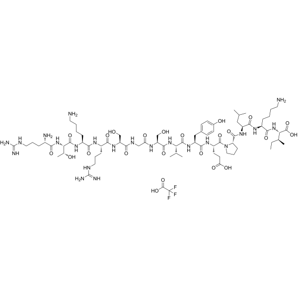
Malantide TFA is a synthetic dodecapeptide derived from the site phosphorylated by cAMP-dependent protein kinase (PKA) on the β-subunit of phosphorylase kinase. Malantide TFA is a highly specific substrate for PKA with a Km of 15 μM and shows protein inhibitor (PKI) inhibition >90% substrate phosphorylation in various rat tissue extracts. Malantide TFA is also an efficient substrate for PKC with a Km of 16 μM.
Nur für Forschungszwecke. Wir verkaufen nicht an Patienten.
- Formel: C74H125F3N22O23
- Molecular Weight:1747.91
-
Speicherung:
Please store the product under the recommended conditions in the Certificate of Analysis.

To place orders, for customer services and technical support, please contact: MedChemExpress USA
Tel: 609-228-6898 E-mail: [email protected] [email protected]


-
 Biologische Aktivität
-
Chemical Information
- Protokoll
- Reinheit & Dokumentation
- Verweise
-
 Help & FAQs


Publications Citing Use of MedChemExpress (MCE) Malantide TFA
More

Biologische Aktivität
Beschreibung
IC50 & Target
In Vitro
The Km values of Malantide are 15 μM and 223 μM for PKA and PKG, respectively. The Vmax values are 23.8 units/mg and 6.6 units/mg for for PKA and PKG, respectively[1].
A statistically significant effect of isoprenaline on the activity ratio of guinea-pig heart was only: observed with Malantide[1].
MedChemExpress (MCE) has not independently confirmed the accuracy of these methods. They are for reference only.


Chemical Information
-
Molecular Weight 1747.91
-
Formel C74H125F3N22O23
-
Sequence
Arg-Thr-Lys-Arg-Ser-Gly-Ser-Val-Tyr-Glu-Pro-Leu-Lys-Ile
-
Sequence Shortening
RTKRSGSVYEPLKI
-
Versand
Room temperature in continental US; may vary elsewhere.
-
Speicherung
Please store the product under the recommended conditions in the Certificate of Analysis.


Publications (1)
-
Journal Impact Factor
-
Most Recent
-
Int J Biol Sci
PKCα Induced the Generation of Extracellular Vesicles in Activated Platelets to Promote Breast Cancer Metastasis. [Abstract]2024 Jul 15;20(10):3956-3971. PMID: 39113702


Reinheit & Dokumentation
Verweise
[1]. K J Murray, et al. Use of a Synthetic Dodecapeptide (Malantide) to Measure the Cyclic AMP-dependent Protein Kinase Activity Ratio in a Variety of Tissues. Biochem J. 1990 May 1;267(3):703-8. [Content Brief]
[2]. Z H Zhao, et al. Characterization of a New Substrate for Protein Kinase C: Assay by Continuous Fluorometric Monitoring and High Performance Liquid Chromatography. Biochem Biophys Res Commun. 1991 May 15;176(3):1454-61. [Content Brief]

Calculators
Konzentration (Stammlösung) × Volumen (Stammlösung) = Konzentration (Ziellösung) × Volumen (Ziellösung)


Malantide TFA Related Classifications
HY-P1597A Related Classifications
 Powered by Bioz