



Navitoclax-d8
Based on 1 publication(s) in Google Scholar
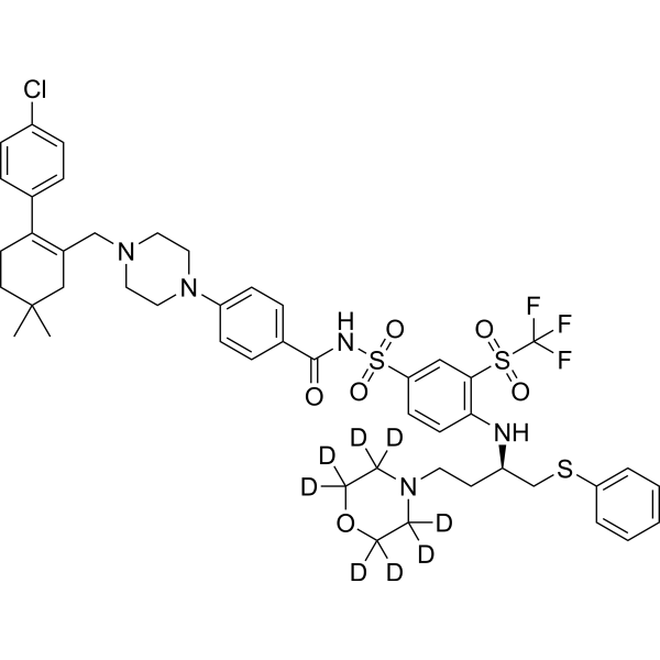
Navitoclax-d8 is the d8-labeled Navitoclax (HY-10087). Navitoclax (ABT-263 ) is an orally active BH3 mimetic and an inhibitor of Bcl-2, Bcl-xL, and Bcl-w, with a Ki value of <1 nM for each target. Navitoclax triggers endogenous Apoptosis and downregulates the expression of Survivin and TGFβ2. Navitoclax alleviates fibrosis and induces thrombocytopenia. Navitoclax exhibits anticancer activity against a variety of tumors and enhances the efficacy of chemotherapy and radiotherapy. Navitoclax can be used in the research of acute lymphoblastic leukemia, ovarian cancer, and fibrotic diseases.
For research use only. We do not sell to patients.
- Purity : 99.71%
- CAS No.: 1217620-38-6
- Formula: C47H47D8ClF3N5O6S3
- Molecular Weight:982.66
-
Storage:Powder -20°C, 3 years ; In solvent -80°C, 6 months , -20°C, 1 month

To place orders, for customer services and technical support, please contact: MedChemExpress USA
Tel: 609-228-6898 E-mail: [email protected] [email protected]


-
 Biological Activity
-
Chemical Information
-
Solvent & Solubility
- Protocols
- Purity & Documentation
- References
-
 Help & FAQs


-
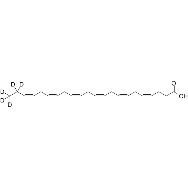
HY-B2167S
Docosahexaenoic acid-d5Stable Isotope -
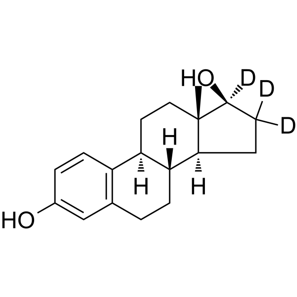
HY-B0141S
Estradiol-d3Stable Isotope -
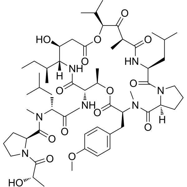
HY-105055
Didemnin BDepsipeptide -
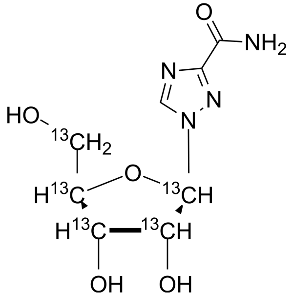
HY-B0434S
Ribavirin-13C5Stable Isotope -
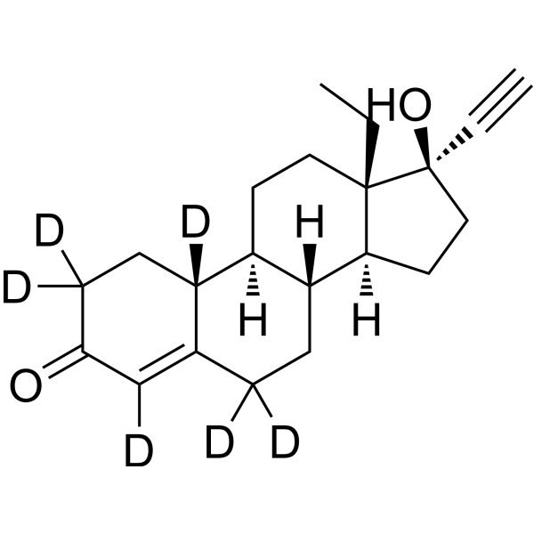
HY-B0257S1
Norgestrel-d6Stable Isotope -

HY-W777335
Zanamivir-13C,15N2Stable Isotope -

HY-112450
Olomoucine IICDK Inhibitor -
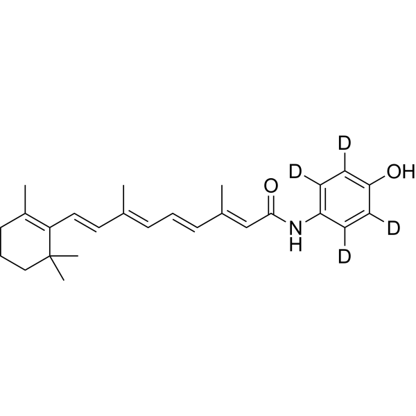
HY-110360
Fenretinide-d4Stable Isotope -

HY-122425
Nylidrin -

HY-W439855
10-NorparvulenoneAntibiotic -

HY-135766
ColletodiolFungal Metabolite -
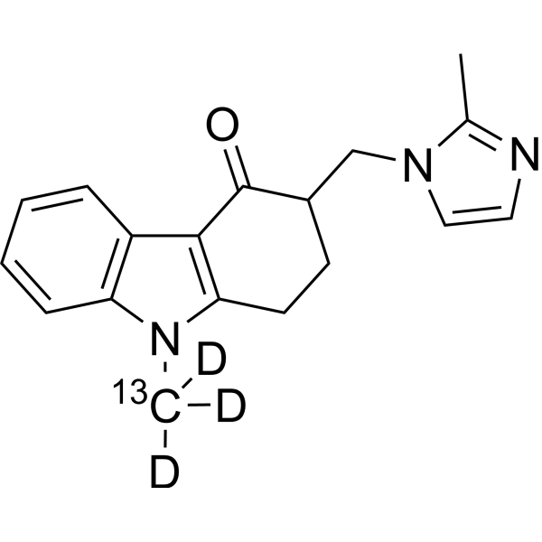
HY-B0002BS2
Ondansetron-13C,d3Stable Isotope
Publications Citing Use of MedChemExpress (MCE) Navitoclax-d8
More

Biological Activity
Description
IC50 & Target
|
Bcl-2 <1 nM (Ki) |
Bcl-xL <1 nM (Ki) |
Bcl-W <1 nM (Ki) |
Application
1. This compound can be used as a tracer
2. This compound can be used as an internal standard for quantitative analysis by NMR, GC-MS, or LC-MS.


Chemical Information
-
CAS No. 1217620-38-6
-
Unlabeled CAS 923564-51-6
-
Appearance Solid
-
Molecular Weight 982.66
-
Formula C47H47D8ClF3N5O6S3
-
Color White to off-white
-
SMILES
CC(C)(C1)CCC(C2=CC=C(C=C2)Cl)=C1CN3CCN(C4=CC=C(C=C4)C(NS(=O)(C5=CC(S(C(F)(F)F)(=O)=O)=C(C=C5)N[C@@H](CSC6=CC=CC=C6)CCN7C([2H])([2H])C([2H])([2H])OC([2H])([2H])C7([2H])[2H])=O)=O)CC3
-
Shipping
Room temperature in continental US; may vary elsewhere.
-
Storage
Powder -20°C 3 years In solvent -80°C 6 months -20°C 1 month


Publications (1)
-
Journal Impact Factor
-
Most Recent
-
Nat Commun
Proapoptotic Bcl-2 inhibitor as potential host directed therapy for pulmonary tuberculosis. [Abstract]2025 Mar 27;16(1):3003. PMID: 40148277


Solvent & Solubility
In Vitro:
DMSO : ≥ 25 mg/mL (25.44 mM; Hygroscopic DMSO has a significant impact on the solubility of product, please use newly opened DMSO)
* "≥" means soluble, but saturation unknown.
Please refer to the solubility information to select the appropriate solvent. Once prepared, please aliquot and store the solution to prevent product inactivation from repeated freeze-thaw cycles.
Storage method and period of stock solution: -80°C, 6 months; -20°C, 1 month. When stored at -80°C, please use it within 6 months. When stored at -20°C, please use it within 1 month.
Please refer to the solubility information to select the appropriate solvent. Once prepared, please aliquot and store the solution to prevent product inactivation from repeated freeze-thaw cycles.
Storage method and period of stock solution: -80°C, 6 months; -20°C, 1 month. When stored at -80°C, please use it within 6 months. When stored at -20°C, please use it within 1 month.
Concentration (start) × Volume (start) = Concentration (final) × Volume (final)

Protocols
-
Fibrosis/Collagen Morphometry
Fibrosis and collagen morphometry is based on the quantitative visualization of fibrillar collagen deposition in tissue sections using histochemical stains such as Sirius Red (Picrosirius Red) or Masson's trichrome, followed by image-based or polarization-enhanced analysis to estimate collagen proportional area as a surrogate of extracellular matrix accumulation during fibrotic remodeling. Sirius Red combined with polarized light microscopy enhances detection of collagen fibers due to birefringence properties, enabling more specific visualization of collagen type I and III fibrils compared to conventional bright-field histology, while whole-section or region-restricted digital morphometry reduces field-selection bias in fibrosis assessment. Alternative quantitative approaches include second harmonic generation (SHG) and two-photon excited fluorescence microscopy, which enable label-free detection of fibrillar collagen and have been validated against histological staining and biochemica
-
RNA extraction experimental
By lysing cells, releasing RNA, and removing impurities such as proteins and DNA, high-purity RNA products are finally obtained. The commonly used traditional method is the guanidine isothiocyanate/phenol/chloroform method (Trizol), which is suitable for a variety of animal materials including animal tissues, microorganisms, cultured cells, etc., and most plant materials.
-
Collagen: Sirius Red Staining
Sirius Red or picrosirius red staining is a histochemical method for visualizing collagen-rich extracellular matrix in tissue sections, and collagen fibers are detected as red-stained structures under bright-field microscopy with enhanced birefringence under polarized light. Picrosirius red is useful for assessing total collagen organization, distribution, and fibrosis burden, but polarized color should not be interpreted as a definitive collagen type I versus type III readout because color is affected by fiber orientation, thickness, and packing.
-
Apoptosis
Apoptosis, also called programmed cell death, is generally characterized by distinct morphological characteristics.
-
TUNEL staining for apoptotic DNA fragmentation
TUNEL staining detects DNA strand breaks by using terminal deoxynucleotidyl transferase to add labeled nucleotides to exposed 3′-OH DNA termini, generating either microscopic staining in fixed cells or tissue sections, or fluorescence/cytometric signal in cell suspensions. TUNEL positivity reflects DNA fragmentation but should not be interpreted alone as definitive apoptosis, because TUNEL can also label necrotic, autolytic, mechanically damaged, or DNA-repair-associated DNA breaks.
-
Annexin V plus membrane-impermeant dye apoptosis staining
Annexin V-based apoptosis assays rely on the detection of phosphatidylserine (PS) externalization from the inner leaflet of the plasma membrane to the outer leaflet, an early biochemical hallmark of apoptosis. Fluorescently labeled Annexin V binds PS in a calcium-dependent manner, enabling identification of early apoptotic cells by flow cytometry or fluorescence microscopy. When combined with a membrane-impermeant DNA-binding dye (e. g. , propidium iodide), this approach allows discrimination between viable (Annexin V−/dye−), early apoptotic (Annexin V+/dye−), and late apoptotic or necrotic (Annexin V+/dye+) cell populations by assessing membrane integrity and PS exposure.
-
Connective Tissue: Masson's Trichrome/Collagen Trichrome Staining
Masson’s Trichrome (collagen/trichrome staining) is a histological technique that differentially stains tissue compartments using sequential acidic dyes to distinguish collagen from muscle and cytoplasmic components based on dye affinity and tissue permeability differences, enabling visualization of fibrosis and connective tissue architecture in histological sections. The classical formulation typically uses Weigert's iron hematoxylin for nuclear staining, Biebrich scarlet-acid fuchsin for cytoplasm and muscle, and aniline blue (or light green variants) for collagen, producing a characteristic blue/green collagen signal contrasted against red cytoplasm and dark nuclei. The staining principle relies on selective displacement of smaller dye molecules by larger anionic dyes in collagen-rich regions under controlled acidified conditions, which enhances collagen-specific dye retention. This property makes the method widely used for fibrosis assessment in organs such as heart, liver, lung, a
-
Apoptosis Solutions
Apoptosis is a regulated, generally non-lytic cell-death pathway that removes unwanted, damaged, infected, or abnormal cells through coordinated morphological changes, caspase activation, DNA fragmentation, and membrane remodeling. The intrinsic apoptosis pathway is controlled mainly by mitochondrial outer membrane permeabilization, BCL-2 family proteins, cytochrome c release, apoptosome formation, caspase-9 activation, and downstream executioner caspase-3/7 activation. The extrinsic apoptosis pathway is initiated by death receptors such as Fas, TNFR, and TRAIL receptors, which recruit adaptor proteins and activate caspase-8 before engaging executioner caspases or mitochondrial amplification through BID cleavage. Apoptosis is linked to many phenotypes, including cancer cell killing, tissue homeostasis, immune regulation, neurodegeneration, infection response, and treatment-induced cytotoxicity; unresolved questions include how apoptosis interacts with necroptosis, pyroptosis, ferroptos


Purity & Documentation
-
Data Sheet (278 KB)
-
SDS (252 KB)
- English - EN (252 KB)
- Français - FR (252 KB)
- Deutsch - DE (252 KB)
- Norwegian - NO (252 KB)
- Español - ES (252 KB)
- Swedish - SV (252 KB)
- Italian - IT (252 KB)
- Korean - KR (252 KB)
- Portuguese - PT (252 KB)
-
Handling Instructions (2659 KB)
References
[1]. Lock R, et al. Initial testing (stage 1) of the BH3 mimetic ABT-263 by the pediatric preclinical testing program. Pediatric blood & cancer. 2008 Jun;50(6):1181-9. [Content Brief]
[2]. Chen J, et al. The Bcl-2/Bcl-X(L)/Bcl-w inhibitor, navitoclax, enhances the activity of chemotherapeutic agents in vitro and in vivo. Mol Cancer Ther. 2011 Dec;10(12):2340-9. [Content Brief]
[3]. Chen J, et al. The Bcl-2/Bcl-X(L)/Bcl-w inhibitor, navitoclax, enhances the activity of chemotherapeutic agents in vitro and in vivo. Molecular cancer therapeutics. 2011 Dec;10(12):2340-9. [Content Brief]
[4]. Mohamad Anuar NN, et al. Clinical Review: Navitoclax as a Pro-Apoptotic and Anti-Fibrotic Agent. Frontiers in pharmacology. 2020;11:564108. [Content Brief]
[5]. Wilson WH, et al. Navitoclax, a targeted high-affinity inhibitor of BCL-2, in lymphoid malignancies: a phase 1 dose-escalation study of safety, pharmacokinetics, pharmacodynamics, and antitumour activity. The Lancet. Oncology. 2010 Dec;11(12):1149-59. [Content Brief]
[6]. Wong M, et al. Navitoclax (ABT-263) reduces Bcl-x(L)-mediated chemoresistance in ovarian cancer models. Molecular cancer therapeutics. 2012 Apr;11(4):1026-35. [Content Brief]


Complete Stock Solution Preparation Table
Please refer to the solubility information to select the appropriate solvent. Once prepared, please aliquot and store the solution to prevent product inactivation from repeated freeze-thaw cycles.
Storage method and period of stock solution: -80°C, 6 months; -20°C, 1 month. When stored at -80°C, please use it within 6 months. When stored at -20°C, please use it within 1 month.
| Optional Solvent | Concentration Solvent Mass | 1 mg | 5 mg | 10 mg | 25 mg |
|---|---|---|---|---|---|
| DMSO | 1 mM | 1.0176 mL | 5.0882 mL | 10.1765 mL | 25.4412 mL |
| 5 mM | 0.2035 mL | 1.0176 mL | 2.0353 mL | 5.0882 mL | |
| 10 mM | 0.1018 mL | 0.5088 mL | 1.0176 mL | 2.5441 mL | |
| 15 mM | 0.0678 mL | 0.3392 mL | 0.6784 mL | 1.6961 mL | |
| 20 mM | 0.0509 mL | 0.2544 mL | 0.5088 mL | 1.2721 mL | |
| 25 mM | 0.0407 mL | 0.2035 mL | 0.4071 mL | 1.0176 mL |


Navitoclax-d8 Related Classifications
HY-10087S Related Classifications
 Powered by Bioz