



Nogalamycin

Based on 1 Customer Validation
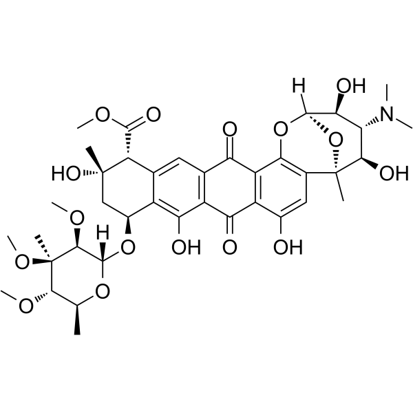
Nogalamycin is an anthracyclinone antibiotic. Nogalamycin is a potent antibiotic against Gram-positive bacteria, also has cytotoxicity against certain tumor cells. Nogalamycin is produced by Streptomyces nogalater var. Nogalater. Nogalamycin selectively inhibits RNA synthesis after binding to DNA template. Nogalamycin can be used for researching anticancer.
For research use only. We do not sell to patients.
- Purity : 97.2%
- CAS No.: 1404-15-5
- Formula: C39H49NO16
- Molecular Weight:787.80
-
Storage:Powder -20°C, 3 years ; In solvent -80°C, 6 months , -20°C, 1 month

To place orders, for customer services and technical support, please contact: MedChemExpress USA
Tel: 609-228-6898 E-mail: [email protected] [email protected]


-
 Biological Activity
-
Chemical Information
-
Solvent & Solubility
- Protocols
- Purity & Documentation
- References
-
 Help & FAQs




Biological Activity
Description
IC50 & Target
RNA Synthesis[2]
Cellular Effect
|
Cell Line

|
Type | Value | Description | References |
|---|---|---|---|---|
| CCRF-CEM | IC50 |
59 μM
Compound: Nogalamycin
|
Cytotoxicity in human leukemic CCRF-CEM cells.
Cytotoxicity in human leukemic CCRF-CEM cells.
|
[PMID: 14667232] |
In Vitro
Nogalamycin (0-10 μg/ml; 2 hours) exhibits lethality against CHO, L1210 and B16 with LD50s of 2.74 μg/ml, 0.25 μg/ml and 0.15 μg/ml[1].
MedChemExpress (MCE) has not independently confirmed the accuracy of these methods. They are for reference only. Further protocols information, click here.
-
Cell Line:CHO, L1210 and B16 cells[1]
-
Concentration:0-10 μg/ml
-
Incubation Time:2 hours
-
Result:Exhibited lethality against CHO, L1210 and B16 with LD50s of 2.74 μg/ml, 0.25 μg/ml and 0.15 μg/ml.
In Vivo
MedChemExpress (MCE) has not independently confirmed the accuracy of these methods. They are for reference only.
-
Animal Model:Rats[1]
-
Dosage:0-1000 mug/kg
-
Administration:IP
-
Result:Regressed chemically-induced fibrosarcoma tumors and tumors of spontaneous origin in rats, and the most effective concentration was 556 mug/kg.


Chemical Information
-
CAS No. 1404-15-5
-
Appearance Solid
-
Molecular Weight 787.80
-
Formula C39H49NO16
-
Color Orange to red
-
SMILES
O=C1C2=C(C(C3=C1C=C4C([C@H](C[C@](C)(O)[C@@H]4C(OC)=O)O[C@@]5([H])[C@@H]([C@@](C)([C@@H](OC)[C@H](C)O5)OC)OC)=C3O)=O)C(O)=CC6=C2O[C@]([C@@H](O)[C@H](N(C)C)[C@H]7O)([H])O[C@]67C
-
Shipping
Room temperature in continental US; may vary elsewhere.
-
Storage
Powder -20°C 3 years In solvent -80°C 6 months -20°C 1 month


Solvent & Solubility
In Vitro:
DMSO : 10 mg/mL (12.69 mM; Need ultrasonic and warming; Hygroscopic DMSO has a significant impact on the solubility of product, please use newly opened DMSO)
Please refer to the solubility information to select the appropriate solvent. Once prepared, please aliquot and store the solution to prevent product inactivation from repeated freeze-thaw cycles.
Storage method and period of stock solution: -80°C, 6 months; -20°C, 1 month. When stored at -80°C, please use it within 6 months. When stored at -20°C, please use it within 1 month.
Please refer to the solubility information to select the appropriate solvent. Once prepared, please aliquot and store the solution to prevent product inactivation from repeated freeze-thaw cycles.
Storage method and period of stock solution: -80°C, 6 months; -20°C, 1 month. When stored at -80°C, please use it within 6 months. When stored at -20°C, please use it within 1 month.
Concentration (start) × Volume (start) = Concentration (final) × Volume (final)

Protocols
-
Cell Cytotoxicity Assay
Cytotoxicity assays are usually based on the assessment of cell membrane damage, which can also be indirectly detected by measuring cell viability. Detection methods include MTT assay, CKK-8 assay, LDH assay and ATP assay, etc.
-
Mammalian live/dead viability and cytotoxicity staining
Live/dead viability and cytotoxicity staining assays are based on the simultaneous detection of intracellular esterase activity in metabolically active (viable) cells and membrane integrity loss in non-viable cells. In commonly used dual-staining approaches, membrane-permeant fluorogenic substrates are converted by intracellular esterases into fluorescent products in live cells, while impermeant DNA-binding dyes selectively enter cells with compromised plasma membranes and label nucleic acids in dead or dying cells, enabling discrimination between viable and non-viable populations by fluorescence microscopy or flow cytometry.
-
Gram Staining of Tissue Sections
Gram staining of tissue sections is a histochemical technique used to differentiate Gram-positive and Gram-negative bacteria within histological specimens based on differences in bacterial cell wall structure and dye retention, adapted from classical bacteriological Gram staining into tissue-compatible “histological Gram stain” variants. In tissue applications, modifications of the Brown-Hopps and Brown-Brenn methods are commonly used to improve differentiation of microorganisms embedded within host connective tissue and to reduce overstaining or loss of Gram-negative signal, which are known limitations of earlier approaches. The principle relies on crystal violet-iodine complex retention in Gram-positive organisms and subsequent decolorization and counterstaining steps that allow contrast visualization of Gram-negative organisms against tissue background.
-
Cell Viability Determination by MTT Colorimetric Assay
The following protocol uses the MTT colorimetric assay as a classic literature-established method for assessing cell viability/metabolic activity in cultured mammalian cells. MTT[3-(4,5-dimethylthiazol-2-yl)-2,5-diphenyltetrazolium bromide] is reduced by metabolically active cells to a colored formazan product; the amount of formazan is quantified spectrophotometrically and provides an indirect measure of metabolically active viable cells. Importantly, MTT reduction reflects cellular oxidoreductase/metabolic activity rather than an absolute direct count of living cells, so changes in cellular metabolism can alter the signal independently of cell number.


Purity & Documentation
-
Data Sheet (279 KB)
-
SDS (558 KB)
- English - EN (558 KB)
- Français - FR (558 KB)
- Deutsch - DE (558 KB)
- Norwegian - NO (558 KB)
- Español - ES (558 KB)
- Swedish - SV (558 KB)
- Italian - IT (558 KB)
- Korean - KR (558 KB)
- Portuguese - PT (558 KB)
-
Handling Instructions (2659 KB)
References
[1]. Bhuyan BK, et al. Cell kill kinetics of several nogalamycin analogs and adriamycin for Chinese hamster ovary, L1210 leukemia, and B16 melanoma cells in culture. Cancer Res. 1981;41(1):18-24. [Content Brief]
[2]. Bhuyan BK, et al. Differential interaction of nogalamycin with DNA of varying base composition. Proc Natl Acad Sci U S A. 1965;54(2):566-572. [Content Brief]
[3]. Bempong MA. Cytological effect of nogalamycin in mammals and regression of methylcholanthrene-induced and spontaneous tumors in rats. Int J Clin Pharmacol Biopharm. 1976;14(1):6-14. [Content Brief]


Complete Stock Solution Preparation Table
Please refer to the solubility information to select the appropriate solvent. Once prepared, please aliquot and store the solution to prevent product inactivation from repeated freeze-thaw cycles.
Storage method and period of stock solution: -80°C, 6 months; -20°C, 1 month. When stored at -80°C, please use it within 6 months. When stored at -20°C, please use it within 1 month.
| Optional Solvent | Concentration Solvent Mass | 1 mg | 5 mg | 10 mg | 25 mg |
|---|---|---|---|---|---|
| DMSO | 1 mM | 1.2694 mL | 6.3468 mL | 12.6936 mL | 31.7339 mL |
| 5 mM | 0.2539 mL | 1.2694 mL | 2.5387 mL | 6.3468 mL | |
| 10 mM | 0.1269 mL | 0.6347 mL | 1.2694 mL | 3.1734 mL |


Nogalamycin Related Classifications
HY-105846 Related Classifications
 Powered by Bioz