



Bleomycin A5
Based on 1 publication(s) in Google Scholar
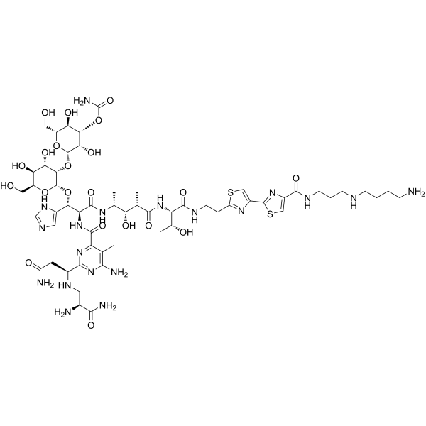
Bleomycin A5 (Pingyangmycin) is a glycopeptide antibiotic with multiple biological activities, which can be isolated from Streptomyces. Bleomycin A5 exerts cytotoxic effects by binding to Fe2+ to form a complex, inducing single-strand and double-strand DNA breaks, and inhibiting DNA replication. Bleomycin A5 inhibits Drp1-mediated mitochondrial fission and suppresses PINK1/Parkin pathway-mediated mitophagy, ultimately triggering mitochondria-mediated cellular apoptosis. Bleomycin A5 can be used in cancer research.
For research use only. We do not sell to patients.
- Purity : 99.81%
- CAS No.: 11116-32-8
- Formula: C57H89N19O21S2
- Molecular Weight:1440.56
-
Storage:
4°C, sealed storage, away from moisture and light
* In solvent : -80°C, 6 months; -20°C, 1 month (sealed storage, away from moisture and light)

To place orders, for customer services and technical support, please contact: MedChemExpress USA
Tel: 609-228-6898 E-mail: [email protected] [email protected]


-
 Biological Activity
-
Chemical Information
-
Solvent & Solubility
- Protocols
- Purity & Documentation
- References
-
 Help & FAQs


Publications Citing Use of MedChemExpress (MCE) Bleomycin A5
MoreAll Antibiotic Isoforms
MoreAll DNA/RNA Synthesis Isoforms
More

Biological Activity
Description
IC50 & Target
|
Glycopeptide |
In Vivo
MedChemExpress (MCE) has not independently confirmed the accuracy of these methods. They are for reference only.


Chemical Information
-
CAS No. 11116-32-8
-
Appearance Solid
-
Molecular Weight 1440.56
-
Formula C57H89N19O21S2
-
Color White to off-white
-
SMILES
O=C(N[C@H](C)[C@@H](O)[C@H](C)C(N[C@@H]([C@H](O)C)C(NCCC1=NC(C2=NC(C(NCCCNCCCCN)=O)=CS2)=CS1)=O)=O)[C@@H](NC(C3=NC([C@H](CC(N)=O)NC[C@H](N)C(N)=O)=NC(N)=C3C)=O)[C@H](C4=CN=CN4)O[C@H](O[C@H]5CO)[C@H]([C@H]([C@@H]5O)O)O[C@H](O[C@@H]6CO)[C@H]([C@H]([C@@H]6O)OC(N)=O)O
-
Synonyms
Pingyangmycin
-
Structure Classification
-
Initial Source
-
Shipping
Room temperature in continental US; may vary elsewhere.
-
Storage
4°C, sealed storage, away from moisture and light
* In solvent : -80°C, 6 months; -20°C, 1 month (sealed storage, away from moisture and light)


Publications (1)
-
Journal Impact Factor
-
Most Recent
-
Exp Ther Med
MicroRNA‑203a‑3p improves bleomycin and pingyangmycin sensitivity by inactivating the PI3K/AKT pathway in hemangioma. [Abstract]2024 Jan 4;27(2):80. PMID: 38274341


Solvent & Solubility
In Vitro:
H2O : ≥ 100 mg/mL (69.42 mM)
* "≥" means soluble, but saturation unknown.
Please refer to the solubility information to select the appropriate solvent. Once prepared, please aliquot and store the solution to prevent product inactivation from repeated freeze-thaw cycles.
Storage method and period of stock solution: -80°C, 6 months; -20°C, 1 month (sealed storage, away from moisture and light). When stored at -80°C, please use it within 6 months. When stored at -20°C, please use it within 1 month.
* Note: If you choose water as the stock solution, please dilute it to the working solution, then filter and sterilize it with a 0.22 μm filter before use.
Please refer to the solubility information to select the appropriate solvent. Once prepared, please aliquot and store the solution to prevent product inactivation from repeated freeze-thaw cycles.
Storage method and period of stock solution: -80°C, 6 months; -20°C, 1 month (sealed storage, away from moisture and light). When stored at -80°C, please use it within 6 months. When stored at -20°C, please use it within 1 month.
* Note: If you choose water as the stock solution, please dilute it to the working solution, then filter and sterilize it with a 0.22 μm filter before use.
Concentration (start) × Volume (start) = Concentration (final) × Volume (final)


Purity & Documentation
-
Data Sheet (275 KB)
-
SDS (252 KB)
- English - EN (252 KB)
- Français - FR (252 KB)
- Deutsch - DE (252 KB)
- Norwegian - NO (252 KB)
- Español - ES (252 KB)
- Swedish - SV (252 KB)
- Italian - IT (252 KB)
- Korean - KR (252 KB)
- Portuguese - PT (252 KB)
-
Handling Instructions (2659 KB)
References
[1]. Yaowu Yang, et al. Bleomycin A5 sclerotherapy for cervicofacial lymphatic malformations. J Vasc Surg. 2011 Jan;53(1):150-5. [Content Brief]
[2]. Wu F, et al. Bleomycin A5 suppresses Drp1‑mediated mitochondrial fission and induces apoptosis in human nasal polyp‑derived fibroblasts. Int J Mol Med. 2021;47(1):346-360. [Content Brief]
[3]. Aouida M, et al. A new twist in cellular resistance to the anticancer drug bleomycin-A5. Curr Drug Metab. 2010;11(7):595-602. [Content Brief]
[4]. Pan JB, et al. Rolipram attenuates bleomycin A5-induced pulmonary fibrosis in rats. Respirology. 2009;14(7):975-982. [Content Brief]
[5]. Lan Y, et al. Pingyangmycin inhibits glycosaminoglycan sulphation in both cancer cells and tumour tissues. J Cell Mol Med. 2020;24(6):3419-3430. [Content Brief]


Complete Stock Solution Preparation Table
Please refer to the solubility information to select the appropriate solvent. Once prepared, please aliquot and store the solution to prevent product inactivation from repeated freeze-thaw cycles.
Storage method and period of stock solution: -80°C, 6 months; -20°C, 1 month (sealed storage, away from moisture and light). When stored at -80°C, please use it within 6 months. When stored at -20°C, please use it within 1 month.
| Optional Solvent | Concentration Solvent Mass | 1 mg | 5 mg | 10 mg | 25 mg |
|---|---|---|---|---|---|
| H2O | 1 mM | 0.6942 mL | 3.4709 mL | 6.9417 mL | 17.3544 mL |
| 5 mM | 0.1388 mL | 0.6942 mL | 1.3883 mL | 3.4709 mL | |
| 10 mM | 0.0694 mL | 0.3471 mL | 0.6942 mL | 1.7354 mL | |
| 15 mM | 0.0463 mL | 0.2314 mL | 0.4628 mL | 1.1570 mL | |
| 20 mM | 0.0347 mL | 0.1735 mL | 0.3471 mL | 0.8677 mL | |
| 25 mM | 0.0278 mL | 0.1388 mL | 0.2777 mL | 0.6942 mL | |
| 30 mM | 0.0231 mL | 0.1157 mL | 0.2314 mL | 0.5785 mL | |
| 40 mM | 0.0174 mL | 0.0868 mL | 0.1735 mL | 0.4339 mL | |
| 50 mM | 0.0139 mL | 0.0694 mL | 0.1388 mL | 0.3471 mL | |
| 60 mM | 0.0116 mL | 0.0578 mL | 0.1157 mL | 0.2892 mL |
* Note: If you choose water as the stock solution, please dilute it to the working solution, then filter and sterilize it with a 0.22 μm filter before use.


Bleomycin A5 Related Classifications
HY-N10470 Related Classifications
 Powered by Bioz